This website uses cookies to ensure you get the best experience on our website.


- Table of Contents
Plan chromogenic PRKCA IHC in paraffin sections with tissue controls, fixation, detection, and scoring guidance. Expect mainly cytoplasmic staining and high CNS expression (HPA tissue IHC).

Expected localisation, validated protocols, controls and antibodies — the at-a-glance facts below, then the full design guide.
| Expected localisation | Cytoplasm in most tissues; Purkinje cell membrane (HPA tissue IHC) | |
| Staining pattern | Mostly cytoplasmic; strong CNS neuronal staining (HPA tissue IHC) | |
| Antigen retrieval | Citrate pH 6 HIER, heat-mediated (datasheet PB9318) | |
| Positive control | Adrenal gland+4 more · see all | |
| Negative control | Breast+4 more · see all |
| Fixation | Keep fixation consistent across sections (standard IHC practice; not target-specific); Matched tissue-IHC evidence does not establish the fixation claim. Validate the specimen-specific method before use. (selected-SKU IHC image PB9318) | |
| Caveat | Staining and RNA show only medium consistency (HPA tissue IHC) | |
| Regulation | Elevated in some cancer cells (UniProt) | |
| Isoform / epitope | 0 annotated isoforms; chain 2–672; no TM segment (UniProt) |
The catalog antibody’s IHC-P protocol is paired with published PRKCA staining protocols for NP tissue, rat brain, and mouse lung (PMC10249188; PMC10707860; PMC3895109).
| Sample | Paraffin-embedded mouse intestine tissues; fixative not specified (datasheet PB9318) |
| Fixation | Image fixative and duration unreported (datasheet PB9318); verify before use. |
| Sectioning | 4–5 µm sections on charged slides (standard) |
| Deparaffinisation | Xylene, graded ethanol series to water (standard) |
| Antigen retrieval | Heat retrieval: Citrate pH 6, 20 min (datasheet PB9318) |
| Peroxidase block | 3% H2O2, 10 min, room temperature (standard) |
| Blocking | 10% goat serum (datasheet PB9318) |
| Primary antibody | Rabbit anti-PRKCA, 0.5-1μg/ml (datasheet PB9318) |
| Primary incubation | Overnight at 4 °C (datasheet PB9318) |
| Detection | Streptavidin-biotin complex (SABC), DAB chromogen (datasheet PB9318) |
| Counterstain | Hematoxylin, blue, dehydrate and mount (standard) |
| Expected result | PRKCA-positive staining in glandular cells of adrenal gland (HPA tissue IHC: High). HPA tissue profile: Cytoplasmic expression in most tissues, high expression in CNS. No signal in the no-primary control. |
PRKCA should appear mainly in the cytoplasm, with membrane staining in some cells; it has no transmembrane segment (HPA: tissue IHC; UniProt P17252: topology). High staining is reported in adrenal glandular cells, cerebellar Purkinje cells and hippocampal neurons (HPA: tissue IHC). HPA rates the tissue IHC pattern Enhanced, with medium consistency between antibody staining and RNA expression (HPA: tissue IHC).
| Strong cytoplasmic staining in Purkinje cells, with some membrane staining. | This matches the reported cerebellar pattern (HPA: Purkinje cells, High, cytoplasm/membrane). Judge the cell and compartment together; membrane staining alone is less informative than the full pattern (HPA: tissue IHC). |
| Strong signal in adrenal glandular cells or hippocampal neurons. | Both are useful known-positive reference populations (HPA: High in adrenal glandular cells and hippocampal neurons). Compare their staining with adjacent cells rather than assigning one intensity threshold to every tissue (HPA: tissue IHC). |
| Predominantly nuclear staining where cytoplasmic staining was expected. | Check for an artefact or a context-dependent distribution before calling it specific: tissue IHC is mainly cytoplasmic (HPA: tissue IHC), while nuclear localisation is recorded for PRKCA (UniProt P17252: subcellular location). |
| Strong staining in breast adipocytes or parathyroid glandular cells. | These populations were not detected in the reported tissue IHC data (HPA: breast adipocytes; parathyroid glandular cells). Investigate cross-reactivity or endogenous chromogen activity (standard IHC practice); the HPA result is a comparator, not proof that every specimen must be negative. |
| Uniform colour across cells and extracellular areas, or no signal in a known-positive tissue. | Uniform colour suggests background from detection or washing (standard IHC practice). A blank positive reference makes a negative test section uninterpretable until the staining run is checked (standard IHC practice; HPA: High reference populations). |
| Tissue and cell choice | HPA reports High staining in Purkinje cells, hippocampal neurons and adrenal glandular cells, but Low staining in endometrial glandular cells (HPA: tissue IHC). A weak section from a Low population is a poor sole positive control. |
| Compartment and topology | HPA tissue staining is mainly cytoplasmic, and Purkinje staining includes membrane signal (HPA: tissue IHC). PRKCA lacks a transmembrane segment; membrane association should not be interpreted as evidence of an integral membrane protein (UniProt P17252: topology). |
| Strength of IHC evidence | HPA assigns Enhanced IHC status to HPA006563, HPA006564, CAB003844 and CAB016290 (HPA: antibodies). The tissue profile has medium staining–RNA consistency, so compare a questionable pattern with expected cell populations and controls (HPA: tissue IHC; standard IHC practice). |
| IF/ICC Q: What localisation is expected? | A: Mainly cytosolic signal, with supported plasma membrane signal; vesicle localisation is uncertain (HPA: subcellular ICC-IF). This is an IF/ICC interpretation cue, not an IHC-P staining condition. |
| Situation | Likely cause | Next action |
|---|---|---|
| A known-positive section has no visible PRKCA signal. | The run may have failed, or the chosen area may lack the reported positive cells (standard IHC practice; HPA: tissue IHC). | Check that Purkinje cells, hippocampal neurons or adrenal glandular cells are present; review the run's controls, antibody application, retrieval and detection steps (HPA: High populations; standard IHC practice). |
| A test section is weak while the positive reference stains clearly. | The sampled population may normally stain at a lower level; HPA reports Low staining in endometrial glandular cells (HPA: tissue IHC). | Score identifiable cells against the positive reference and the relevant HPA population; avoid increasing detection solely to force a High-like result in a Low population (HPA: tissue IHC; standard IHC practice). |
| The slide has widespread diffuse brown background. | Nonspecific detection, endogenous activity or inadequate washing can produce diffuse colour (standard chromogenic IHC practice). | Review reagent and no-primary controls, blocking and wash steps; assess whether signal remains confined to cells with an expected pattern (standard IHC practice; HPA: tissue IHC). |
| Signal is predominantly nuclear. | The tissue profile is mainly cytoplasmic, although UniProt also records nuclear localisation (HPA: tissue IHC; UniProt P17252: subcellular location). | Compare the same run's positive reference and controls before treating nuclear signal as specific; record the specimen context if the pattern persists (standard IHC practice). |
| Reported negative cell populations stain strongly. | Cross-reactivity or endogenous detection activity is possible (standard IHC practice); HPA reports no detection in breast adipocytes and parathyroid glandular cells (HPA: tissue IHC). | Confirm cell identity and review no-primary and detection controls. Treat the HPA negatives as reference observations rather than universal absence claims (HPA: tissue IHC; standard IHC practice). |
| Only slide edges or isolated damaged areas stain intensely. | Uneven reagent coverage or local section damage can distort a chromogenic pattern (standard IHC practice). | Inspect intact central tissue and control sections, then repeat with even reagent coverage if the pattern cannot be scored reliably (standard IHC practice). |


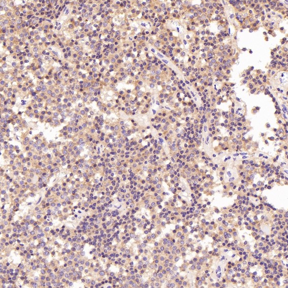
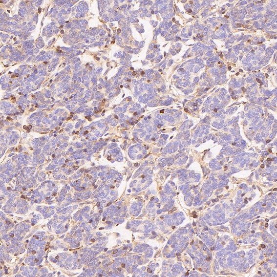
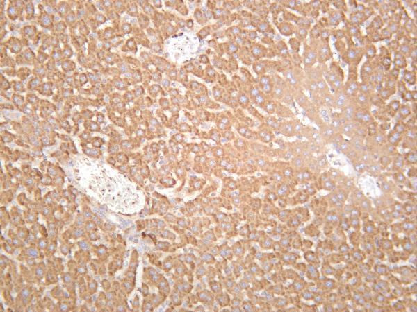
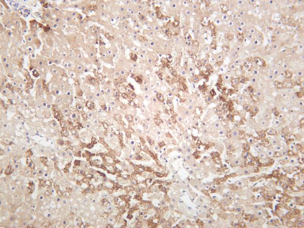
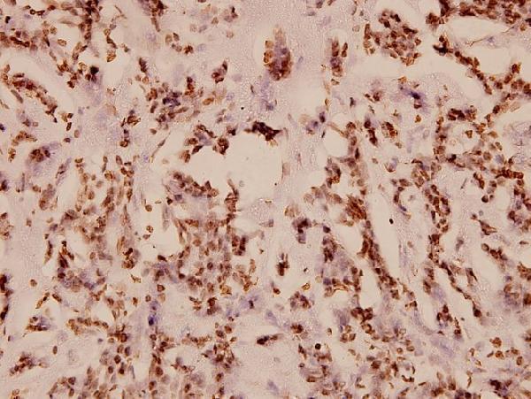
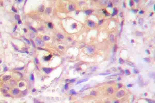
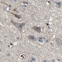
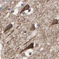


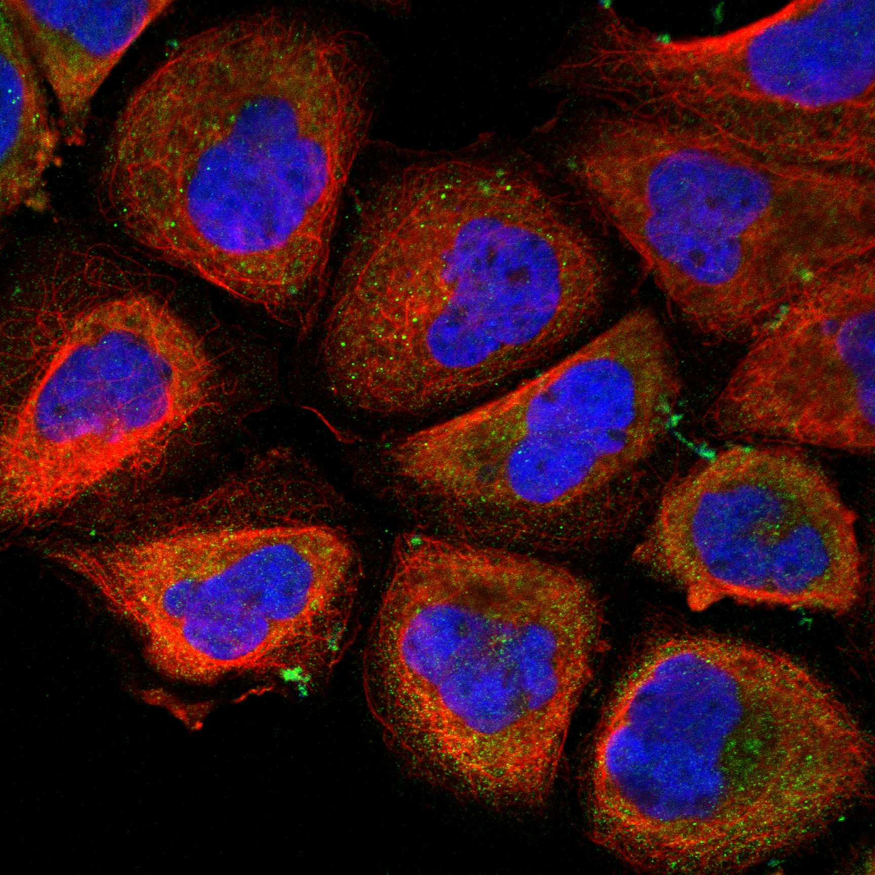
Comprehensive Human Protein Atlas IHC scoring per tissue (reliability: Enhanced — Medium consistency between antibody staining and RNA expression data.). Rows are taken directly from the HPA tissue chart — click any row's HPA link to view the source.
| Tissue | Cell type | Level | Evidence | Source |
|---|---|---|---|---|
| Adrenal gland | Glandular cells | High | Protein (IHC) | HPA → |
| Cerebellum | Purkinje cells - cytoplasm/membrane | High | Protein (IHC) | HPA → |
| Hippocampus | Neuronal cells | High | Protein (IHC) | HPA → |
| Appendix | Lymphoid tissue | Medium | Protein (IHC) | HPA → |
| Bronchus | Respiratory epithelial cells | Medium | Protein (IHC) | HPA → |
| Tissue | Cell type | Level | Evidence | Source |
|---|---|---|---|---|
| Breast | Adipocytes | Not detected | Protein (IHC) | HPA → |
| Parathyroid gland | Glandular cells | Not detected | Protein (IHC) | HPA → |
| Salivary gland | Glandular cells | Not detected | Protein (IHC) | HPA → |
| Skin | Fibroblasts | Not detected | Protein (IHC) | HPA → |
| Smooth muscle | Smooth muscle cells | Not detected | Protein (IHC) | HPA → |
Troubleshoot PRKCA staining in paraffin sections by checking retrieval, compartment, cell type, detection background, and scoring against the available tissue evidence.
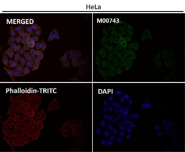
IHC images cover human and mouse tissues; the catalog lists human, mouse, and rat reactivity, and M00743 has IF images (catalog image captions; catalog reactivity and IF images).
PB9318 shows IHC in mouse intestine, PA1065 in human mammary cancer, and M00743 in human pituitary tumor (respective IHC image captions). M00743-6 shows mouse liver, A00743T495 shows human breast carcinoma, and A00743T497 shows human breast carcinoma with a phospho-PKC-pan antibody (respective IHC image captions).
Which to pick: For paraffin-section IHC, choose PB9318 for its mouse intestine example or PA1065 for its human mammary cancer example; neither caption reports the fixative (respective IHC image captions). For IF/ICC, choose M00743: its application list includes both, it is a rabbit monoclonal, and it has IF images (M00743 catalog applications, clone, and IF image captions). For work across human, mouse, and rat, PB9318 has IHC captions for all three species; M00743-6 also has human, mouse, and rat reactivity and is listed as a recombinant rabbit monoclonal (PB9318 IHC image captions; M00743-6 catalog reactivity and description).