
Click image to see more details
-

-
-
-
-
+1
Product Info Summary
| SKU: | PB9318 |
|---|---|
| Size: | 100 μg/vial |
| Reactive Species: | Human, Mouse, Rat |
| Host: | Rabbit |
| Application: | IHC, WB |
Customers Who Bought This Also Bought

Product info
Product Name
Anti-PKC alpha/PRKCA Antibody Picoband®
SKU/Catalog Number
PB9318
PB0354 is an alternative SKU for this antibody, used in previous lots.
Size
100 μg/vial
Form
Lyophilized
Description
Boster Bio Anti-PKC alpha/PRKCA Antibody Picoband® catalog # PB9318. Tested in IHC, WB applications. This antibody reacts with Human, Mouse, Rat. The brand Picoband indicates this is a premium antibody that guarantees superior quality, high affinity, and strong signals with minimal background in Western blot applications. Only our best-performing antibodies are designated as Picoband, ensuring unmatched performance.
Storage & Handling
Store at -20˚C for one year from date of receipt. After reconstitution, at 4˚C for one month. It can also be aliquotted and stored frozen at -20˚C for six months. Avoid repeated freeze-thaw cycles.
Cite This Product
Anti-PKC alpha/PRKCA Antibody Picoband® (Boster Biological Technology, Pleasanton CA, USA, Catalog # PB9318)
Host
Rabbit
Contents
Each vial contains antibody formulated with stabilizing components, 0.9 mg NaCl, 0.2 mg Na2HPO4, and 0.05 mg NaN3.
*This antibody is supplied in a stabilized formulation.
Compatibility with conjugation reactions depends on the chemistry of the conjugation method used.
For conjugation methods that are not compatible with the stabilizing components present in this formulation, a carrier-free antibody format is required.
Clonality
Polyclonal
Isotype
Rabbit IgG
Immunogen
E.coli-derived human PKC alpha recombinant protein (Position: M153-L342). Human PKC alpha shares 97% and 99% amino acid (aa) sequence identity with mouse and rat PKC alpha, respectively.
Cross-reactivity
No cross-reactivity with other proteins
Reactive Species
PB9318 is reactive to PRKCA in Human, Mouse, Rat
Observed Molecular Weight
77 kDa
Calculated molecular weight
76.8 kDa
Background of PRKCA
Protein kinase C (PKC) is the major phorbol ester receptor. Activation of PKC by calcium ions and the second messenger diacylglycerol is thought to play a central role in the induction of cellular responses to a variety of ligand-receptor systems and in the regulation of cellular responsiveness to external stimuli. Three of these, termed alpha, beta and gamma, are highly homologous. PRKCA1 is mapped to 17q22-q23.2. PRKCA1 regulates cardiac contractility and propensity toward heart failure.
Antibody Validation
Boster validates all antibodies on WB, IHC, ICC, Immunofluorescence, and ELISA with known positive control and negative samples to ensure specificity and high affinity, including thorough antibody incubations.
Application & Images
Applications
PB9318 is guaranteed for IHC, WB Boster Guarantee
Recommend Dilution
| Application | Dilution | Species |
|---|---|---|
| Western blot | 0.1-0.5μg/ml | Human, Mouse, Rat |
| Immunohistochemistry (Paraffin-embedded Section) | 0.5-1μg/ml | Human, Mouse, Rat |
Tested application
Suggested blocking solution with 5% non-fat milk or BSA; (*)Recommended protein loading: 20-40 µg per lane
Use TE buffer pH 9.0 for antigen retrieval; (*) citrate buffer pH 6.0 is an alternative.
Validation Images & Assay Conditions
Click image to see more details
Western blot analysis of PKC alpha using anti-PKC alpha antibody (PB9318).
Electrophoresis was performed on a 5-20% SDS-PAGE gel at 70V (Stacking gel) / 90V (Resolving gel) for 2-3 hours. The sample well of each lane was loaded with 30 ug of sample under reducing conditions.
Lane 1: human U87 whole cell lysates,
Lane 2: human HEL whole cell lysates,
Lane 3: human A549 whole cell lysates,
Lane 4: human U20S whole cell lysates,
Lane 5: rat brain tissue lysates,
Lane 6: mouse brain tissue lysates.
After electrophoresis, proteins were transferred to a nitrocellulose membrane at 150 mA for 50-90 minutes. Blocked the membrane with 5% non-fat milk/TBS for 1.5 hour at RT. The membrane was incubated with rabbit anti-PKC alpha antigen affinity purified polyclonal antibody (Catalog # PB9318) at 0.5 μg/mL overnight at 4°C, then washed with TBS-0.1%Tween 3 times with 5 minutes each and probed with a goat anti-rabbit IgG-HRP secondary antibody at a dilution of 1:5000 for 1.5 hour at RT. The signal is developed using an Enhanced Chemiluminescent detection (ECL) kit (Catalog # EK1002) with Tanon 5200 system. A specific band was detected for PKC alpha at approximately 77 kDa. The expected band size for PKC alpha is at 80 kDa.

Click image to see more details
IHC analysis of PKC alpha using anti-PKC alpha antibody (PB9318).
PKC alpha was detected in paraffin-embedded section of mouse intestine tissues. Heat mediated antigen retrieval was performed in citrate buffer (pH6, epitope retrieval solution) for 20 mins. The tissue section was blocked with 10% goat serum. The tissue section was then incubated with 1μg/ml rabbit anti-PKC alpha Antibody (PB9318) overnight at 4°C. Biotinylated goat anti-rabbit IgG was used as secondary antibody and incubated for 30 minutes at 37°C. The tissue section was developed using Strepavidin-Biotin-Complex (SABC)(Catalog # SA1022) with DAB as the chromogen.

Click image to see more details
IHC analysis of PKC alpha using anti-PKC alpha antibody (PB9318).
PKC alpha was detected in paraffin-embedded section of rat intestine tissues. Heat mediated antigen retrieval was performed in citrate buffer (pH6, epitope retrieval solution) for 20 mins. The tissue section was blocked with 10% goat serum. The tissue section was then incubated with 1μg/ml rabbit anti-PKC alpha Antibody (PB9318) overnight at 4°C. Biotinylated goat anti-rabbit IgG was used as secondary antibody and incubated for 30 minutes at 37°C. The tissue section was developed using Strepavidin-Biotin-Complex (SABC)(Catalog # SA1022) with DAB as the chromogen.

Click image to see more details
IHC analysis of PKC alpha using anti-PKC alpha antibody (PB9318).
PKC alpha was detected in paraffin-embedded section of human mammary cancer tissues. Heat mediated antigen retrieval was performed in citrate buffer (pH6, epitope retrieval solution) for 20 mins. The tissue section was blocked with 10% goat serum. The tissue section was then incubated with 1μg/ml rabbit anti-PKC alpha Antibody (PB9318) overnight at 4°C. Biotinylated goat anti-rabbit IgG was used as secondary antibody and incubated for 30 minutes at 37°C. The tissue section was developed using Strepavidin-Biotin-Complex (SABC)(Catalog # SA1022) with DAB as the chromogen.

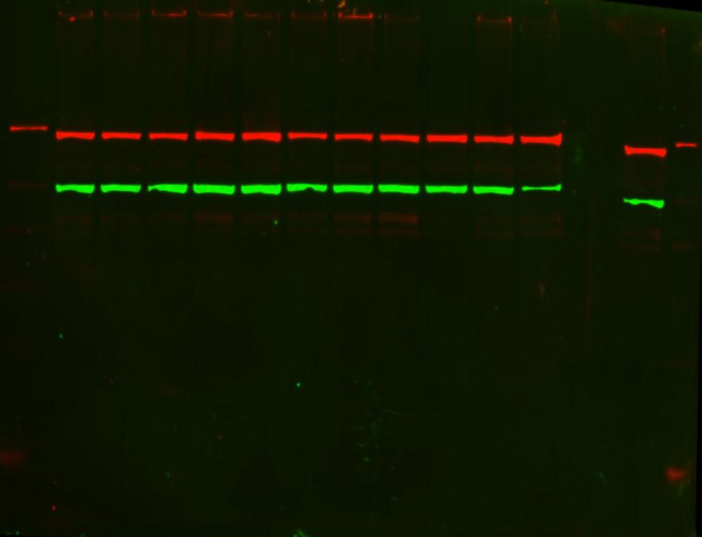
Click image to see more details
Western blot analysis of PKC alpha/PRKCA using anti-PKC alpha/PRKCA Antibody (PB9318).
Electrophoresis was performed on a 5-20% SDS-PAGE gel at 70V (Stacking gel) / 90V (Resolving gel) for 2-3 hours. The sample well of each lane was loaded with 30 ug of sample under reducing conditions.
After electrophoresis, proteins were transferred to a nitrocellulose membrane at 150 mA for 50-90 minutes. Blocked the membrane with 2.5% BSA on 1X-TBST 1.5 hour at RT. The membrane was incubated with rabbit anti-PKC alpha/PRKCA Antibody (PB9318) at 1:1000 (in 0.5%l BSA containing 1X-TBST) and overnight at 4°C, then washed with TBS-0.1%Tween 3 times with 5 minutes each and probed with a donkey anti-rabbit Ig-Cy5 secondary antibody at a dilution of 1:1000 (in 0.5%l BSA containing 1X-TBST) at 25°C for 2 h. The signal is developed using an CyDye. A specific band was detected for PKC alpha/PRKCA at approximately 76 kDa. The expected band size for PKC alpha/PRKCA is at 76 kDa.
Specific Publications For Anti-PKC alpha/PRKCA Antibody Picoband® (PB9318)
Loading publications
Recommended Resources
Here are featured tools and databases that you might find useful.
- Boster's Pathways Library
- Protein Databases
- Bioscience Research Protocol Resources
- Data Processing & Analysis Software
- Photo Editing Software
- Scientific Literature Resources
- Research Paper Management Tools
- Molecular Biology Software
- Primer Design Tools
- Bioinformatics Tools
- Phylogenetic Tree Analysis
Customer Reviews
Have you used Anti-PKC alpha/PRKCA Antibody Picoband®?
Share your experimental results or join a short interview to earn up to $1,000 in product credits or other rewards.
2 Reviews For Anti-PKC alpha/PRKCA Antibody Picoband®
Great PKC antibody for IHC
Excellent
C Parson
Verified customer
Submitted 2020-03-23
A Good PKC Alpha Antibody for IP and WB--Ramaz Geguchadze, University of New England, Biomedical Sciences, Research Scientist
Excellent
Source: Biocompare.com
| SKU | PB9318 |
|---|---|
| Application | Western Blot |
| Sample | Dorsal root ganglia (neuronal cells) |
| Detection | CyDye |
"We used Anti-PKC Alpha Picoband™ Antibody for immunoprecipitation experiment from neuronal cells in order to characterize PKC Alpha expression in different conditions. Strong positive band with right mass. Works great without optimization! Good quality and ready for publish Ab."
Ramaz Geguchadze
Verified customer
Submitted 2020-01-10
Customer Q&As
Have a question?
Find answers in Q&As, reviews.
Can't find your answer?
Submit your question
1 Customer Q&As for Anti-PKC alpha/PRKCA Antibody Picoband®
Question
We are currently using anti-PKC alpha/PRKCA antibody PB9318 for rat tissue, and we are content with the IHC-P results. The species of reactivity given in the datasheet says human, mouse, rat. Is it true that the antibody can work on primate tissues as well?
Verified Customer
Verified customer
Asked: 2020-04-29
Answer
The anti-PKC alpha/PRKCA antibody (PB9318) has not been validated for cross reactivity specifically with primate tissues, but there is a good chance of cross reactivity. We have an innovator award program that if you test this antibody and show it works in primate you can get your next antibody for free. Please contact me if I can help you with anything.
Boster Scientific Support
Answered: 2020-04-29
