This website uses cookies to ensure you get the best experience on our website.
- Table of Contents
17 Q&As
4 Q&As
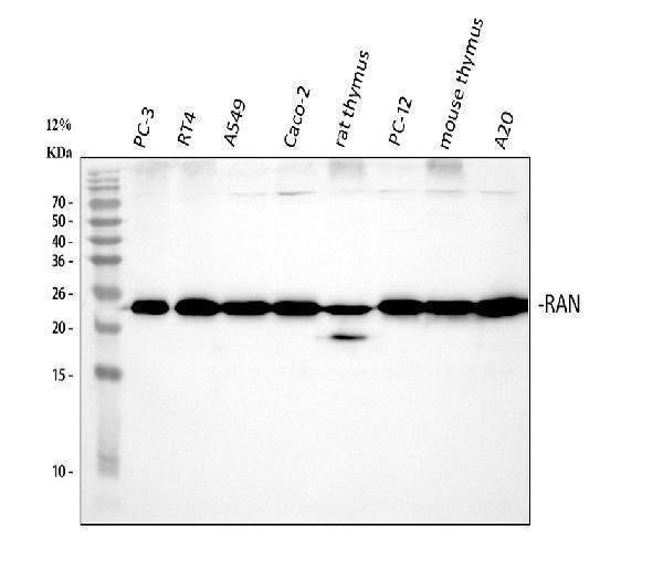
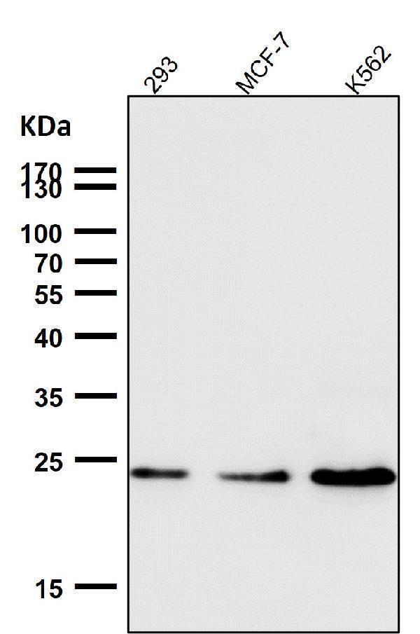
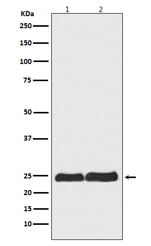
Facts about GTP-binding nuclear protein Ran.
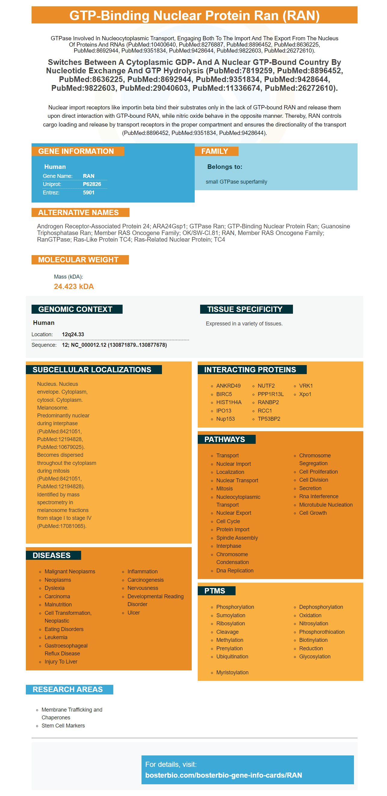
GTPase involved in nucleocytoplasmic transport, Engaging both to the import and the export from the nucleus of proteins and RNAs (PubMed:10400640, PubMed:8276887, PubMed:8896452, PubMed:8636225, PubMed:8692944, PubMed:9351834, PubMed:9428644, PubMed:9822603, PubMed:26272610).
Switches between a cytoplasmic GDP- and a nuclear GTP-bound country by nucleotide exchange and GTP hydrolysis (PubMed:7819259, PubMed:8896452, PubMed:8636225, PubMed:8692944, PubMed:9351834, PubMed:9428644, PubMed:9822603, PubMed:29040603, PubMed:11336674, PubMed:26272610).Nuclear import receptors like importin beta bind their substrates only in the lack of GTP-bound RAN and release them upon direct interaction with GTP-bound RAN, while nitric oxide behave in the opposite manner. Thereby, RAN controls cargo loading and release by transport receptors in the proper compartment and ensures the directionality of the transport (PubMed:8896452, PubMed:9351834, PubMed:9428644).
Human | |
---|---|
Gene Name: | RAN |
Uniprot: | P62826 |
Entrez: | 5901 |
Belongs to: |
---|
small GTPase superfamily |
Androgen receptor-associated protein 24; ARA24Gsp1; GTPase Ran; GTP-binding nuclear protein Ran; guanosine triphosphatase Ran; member RAS oncogene family; OK/SW-cl.81; RAN, member RAS oncogene family; RanGTPase; Ras-like protein TC4; Ras-related nuclear protein; TC4
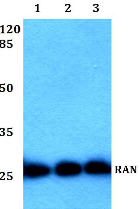
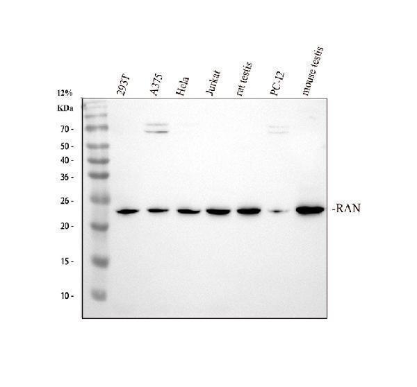
Mass (kDA):
24.423 kDA
Human | |
---|---|
Location: | 12q24.33 |
Sequence: | 12; NC_000012.12 (130871879..130877678) |
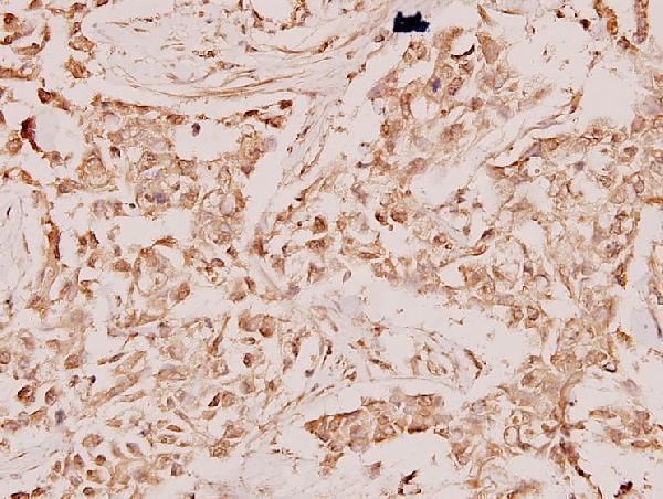
Expressed in a variety of tissues.
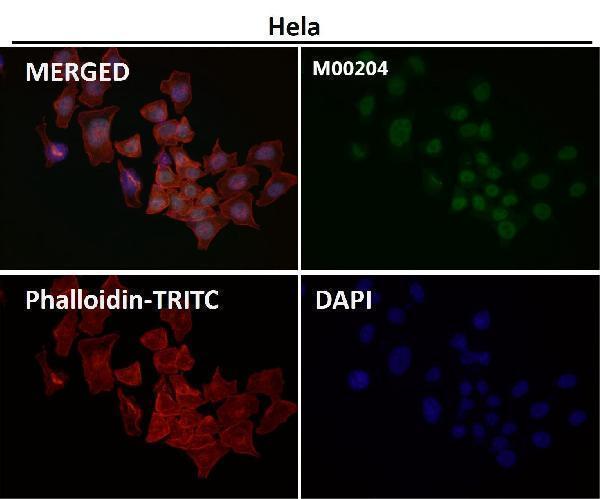
Nucleus. Nucleus envelope. Cytoplasm, cytosol. Cytoplasm. Melanosome. Predominantly nuclear during interphase (PubMed:8421051, PubMed:12194828, PubMed:10679025). Becomes dispersed throughout the cytoplasm during mitosis (PubMed:8421051, PubMed:12194828). Identified by mass spectrometry in melanosome fractions from stage I to stage IV (PubMed:17081065).
If you are using a Boster product for your research, you've probably asked yourself how to get the best results. After all, it's important to know the best uses for the RAN Marker, right? Here are a few tips. You can use the RAN Marker for your research purposes, and it's possible to get product credits by submitting results of species and applications.
The RAN Marker is a highly specific biomarker for the cytokine IL-17A. It has many applications in cancer research, and many studies rely on the results of these tests. Boster Bio offers a wide array of antibodies and test kits for a variety of applications, including cancer research, pharmacology, and immunology. The company's antibodies are tested using human, monkey, and rabbit samples to ensure their quality and safety.
If you're looking for high-affinity primary antibodies, look no further than Boster Bio. These antibodies are highly cited and have been validated using ELISA, immunohistochemistry, and Western Blotting. Boster's high-affinity primary antibodies are perfect for a variety of applications. And if you're using them in a ELISA kit, they can be even more powerful.
When you use primary antibodies, you're actually using immunoglobulins that are only specific to your target antigen. This is known as a monoclonal antibody, and its quality is generally measured in two ways: its affinity and specificity. A high affinity primary antibody will be highly specific and will bind only to the antigen of interest. Good primary antibodies will purify, measure, and detect the antigen of interest.
MBP-Linker-scFv was tested against IFN-g via ELISA. This was carried out to determine its specificity. It also showed no cross-reactivity with other antigens. This affinity constant was calculated from measured data, which indicated that the MBP-Lk-scFv is highly specific. In a second step, MBP-Linker-scFv was used to perform affinity measurements by ELISA.
Antibody titers against influenza were determined by measuring inhibition of cell death in MDCK cells after 50% Tissue culture infectious doses. This was a robust method to confirm that these antibodies are highly specific to influenza virus. The results are based on preliminary data, and the results will likely differ in future research. However, these results suggest the potential value of high-affinity primary antibodies for OAS. There are other benefits of high-affinity primary antibodies as well.
In this method, 8 HAU of virus were lysed and incubated with 10ug/ml of the mAb. Protein-A-Sepharose was then used to purify the mAb. The eluted mAb was then separated from protein-A by boiling in Laemmli buffer. Next, the mAb was tested on 12% Tris-Glycine polyacrylamide gels for immunoreactivity. After this, the positive supernatants underwent limiting-dilution subcloning. Finally, the selected antibodies were subjected to further application testing.
If you want to use secondary antibodies, you may want to choose one from Boster Bio. These antibodies are purified from antiserum, and are conjugated with Cy3 to allow for detection, quantification, and localization. They are suitable for IHC and ICC/IF as well as Western Blotting. These antibodies are suitable for many research applications, including those in immunohistochemistry, cancer research, and developmental biology.
Secondary antibodies are made by immunizing a host animal with an antibody from a different species. The primary antibody is a rabbit polyclonal antibody. The secondary antibody is then raised in a different species. The specificity of the antibody depends on the characteristics of the immunization animal. Boster Bio Secondary antibodies using the RAN marker are suitable for immunolabeling research. These are suitable for both animal and human research.
Using a secondary antibody in multiplex fluorescent western blot experiments requires appropriate secondary antibodies. In addition, it is important to select the correct fluorophore for the imager. The brightest fluorophore should be used for the target protein that is least abundant. The most fluorophores should be used for highly abundant targets. Autofluorescence may be an issue at shorter wavelengths, but is less of a problem above 550 nm. It can decrease the signal-to-noise ratio.
Indirect staining involves the use of fluorescent secondary antibodies to detect target proteins. For instance, if a primary antibody was raised in a rabbit, then the secondary antibody must be raised against a specific anti-rabbit immunoglobulin. If the antibody is conjugated with an enzyme, it must be used with the right substrate. If it is conjugated with an enzyme, it must use a buffer that is free of phosphate.
To troubleshoot the RAN Marker, you need to know how it works. It can be damaged if the LED doesn't function. However, there are some simple fixes for this problem. These solutions will work in most cases. Troubleshooting the RAN Marker can be simple. Keep reading to learn more. The marker is equipped with a LED, which can be easily fixed. Troubleshooting the RAN Marker will prevent it from malfunctioning.
Open RAN is a key technology for 5G networks, as it removes the need for proprietary hardware infrastructure. With Open RAN, operators can source radio components from a variety of vendors, creating a powerful, vendor-agnostic network. Read on for more information on Open RAN and how it will benefit mobile operators. Read on to discover how Open RAN is helping mobile operators meet the demands of the 21st century.
The RAN's employee benefits program provides health insurance coverage at no cost to employees and their dependents. This includes children, spouses, and domestic partners. Furthermore, RAN contributes to an Employee's Health Savings Account, and enables employees to set up a Dependent Care Reimbursement account. While these programs are limited to mobile users, they do provide substantial coverage and reduce costs for RAN operators.
PMID: 2108320 by Drivas G.T., et al. Characterization of four novel ras-like genes expressed in a human teratocarcinoma cell line.
PMID: 1855255 by Matsumoto T., et al. Premature initiation of mitosis in yeast lacking RCC1 or an interacting GTPase.