This website uses cookies to ensure you get the best experience on our website.
- Table of Contents
4 Citations 15 Q&As
1 Citations 6 Q&As
2 Citations 17 Q&As
1 Citations
Facts about Mothers against decapentaplegic homolog 3.
Also can form a SMAD3/SMAD4/JUN/FOS complex at the AP-1/SMAD site to regulate TGF-beta-mediated transcription. Has an inhibitory effect on wound healing probably by modulating both growth and migration of primary keratinocytes and by changing the TGF- mediated chemotaxis of monocytes.
Human | |
---|---|
Gene Name: | SMAD3 |
Uniprot: | P84022 |
Entrez: | 4088 |
Belongs to: |
---|
dwarfin/SMAD family |
DKFZp586N0721; DKFZp686J10186; hMAD-3; hSMAD3; HsT17436; JV15-2MGC60396; MAD homolog 3; mad protein homolog; MAD, mothers against decapentaplegic homolog 3 (Drosophila); MAD, mothers against decapentaplegic homolog 3; mad3; MADH3mad homolog JV15-2; mothers against decapentaplegic homolog 3; Mothers against DPP homolog 3; SMA- and MAD-related protein 3; SMAD 3; SMAD family member 3HSPC193; SMAD, mothers against DPP homolog 3 (Drosophila); SMAD, mothers against DPP homolog 3; Smad3
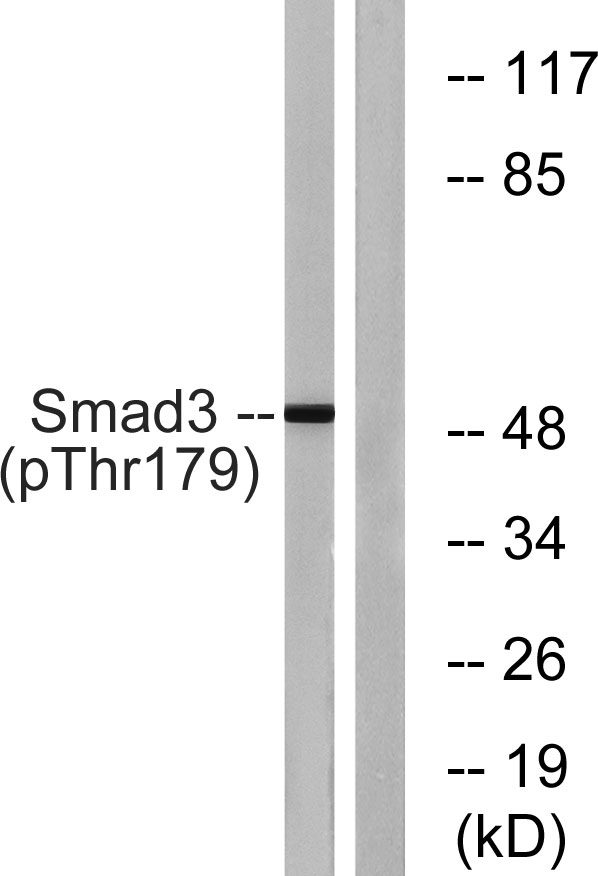
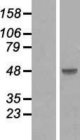
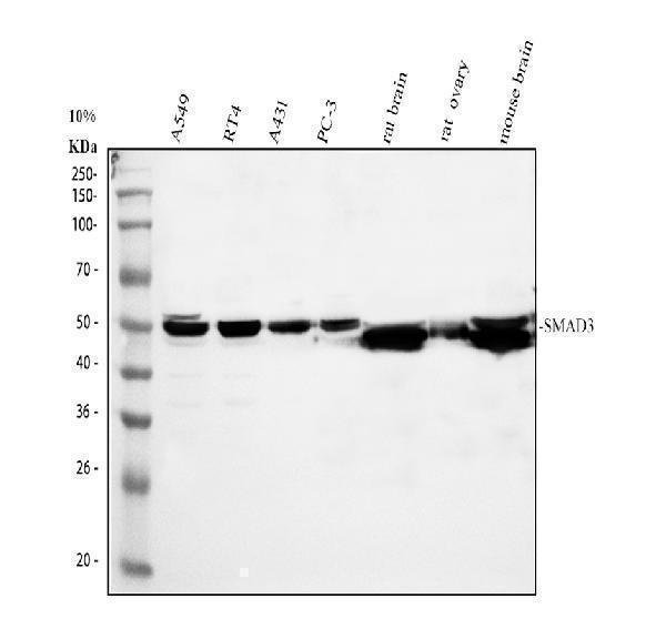
Mass (kDA):
48.081 kDA
Human | |
---|---|
Location: | 15q22.33 |
Sequence: | 15; NC_000015.10 (67065602..67195195) |
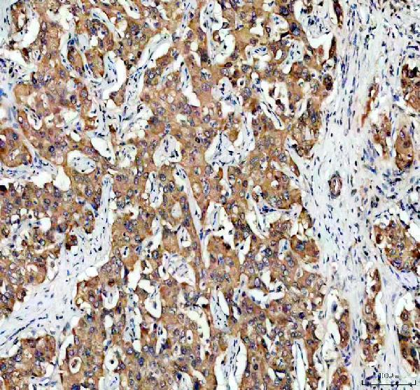
Cytoplasm. Nucleus. Cytoplasmic and nuclear in the absence of TGF-beta. On TGF-beta stimulation, migrates to the nucleus when complexed with SMAD4 (PubMed:15799969). Through the action of the phosphatase PPM1A, released from the SMAD2/SMAD4 complex, and exported out of the nucleus by interaction with RANBP1 (PubMed:16751101, PubMed:19289081). Co-localizes with LEMD3 at the nucleus inner membrane (PubMed:15601644). MAPK-mediated phosphorylation appears to have no effect on nuclear import (PubMed:19218245). PDPK1 prevents its nuclear translocation in response to TGF-beta (PubMed:17327236).
The SMAD3 Marker is a highly specific antibody that binds to SMAD3 receptors in cells. It is particularly useful for the analysis of cell membrane proteins, such as fibroblasts. The SMAD3 marker can be used for a variety of research purposes, including the diagnosis and treatment of various diseases. Researchers can submit their samples and results for species, applications, and special samples for publication on the Boster website, which gives them credit for their work. In addition to enabling researchers to submit their data for publication, Boster Bio offers product credits for their work.
If you want to test the SMAD3 marker in your samples, you should use a kit from Boster Bio. These kits come with a 96-well plate for the relevant biomarker panel, and they include all the reagents you need for the test. One kit is capable of testing 80 samples in 2.5 hours. The SMAD3 marker is involved in protein, lipid, and cellular energy production. It also participates in carbon and nitrogen donation.
The SMAD3 marker is a protein encoded by the SMAD3 gene. The protein transmits chemical signals from the cell's surface to its nucleus. TGF-b activates a group of SMAD proteins, including SMAD3. The SMAD protein complex then moves to the nucleus where it binds to specific regions of DNA, controlling specific genes.
SMAD3 interacts with its transcriptional corepressor SKI, which controls the expression of SMAD3 and its cofactor FOXh2. Its hydrophobic patches are composed of multiple subsets, and the tethers bind to specific hydrophobic patches to regulate SMAD3 activity. The resulting methylation is responsible for controlling the output of TGF-b signaling.
The SMAD3 marker is expressed in the nuclei of GC-enveloped oocytes, including in primordial follicles. The mechanism of this arrest is unknown, but the TGF-b-activated transcription factor SMAD3 is expressed in primordial GC nuclei along with cell cycle proteins. SMAD3 colocalises with P27 and is co-precipitated with DNA sequences that are upstream of Ccnd2 and Myc transcription start sites.
The Cell-based SMAD3 ELSA kit from Boster Bio recognizes endogenous levels of total Smad3 protein in human cells. The kit identifies Smad3 proteins from indicated species, as well as homologous proteins from other species. The kit can be used to screen for inhibitors, activators, and their effects on human cells. Among the many uses of the kit is to measure the concentration of Smad3 protein.
The sensitivity of the ELISA kit for SMAD3 is very high. Its detection limit is 1.5ng per lane under reducing conditions. The results are positive for tested and predicted species, based on sequence similarities. For staining formalin/paraffin sections, it is required to boil the section for 20 minutes in 10mM citrate buffer, pH 6.0. Other applications are not yet tested. Therefore, the end-user should determine dilutions.
The test for the cell-based SMAD3 ELISA from Boster Bio is extremely sensitive and accurate. Using this kit, researchers can detect the level of SMAD3 protein in human cell membranes in a matter of minutes. The method requires minimal set-up and has a low false-positive rate, which may be a sign of a faulty ELISA.
The method of incubating the Boster Bio SMAD3 marker requires the use of primary and secondary antibodies. The primary antibody is a rabbit polyclonal antibody raised in a different species. The secondary antibody is generated by immunizing a different host with the primary antibody. The resulting secondary antibody has a specificity that is dependent on the characteristics of the primary antibody.
The sample preparation and incubation methods involve the use of fluorescent dyes and primary antibodies. Flow cytometry is an advanced cell biology technique that uses a laser-based system to profile cells in a heterogeneous fluid mixture. The technique is also known as FACS (fluorescence-activated cell sorting). The Boster Bio SMAD3 marker incubation with secondary antibodies has been designed to facilitate this process by offering comprehensive step-by-step protocols for flow cytometry. The Boster Bio protocol also includes product datasheets and sample preparation guides.
The rabbit anti-TJP1 antibody (PB9234) was used to detect TJP1 in human intestinal cancer tissue. Antibody retrieval was carried out using a citrate buffer and pH6 epitope retrieval solution. Then, a tissue section was blocked with 10% goat serum. The next step was incubation with the rabbit anti-TJP1 antibody and biotinylated goat anti-rabbit IgG as the secondary antibody. After incubation with the secondary antibody, the signal was visualized by an enhanced chemiluminescence (EC) detection kit. The western band density was quantified using the Quantity One 4.6 software. In this step, the specific band was detected at approximately 220-240KD, which was significantly higher than expected.
We have found that immunoreactive traces on the membrane can be quantified. The protein levels of these antigens are proportional to the concentration of MDA. Thus, immunoreactive traces on the membrane are a quantitative method to determine lipoperoxidative alterations. The immunological probe detects traces of membrane damages regardless of the cell type and pro-oxidant condition. This article summarizes the main findings of this study.
The SMAD3 gene encodes an important component of the TGF-beta signaling pathway, which plays a key role in bone formation. Overexpression of Smad3 inhibits bone morphogenetic protein-2 (BMP2)-induced ALP activity in ST-2 cells. Furthermore, Smad3 expression inhibits the commitment of pluripotent mesenchymal stem cells (PMSCs) to osteoblasts.
In addition to its role in bone formation, myostatin has a role in the regulation of bone resorption. When inhibition of this gene occurs, bone resorption is reduced and bone formation improves. Inactivation of the anti-myostatin decoy receptor results in increased bone mass. Moreover, osteoclasts differentiate from tissue resident macrophages. RANKL, a gene involved in osteoclast differentiation, is highly expressed in pannus tissue. Several other factors also play a role in osteoclast differentiation.
Regulation of bone formation by the SMAD-3 marker may occur by inhibiting the expression of Cdk6. This gene is required for osteoblast differentiation. However, overexpression of Cdk6 inhibits osteoblast differentiation. Thus, downregulation of Cdk6 may be required for efficient bone formation. However, further studies are needed to find the specific transcriptional repressor that regulates Cdk6.
SMAD3 is a nuclear receptor that regulates gene expression through binding to its promoter region. This binding activity is mediated by Smad2, Smad3, and MeCP2 proteins. Inhibition of coactivator recruitment can be used to target specific target genes or pathways in a cell. This antibody has several applications. It can be used to identify genes that express SMADs and their coactivators.
The research team includes Dorfman, Mauricio D.; Kerr, Bredford; Garcia-Rudaz, Cecilia; Paredes, Alfonso H.; and Dissen, Gregory A. All of these researchers are involved in various aspects of drug development. They believe the SMAD3 marker has potential applications in inhibiting coactivator recruitment.
PMID: 8774881 by Zhang Y., et al. Receptor-associated Mad homologues synergize as effectors of the TGF- beta response.
PMID: 8673135 by Riggins G.J., et al. Mad-related genes in the human.
*More publications can be found for each product on its corresponding product page