Click image to see more details
-

-
-
-
-
+6
Product Info Summary
| SKU: | M00059 |
|---|---|
| Size: | 100 μl |
| Reactive Species: | Human, Mouse, Rat |
| Host: | Rabbit |
| Application: | Flow Cytometry, IF, IHC, ICC, WB |
Customers Who Bought This Also Bought

Product info
Product Name
Anti-Smad3 Rabbit Monoclonal Antibody
SKU/Catalog Number
M00059
BM3919 is an alternative SKU for this antibody, used in previous lots.
Size
100 μl
Form
Liquid
Description
Boster Bio Anti-Smad3 Rabbit Monoclonal Antibody catalog # M00059. Tested in WB, IHC, ICC/IF, Flow Cytometry applications. This antibody reacts with Human, Mouse, Rat.
Storage & Handling
Store at -20°C for one year. For short term storage and frequent use, store at 4°C for up to one month. Avoid repeated freeze-thaw cycles.
Cite This Product
Anti-Smad3 Rabbit Monoclonal Antibody (Boster Biological Technology, Pleasanton CA, USA, Catalog # M00059)
Host
Rabbit
Contents
Rabbit IgG in stabilizing components, phosphate buffered saline, pH 7.4, 150mM NaCl, 0.02% sodium azide and 50% glycerol.
*This antibody is supplied in a stabilized formulation.
Compatibility with conjugation reactions depends on the chemistry of the conjugation method used.
For conjugation methods that are not compatible with the stabilizing components present in this formulation, a carrier-free antibody format is required.
Clonality
Monoclonal
Clone Number
FG-19
Isotype
Rabbit IgG
Immunogen
A synthesized peptide derived from human Smad3
Reactive Species
M00059 is reactive to SMAD3 in Human, Mouse, Rat
Observed Molecular Weight
50 kDa
Calculated molecular weight
48.1 kDa
Antibody Validation
Boster validates all antibodies on WB, IHC, ICC, Immunofluorescence, and ELISA with known positive control and negative samples to ensure specificity and high affinity, including thorough antibody incubations.
Application & Images
Applications
M00059 is guaranteed for Flow Cytometry, IF, IHC, ICC, WB Boster Guarantee
Recommend Dilution
WB 1:500-2000
IHC 1:50-200
ICC/IF 1:50-200
FC 1:50
Tested application
Suggested blocking solution with 5% non-fat milk or BSA; (*)Recommended protein loading: 20-40 µg per lane
Use TE buffer pH 9.0 for antigen retrieval; (*) citrate buffer pH 6.0 is an alternative.
Validation Images & Assay Conditions
Click image to see more details
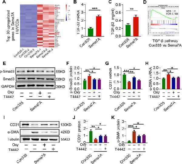
TGF-β2 gene expression and TGF-Smad signaling are augmented in Sema7A-HUVECs. a The top 30 upregulated genes in Sema7A-HUVECs compared with Con335-HVUECs. b TGF-β2 mRNA level was analyzed by qPCR normalized to GAPDH. Fold changes are shown. Data are mean ± SEM, N = 3, *** p < 0.001. c The concentration of TGF-β2 in cell supernatant was detected by ELISA. N = 10. Unpaired two-tailed Student’s t tests was used to analysis the data. Data are mean ± SEM, ** p < 0.01. d GSEA based on gene ontology (GO) pathway database showed TGF-β signaling pathway was enrich in Sema7A-HUVECs. e , f Cells were treated with Oxymatrine (Oxy) (20 μmol/l) or T4442 (1 μg/ml) and the lysates were analyzed by western blotting for Smad3 phosphorylation, normalized to total Smad3. Data are mean ± SEM, N = 3, * p < 0.05; ** p < 0.01. g , h CD31 and α-SMA mRNA in cells treated with inhibitors were analyzed by qPCR normalized to GAPDH. Fold changes are shown. Data are mean ± SEM, N = 3, * p < 0.05; ** p < 0.01; *** p < 0.001. i – k CD31 and α-SMA proteins in cells treated with inhibitors were analysis by western blotting, normalized to tubulin. Data are mean ± SEM, N = 3, * p < 0.05; ** p < 0.01; *** p < 0.001. T4442: TGF-β2 blocking antibody; Oxymatrine (Oxy): TGF/Smad signaling pathway inhibitor.
Index in PubMed under a CC BY license. PMID: 32826874

Click image to see more details
Inhibition of ATF3 reduced TGF-β2 expression and Sema7A-induced EndMT. a ATF3 mRNA level was analyzed by qPCR normalized to GAPDH. Fold changes are shown. Data are mean ± SEM, N = 3, *** p < 0.001 b ATF3 protein expression were analyzed by western blotting, normalized to tubulin. Data are mean ± SEM, N = 3, *** p < 0.001. c TGF-β2 mRNA level was analyzed by qPCR normalized to GAPDH. Fold changes are shown. Data are mean ± SEM, N = 3, * p < 0.05; ** p < 0.01. d The concentration of TGF-β2 in cell supernatant was detected by ELISA among Con335-HUVECs, Sema7A-HUVECs, and Sema7A-HUVECs + ATF3-siRNA. N = 10. Data are mean ± SEM, * p < 0.05; *** p < 0.001. e Chip-qPCR product in agarose gel electrophoresis. f Chip-qPCR TGF-β2 percentage of input in con335-HUVECs and Sema7A - HUVECs were analyzed by qPCR normalized to IgG. Data are mean ± SEM, N = 3, ** p < 0.01. g Schematic graph of the constructed reporter plasmid. TGF-β2 mut indicates the TGF-β2 mutation promoter region in ATF3 binding site. The mutated nucleotides in TGF-β2 fragments are in red letters. h Luciferase reporter assays were performed on HEK 293 T cells. Data are mean ± SEM, N = 3, ** p < 0.01 vs negative control. i , j ATF3-overexpression plasmid was transfected to HUVECs, and the mRNA levels of TGF - β2 and TGF-β1 were performed by qPCR normalized to GAPDH. Fold changes are shown. Data are mean ± SEM, N = 3, ** p < 0.01. k , l P-Smad3 protein in cells treated with siRNA was analyzed by Western blotting, normalized to total Smad3. Data are mean ± SEM, N = 3, * p < 0.05; ** p < 0.01. m – q CD31 and α-SMA RNA and proteins in cells treated with siRNA or control was analyzed by qPCR and western blotting. Data are mean ± SEM, N = 3, * p < 0.05; ** p < 0.01; *** p < 0.001.
Index in PubMed under a CC BY license. PMID: 32826874

Click image to see more details
Β1 integrin mediates Sema7A signal to TGF-β2 via ATF3. a Cells were treated with β1 integrin antibody (P5D2), and ATF3 mRNA level was analyzed by qPCR normalized to GAPDH. Fold changes are shown. Data are mean ± SEM, N = 3, * p < 0.05; ** p < 0.0.1. b ATF3 protein expression was analyzed by western blotting, normalized to tubulin. Data are mean ± SEM, N = 3, * p < 0.05; ** p < 0.01. c TGF-β2 mRNA level was analyzed by qPCR normalized to GAPDH. Fold changes are shown. Data are mean ± SEM, N = 3, * p < 0.05; ** p < 0.0.1. d The concentration of TGF-β2 in cell supernatant was detected by ELISA for Con335-HUVECs, Sema7A-HUVECs, and Sema7A-HUVECs + P5D2. Data are mean ± SEM, N = 10, * p < 0.05; *** p < 0.001. e , f Smad3 phosphorylation were analyzed by western blotting, normalized to total Smad3. Data are mean ± SEM, N = 3, * p < 0.05. g , h CD31 and α-SMA mRNA level were analyzed by qPCR normalized to GAPDH. Fold changes are shown. Data are mean ± SEM, N = 3, * p < 0.05; ** p < 0.01; *** p < 0.001. i – k CD31 and α-SMA proteins were analyzed by western blotting, normalized to tubulin. Data are mean ± SEM, N = 3, * p < 0.05; ** p < 0.01; *** p < 0.001. l ATF3 overexpression plasmid was transfected into P5D2 incubated Sema7A-HUVECs, and TGF-β2 mRNA expression in Sema7A-HUVECs + P5D2 and Sema7A-HUVECs + P5D2 + ATF3 were analyzed by qPCR normalized to GAPDH. Fold changes are shown. Data are mean ± SEM, N = 3, ** p < 0.01. m – o CD31 and α-SMA protein expressions were analyzed by western blotting, normalized to GAPDH. Data are mean ± SEM, N = 3, ** p < 0.01 (Sema7A-HUVECs + P5D2 vs Sema7A-HUVECs + P5D2 + ATF3). P5D2 β1 integrin antibody.
Index in PubMed under a CC BY license. PMID: 32826874

Click image to see more details
Immunohistochemical analysis of paraffin-embedded Human lung large cell cancer, using the Antibody at 1:500 dilution.

Click image to see more details
Western blot analysis of Smad3 using anti-Smad3 antibody (M00059).
Electrophoresis was performed on a 10% SDS-PAGE gel at 80V (Stacking gel) / 120V (Resolving gel) for 2 hours. The sample well of each lane was loaded with 30 ug of sample under reducing conditions.
Lane 1: human Jurkat whole cell lysates,
Lane 2: human SW620 whole cell lysates,
Lane 3: human A431 whole cell lysates,
Lane 4: human Hela whole cell lysates,
Lane 5: rat lung tissue lysates,
Lane 6: rat liver tissue lysates,
Lane 7: mouse lung tissue lysates,
Lane 7: mouse liver tissue lysates.
After electrophoresis, proteins were transferred to a nitrocellulose membrane at 150 mA for 50-90 minutes. Blocked the membrane with 5% non-fat milk/TBS for 1.5 hour at RT. The membrane was incubated with rabbit anti-Smad3 antigen affinity purified monoclonal antibody (M00059) at 1:1000 overnight at 4°C, then washed with TBS-0.1%Tween 3 times with 5 minutes each and probed with a goat anti-rabbit IgG-HRP secondary antibody at a dilution of 1:5000 for 1.5 hour at RT. The signal is developed using an ECL Plus Western Blotting Substrate (Catalog # AR1196-200) with Tanon 5200 system. A specific band was detected for Smad3 at approximately 50 kDa. The expected band size for Smad3 is at 48 kDa.

Click image to see more details
Immunohistochemical analysis of paraffin-embedded Human testis cancer, using the Antibody at 1:500 dilution.

Click image to see more details
Immunohistochemical analysis of paraffin-embedded Human breast cancer, using the Antibody at 1:500 dilution.

Click image to see more details
Immunohistochemical analysis of paraffin-embedded mouse kidney, using Smad3 Antibody.

Click image to see more details
Immunofluorescent analysis using the Antibody at 1:150 dilution.

Click image to see more details
Immunofluorescent analysis of Hela cells, using Smad3 Antibody.
Specific Publications For Anti-Smad3 Rabbit Monoclonal Antibody (M00059)
Loading publications
Recommended Resources
Here are featured tools and databases that you might find useful.
- Boster's Pathways Library
- Protein Databases
- Bioscience Research Protocol Resources
- Data Processing & Analysis Software
- Photo Editing Software
- Scientific Literature Resources
- Research Paper Management Tools
- Molecular Biology Software
- Primer Design Tools
- Bioinformatics Tools
- Phylogenetic Tree Analysis
Customer Reviews
Have you used Anti-Smad3 Rabbit Monoclonal Antibody?
Share your experimental results or join a short interview to earn up to $1,000 in product credits or other rewards.
0 Reviews For Anti-Smad3 Rabbit Monoclonal Antibody
Customer Q&As
Have a question?
Find answers in Q&As, reviews.
Can't find your answer?
Submit your question
17 Customer Q&As for Anti-Smad3 Rabbit Monoclonal Antibody
Question
Our lab were well pleased with the WB result of your anti-Smad3 Rabbit Monoclonal antibody. However we have observed positive staining in cervix carcinoma cytoplasm using this antibody. Is that expected? Could you tell me where is SMAD3 supposed to be expressed?
Verified Customer
Verified customer
Asked: 2020-04-20
Answer
From what I have seen in literature, cervix carcinoma does express SMAD3. Generally SMAD3 expresses in cytoplasm. Regarding which tissues have SMAD3 expression, here are a few articles citing expression in various tissues:
Brain, Pubmed ID: 14702039
Cervix carcinoma, Pubmed ID: 18669648
Colon carcinoma, Pubmed ID: 9464505
Erythroleukemia, Pubmed ID: 23186163
Pancreas, Pubmed ID: 15489334
Placenta, Pubmed ID: 8774881
Boster Scientific Support
Answered: 2020-04-20
Question
Is there a BSA free version of anti-Smad3 Rabbit Monoclonal antibody M00059 available?
Verified Customer
Verified customer
Asked: 2020-03-10
Answer
Thanks for your recent telephone inquiry. I can confirm that some lots of this anti-Smad3 Rabbit Monoclonal antibody M00059 are BSA free. For now, these lots are available and we can make a BSA free formula for you free of charge. It will take 3 extra days to prepare. If you require this antibody BSA free again in future, please do not hesitate to contact me and I will be pleased to check which lots we have in stock that are BSA free.
Boster Scientific Support
Answered: 2020-03-10
Question
I was wanting to use your anti-Smad3 Rabbit Monoclonal antibody for IHC for rat placenta on frozen tissues, but I want to know if it has been validated for this particular application. Has this antibody been validated and is this antibody a good choice for rat placenta identification?
Verified Customer
Verified customer
Asked: 2020-03-03
Answer
As indicated on the product datasheet, M00059 anti-Smad3 Rabbit Monoclonal antibody has been validated for IF, IHC, WB on human, mouse, rat tissues. We have an innovator award program that if you test this antibody and show it works in rat placenta in IHC-frozen, you can get your next antibody for free.
Boster Scientific Support
Answered: 2020-03-03
Question
Our lab want to know about to test anti-Smad3 Rabbit Monoclonal antibody M00059 on rat placenta for research purposes, then I may be interested in using anti-Smad3 Rabbit Monoclonal antibody M00059 for diagnostic purposes as well. Is the antibody suitable for diagnostic purposes?
Verified Customer
Verified customer
Asked: 2020-01-28
Answer
The products we sell, including anti-Smad3 Rabbit Monoclonal antibody M00059, are only intended for research use. They would not be suitable for use in diagnostic work. If you have the means to develop a product into diagnostic use, and are interested in collaborating with us and develop our product into an IVD product, please contact us for more discussions.
Boster Scientific Support
Answered: 2020-01-28
Question
Would anti-Smad3 Rabbit Monoclonal antibody M00059 work for IHC with placenta?
Verified Customer
Verified customer
Asked: 2020-01-21
Answer
According to the expression profile of placenta, SMAD3 is highly expressed in placenta. So, it is likely that anti-Smad3 Rabbit Monoclonal antibody M00059 will work for IHC with placenta.
Boster Scientific Support
Answered: 2020-01-21
Question
I see that the anti-Smad3 Rabbit Monoclonal antibody M00059 works with IHC, what is the protocol used to produce the result images on the product page?
Verified Customer
Verified customer
Asked: 2019-12-24
Answer
You can find protocols for IHC on the "support/technical resources" section of our navigation menu. If you have any further questions, please send an email to support@bosterbio.com
Boster Scientific Support
Answered: 2019-12-24
Question
We have been able to see staining in mouse cervix carcinoma. Do you have any suggestions? Is anti-Smad3 Rabbit Monoclonal antibody supposed to stain cervix carcinoma positively?
Verified Customer
Verified customer
Asked: 2019-12-09
Answer
From literature cervix carcinoma does express SMAD3. From Uniprot.org, SMAD3 is expressed in amniotic fluid, placenta, colon carcinoma, brain, pancreas, cervix carcinoma, erythroleukemia, among other tissues. Regarding which tissues have SMAD3 expression, here are a few articles citing expression in various tissues:
Brain, Pubmed ID: 14702039
Cervix carcinoma, Pubmed ID: 18669648
Colon carcinoma, Pubmed ID: 9464505
Erythroleukemia, Pubmed ID: 23186163
Pancreas, Pubmed ID: 15489334
Placenta, Pubmed ID: 8774881
Boster Scientific Support
Answered: 2019-12-09
Question
Is a blocking peptide available for product anti-Smad3 Rabbit Monoclonal antibody (M00059)?
B. Bhatt
Verified customer
Asked: 2019-12-03
Answer
We do provide the blocking peptide for product anti-Smad3 Rabbit Monoclonal antibody (M00059). If you would like to place an order for it please contact support@bosterbio.com and make a special request.
Boster Scientific Support
Answered: 2019-12-03
Question
I am looking for using your anti-Smad3 Rabbit Monoclonal antibody for activation of cysteine-type endopeptidase activity involved in apoptotic process studies. Has this antibody been tested with western blotting on jurkat cell lysate? We would like to see some validation images before ordering.
Verified Customer
Verified customer
Asked: 2019-10-08
Answer
We appreciate your inquiry. This M00059 anti-Smad3 Rabbit Monoclonal antibody is validated on jurkat cell lysate, mouse kidney. It is guaranteed to work for IF, IHC, WB in human, mouse, rat. Our Boster guarantee will cover your intended experiment even if the sample type has not been be directly tested.
Boster Scientific Support
Answered: 2019-10-08
Question
Thanks for helping with my inquiry over the phone. Here are the WB image, lot number and protocol we used for placenta using anti-Smad3 Rabbit Monoclonal antibody M00059. Let me know if you need anything else.
Verified Customer
Verified customer
Asked: 2019-08-16
Answer
We appreciate the data. You have provided everything we needed. Our lab team are working to resolve your inquiry as quickly as possible, and we appreciate your patience and understanding! Please let me know if there is anything you need in the meantime.
Boster Scientific Support
Answered: 2019-08-16
Question
We are currently using anti-Smad3 Rabbit Monoclonal antibody M00059 for human tissue, and we are happy with the WB results. The species of reactivity given in the datasheet says human, mouse, rat. Is it possible that the antibody can work on dog tissues as well?
R. Moore
Verified customer
Asked: 2019-04-17
Answer
The anti-Smad3 Rabbit Monoclonal antibody (M00059) has not been tested for cross reactivity specifically with dog tissues, but there is a good chance of cross reactivity. We have an innovator award program that if you test this antibody and show it works in dog you can get your next antibody for free. Please contact me if I can help you with anything.
Boster Scientific Support
Answered: 2019-04-17
Question
I have a question about product M00059, anti-Smad3 Rabbit Monoclonal antibody. I was wondering if it would be possible to conjugate this antibody with biotin. I would need it to be without BSA or sodium azide. I am planning on using a buffer exchange of sodium azide with PBS only. Would there be problems for me to conjugate the antibody and store it in -20 degrees in small aliquots?
Verified Customer
Verified customer
Asked: 2017-11-21
Answer
We suggest not storing this antibody with PBS buffer only in -20 degrees. If you want to store it in -20 degrees it is best to add some cryoprotectant like glycerol. If you want carrier free M00059 anti-Smad3 Rabbit Monoclonal antibody, we can provide it to you in a special formula with trehalose and/or glycerol. These molecules will not interfere with conjugation chemistry and provide a good level of protection for the antibody from degradation. Please be sure to specify this in your purchase order.
Boster Scientific Support
Answered: 2017-11-21
Question
Does M00059 anti-Smad3 Rabbit Monoclonal antibody work on parafin embedded sections? If so, which fixation method do you recommend we use (PFA, paraformaldehyde, other)?
H. Collins
Verified customer
Asked: 2015-09-11
Answer
You can see on the product datasheet, M00059 anti-Smad3 Rabbit Monoclonal antibody as been tested on IHC. It is best to use PFA for fixation because it has better tissue penetration ability. PFA needs to be prepared fresh before use. Long term stored PFA turns into formalin, as the PFA molecules congregate and become formalin.
Boster Scientific Support
Answered: 2015-09-11
Question
Is this M00059 anti-Smad3 Rabbit Monoclonal antibody reactive to the isotypes of SMAD3?
S. Carter
Verified customer
Asked: 2014-10-27
Answer
The immunogen of M00059 anti-Smad3 Rabbit Monoclonal antibody is A synthesized peptide derived from human Smad3. Could you tell me which isotype you are interested in so I can help see if the immunogen is part of this isotype?
Boster Scientific Support
Answered: 2014-10-27
Question
See attached the WB image, lot number and protocol we used for placenta using anti-Smad3 Rabbit Monoclonal antibody M00059. Please let me know if you require anything else.
B. Wu
Verified customer
Asked: 2014-01-07
Answer
Thank you very much for the data. Our lab team are working to resolve this as quickly as possible, and we appreciate your patience and understanding! You have provided everything we needed. Please let me know if there is anything you need in the meantime.
Boster Scientific Support
Answered: 2014-01-07
Question
We purchased anti-Smad3 Rabbit Monoclonal antibody for IHC on erythroleukemia in a previous project. I am using mouse, and We are going to use the antibody for IF next. We want examining erythroleukemia as well as amniotic fluid in our next experiment. Could give a recommendation on which antibody would work the best for IF?
J. Evans
Verified customer
Asked: 2013-10-18
Answer
I have checked the website and datasheets of our anti-Smad3 Rabbit Monoclonal antibody and it appears that M00059 has been tested on mouse in both IHC and IF. Thus M00059 should work for your application. Our Boster satisfaction guarantee will cover this product for IF in mouse even if the specific tissue type has not been validated. We do have a comprehensive range of products for IF detection and you can check out our website bosterbio.com to find out more information about them.
Boster Scientific Support
Answered: 2013-10-18
Question
Would anti-Smad3 Rabbit Monoclonal antibody M00059 work on pig IF with colon carcinoma?
P. Patel
Verified customer
Asked: 2013-03-04
Answer
Our lab technicians have not validated anti-Smad3 Rabbit Monoclonal antibody M00059 on pig. You can run a BLAST between pig and the immunogen sequence of anti-Smad3 Rabbit Monoclonal antibody M00059 to see if they may cross-react. If the sequence homology is close, then you can perform a pilot test. Keep in mind that since we have not validated pig samples, this use of the antibody is not covered by our guarantee. However we have an innovator award program that if you test this antibody and show it works in pig colon carcinoma in IF, you can get your next antibody for free.
Boster Scientific Support
Answered: 2013-03-04
