SMAD5 / SMAD family member 5 · IHC design guide

Design Immunohistochemistry for SMAD5

Plan chromogenic SMAD5 IHC on paraffin sections using the observed cytoplasmic and membranous tissue pattern (HPA tissue IHC). Use high staining in testis Leydig cells as a reference (HPA tissue IHC), and interpret nuclear signal in light of BMP-dependent SMAD5 translocation (UniProt).

Evidence assembled Oct 2026 · For research use; verify linked source records and product datasheet before use
Immunohistochemistry protocol sheet for SMAD5 (IHC for SMAD5): expected localisation Cytoplasmic and membranous tissue staining (HPA tissue IHC), antibody M01423, validated IHC image, and IHC protocol steps
Printable SMAD5 IHC protocol sheet — expected localisation Cytoplasmic and membranous tissue staining (HPA tissue IHC), antibody M01423, controls and protocol steps. Open the full SMAD5 IHC guide →

SMAD5 Immunohistochemistry Experimental Design Guide

Expected localisation, validated protocols, controls and antibodies — the at-a-glance facts below, then the full design guide.

Must know before staining
Expected localisation Cytoplasmic and membranous tissue staining (HPA tissue IHC)
Staining pattern Mostly epithelial cells; cytoplasmic and membranous staining (HPA tissue IHC)
Antigen retrieval Tris-EDTA pH 9.0 HIER, 95–98 °C, 20 min (rule: nuclear antigen)
Positive control ⓘ Testis+4 more · see all
Negative control ⓘ Adipose tissue+4 more · see all
Important caveats
Reasons your staining may differ from the expected pattern.
Fixation Keep fixation consistent across paraffin sections (standard IHC practice; not target-specific); optimize empirically.
Caveat BMP signaling may shift SMAD5 signal toward nuclei (UniProt)
Regulation BMP activation phosphorylates SMAD5 (UniProt)
Isoform / epitope 0 annotated isoforms; no propeptide (UniProt)
Section 1

Recommended SMAD5 IHC & IF Protocols

The catalog antibody’s IHC-P protocol is accompanied by four published SMAD5 IHC methods: three paraffin-section protocols and one frozen-section protocol (PMC5239538; PMC12368585; PMC7077029; PMC6923203).

Recommended immunohistochemistry (IHC-P) protocol parameters
SampleParaffin-embedded tissue sections; fixative not specified (datasheet M01423; sample unspecified)
FixationImage fixative and duration unreported (datasheet M01423); verify before use.
Sectioning4–5 µm sections on charged slides (standard)
DeparaffinisationXylene, graded ethanol series to water (standard)
Antigen retrievalHeat-induced epitope retrieval in Tris-EDTA buffer, pH 9.0, 20 min at 95–98 °C (standard rule: nuclear antigen)
Peroxidase block3% H2O2, 10 min, room temperature (standard)
Blocking10% normal serum of the secondary host, 30 min, room temperature (standard)
Primary antibodyRabbit monoclonal (clone BDB-19) anti-SMAD5, 1:50 (datasheet M01423)
Primary incubationOvernight at 4 °C (standard)
DetectionHRP-polymer secondary, DAB chromogen 5–10 min (standard)
CounterstainHematoxylin, blue, dehydrate and mount (standard)
Expected resultSMAD5-positive staining in leydig cells of testis (HPA tissue IHC: High). HPA tissue profile: Cytoplasmic and membranous expression in several tissues, most abundant in epithelial cells. No signal in the no-primary control.
💡Decision noteFor paraffin sections, start with Tris-EDTA pH 9.0 retrieval at 95–98 °C for 20 min (page antigen retrieval).
Section 2

What Is the Expected SMAD5 Staining Pattern?

SMAD5 is cytoplasmic without ligand and can migrate to the nucleus with SMAD4 after BMP signaling (UniProt Q99717). HPA tissue IHC reports cytoplasmic and membranous staining, most abundant in epithelial cells, with high staining in testicular Leydig cells (HPA tissue IHC). SMAD5 has no transmembrane segment (UniProt Q99717 topology). HPA rates its tissue IHC pattern Approved but reports low consistency between antibody staining and RNA expression (HPA tissue IHC).

What am I looking at on my slide?
Clear cytoplasmic staining in epithelial cells, or strong staining in testicular Leydig cells (HPA tissue IHC).This matches reported tissue IHC: several epithelial populations stain at medium intensity, while Leydig cells stain at high intensity (HPA tissue IHC). Assess the named cells rather than judging the whole section as positive.
Signal is confined to an unexpected compartment, such as nuclei in every cell, with no cell-specific pattern.Check specificity before calling this positive. Nuclear SMAD5 is biologically possible after BMP signaling (UniProt Q99717), and nucleoplasmic ICC-IF signal is supported (HPA subcellular); nuclear staining alone is not proof of an artefact.
Comparable staining appears in reported positive cells and reported unstained cells (HPA tissue IHC).Cross-reactivity or detection background becomes plausible. For example, HPA reports medium staining in endometrial glandular cells but no detection in appendix glandular cells (HPA tissue IHC). Tissue context and matched controls matter.
Diffuse chromogen covers cells, stroma, and the section background without clear boundaries.Treat this as uninterpretable background under general IHC practice. Check the detection-only control and examine whether blocking, washing, or chromogen development produced widespread signal before assigning SMAD5 localisation.
No staining is seen in testicular Leydig cells, a reported high-staining population (HPA tissue IHC).A technical failure or unsuitable antibody condition is possible; confirm tissue preservation and the IHC detection controls. HPA's Approved rating carries a low antibody-staining/RNA-consistency caveat, so one negative section is not decisive (HPA tissue IHC).
💡Expected SMAD5 appearanceCall a convincing positive when identifiable Leydig cells show strong staining or reported epithelial cells show moderate, chiefly cytoplasmic staining (HPA tissue IHC); indiscriminate chromogen across unrelated cells and background is suspect under general IHC practice.
How each factor affects the staining
Tissue and cell selectionHPA reports high staining in testicular Leydig cells and medium staining in several epithelial populations (HPA tissue IHC). Adipocytes and appendix glandular cells are reported as not detected (HPA tissue IHC), providing cell-specific comparisons.
Strength of the tissue evidenceThe tissue profile is Approved, yet antibody staining has low consistency with RNA expression (HPA tissue IHC). Use the reported pattern as an expectation to check with controls, rather than an absolute rule for every specimen.
Signaling state and compartmentSMAD5 is cytoplasmic without ligand; BMP receptor activation promotes phosphorylation, SMAD4 association, and nuclear translocation (UniProt Q99717). Tissue IHC may therefore require compartment-aware interpretation, while the supplied evidence does not predict a nuclear fraction for a given section.
Topology and processingSMAD5 has no transmembrane segment or signal peptide (UniProt Q99717 topology and processing). HPA nevertheless describes membranous tissue IHC staining (HPA tissue IHC); that appearance alone does not establish SMAD5 as a membrane-spanning protein.
IF/ICC Q&A: Where is SMAD5 seen?Mainly in the nucleoplasm, with additional cytosolic signal in ICC-IF (HPA subcellular). This supports possible nuclear localisation, but it does not replace the cell-specific expectations or reliability caveat from tissue IHC (HPA tissue IHC).
Antibody validationHPA058931 is listed as IHC Approved and ICC Supported (HPA antibodies). Those ratings describe application-specific evidence; they do not establish a retrieval method, dilution, or target-specific fixation sensitivity for this guide.
Why is my staining missing, weak or wrong?
SituationLikely causeNext action
Reported high-staining Leydig cells show no chromogen (HPA tissue IHC).A failed detection step, unsuitable staining conditions, or specimen variation may explain the result; the tissue profile also has low staining/RNA consistency (HPA tissue IHC).Check section integrity and detection controls, then review the catalog antibody's IHC-P instructions. Reassess the result against more than one identifiable cell population.
The entire section shows brown haze, including areas outside cells.General IHC possibilities include excess detection reagent, insufficient washing, or endogenous enzyme activity; no SMAD5-specific cause is established by the supplied sources.Inspect a detection-only control. Adjust the relevant blocking, washing, or chromogen-development step using standard chromogenic IHC practice before scoring cells.
An expected negative cell population stains as strongly as a reported positive one (HPA tissue IHC).Cross-reactivity or nonspecific detection is possible. HPA reports no detection in adipocytes and appendix glandular cells, alongside staining in specified positive populations (HPA tissue IHC).Verify cell identity on the counterstained section and compare matched controls. Interpret the signal cautiously if cell-specific contrast cannot be reproduced.
Only membrane-like staining is apparent, with no convincing cell-body signal.HPA describes membranous tissue IHC staining, but SMAD5 has no transmembrane segment (HPA tissue IHC; UniProt Q99717 topology). The appearance needs corroboration.Check whether the same section shows the reported cytoplasmic, cell-specific pattern (HPA tissue IHC). Review controls and avoid inferring membrane residence from chromogen placement alone.
Nuclear staining seems to conflict with the cytoplasmic tissue IHC profile (HPA tissue IHC).Nuclear translocation can follow BMP signaling (UniProt Q99717), and nucleoplasmic ICC-IF localisation is supported (HPA subcellular). The supplied evidence does not define the signaling state of this section.Record nuclear and cytoplasmic staining separately in identifiable cells. Compare detection controls and the tissue pattern before treating nuclear signal as either specific or artifactual.

Sample controls for SMAD5 IHC & IF

🧪Run testis first and confirm staining in Leydig cells (HPA: High in Leydig cells). Use adipose tissue adipocytes as the negative comparison (HPA: Not detected in adipocytes); on the testis slide, compare Leydig cells with neighboring cells that lack staining, without assuming those cells are SMAD5-negative (HPA: High in Leydig cells).
Positive control tissue: Testis (Leydig cells, HPA High)
Negative control tissue: Adipose tissue (HPA Not detected)
ICC-IF cell lines (HPA subcellular resource): HPA ICC-IF images show SMAD5 in A-431, U-251MG, U2OS, ASC52telo, with annotated localisation: Nucleoplasm (supported) (HPA subcellular).
Technical controls: Include no-primary (secondary-only) and primary-antibody host- and clonality-appropriate isotype controls, plus a SMAD5 knockout section or a validated peptide-block control (standard IHC practice). On the testis slide, quench endogenous peroxidase for chromogenic detection and check the no-primary control for tissue background (standard IHC practice).
⚠️Feasibility: A target-specific fixation window or fixation effect is unreported, and the selected M01423 paraffin-section caption does not state a fixative (caption: fixative not stated). Antigen-retrieval dependence is unreported, so optimize retrieval empirically; the caption documents staining at 1:50 on a paraffin section, while the supplied evidence does not establish whether frozen sections or IF are easier (caption: paraffin section, 1:50; HPA: ICC-IF images). No testis-specific artefact is reported; assess background on the no-primary slide before scoring Leydig-cell staining (HPA: High in Leydig cells; standard IHC practice).

HPA tissue IHC evidence for SMAD5

Comprehensive Human Protein Atlas IHC scoring per tissue (reliability: Approved — Low consistency between antibody staining and RNA expression data.). Rows are taken directly from the HPA tissue chart — click any row's HPA link to view the source.

Positive expression · recommended positive controls

TissueCell typeLevelEvidenceSource
Testis Leydig cells High Protein (IHC) HPA →
Bronchus Ciliated cells (cell body) Medium Protein (IHC) HPA →
Cervix Squamous epithelial cells Medium Protein (IHC) HPA →
Endometrium Glandular cells Medium Protein (IHC) HPA →
Epididymis Glandular cells Medium Protein (IHC) HPA →

Undetected expression · recommended negative controls

TissueCell typeLevelEvidenceSource
Adipose tissue Adipocytes Not detected Protein (IHC) HPA →
Appendix Glandular cells Not detected Protein (IHC) HPA →
Bone marrow Hematopoietic cells Not detected Protein (IHC) HPA →
Breast Adipocytes Not detected Protein (IHC) HPA →
Caudate Glial cells Not detected Protein (IHC) HPA →
Section 3

Advanced SMAD5 IHC Tips

Troubleshoot SMAD5 staining in paraffin sections by assessing retrieval, cell type, compartment, controls and scoring together (UniProt Q99717; HPA tissue IHC).

Which retrieval conditions should I start with for SMAD5 paraffin sections?
Start with Tris-EDTA at pH 9.0, heated to 95–98 °C for 20 min (page retrieval rule: nuclear antigen). Allow sections to cool consistently, then compare staining with a matched control section processed in the same run (standard IHC practice). The catalog image shows staining in paraffin-embedded Hodgkin's lymphoma at 1:50, but its caption does not report retrieval conditions or fixative (catalog caption: M01423). If staining remains weak, compare a citrate buffer at pH 6.0 as a fallback while holding antibody concentration and detection constant; excessive retrieval can damage morphology and complicate nuclear scoring (standard IHC practice).
Could fixation explain weak or uneven SMAD5 staining?
Target-specific fixation sensitivity is unknown because the supplied catalog caption identifies paraffin embedding but does not state a fixative (catalog caption: M01423). Record the fixative, fixation duration, section age and processing history for each case, then compare weak and strong sections under the same retrieval and detection conditions (standard IHC practice). Where available, use consistently processed control tissue to distinguish a case-level processing problem from a staining-run problem (standard IHC practice). Do not infer a particular fixation effect from SMAD5's cytoplasmic and nuclear distribution or its phosphorylation sites; those annotations establish neither formalin tolerance nor antigen loss (UniProt Q99717: localisation and modified residues).
How should I assess cytoplasmic, nuclear and apparent membranous staining?
Score nuclear and cytoplasmic signal separately: SMAD5 is cytoplasmic without ligand and can enter the nucleus with SMAD4 after BMP receptor activation (UniProt Q99717: localisation and function). HPA reports supported nucleoplasmic and cytosolic localisation in its cell imaging data, while its tissue IHC profile describes cytoplasmic and membranous expression (HPA subcellular; HPA tissue IHC). Because SMAD5 has no transmembrane segment, a membrane-like outline alone should not establish a membrane-associated pool (UniProt Q99717: topology). Check whether the outline persists in negative controls and whether it follows tissue edges or extracellular deposits; record it separately from confidently intracellular staining (standard IHC practice).
Can epitope choice change what SMAD5 IHC appears to measure?
Check the antibody's stated immunogen and epitope before interpreting compartment-specific staining, because the supplied record does not identify either for M01423 (catalog caption: M01423; UniProt Q99717). SMAD5 contains an MH1 domain at residues 13–137 and an MH2 domain at 271–465, with annotated phosphoserines at 463 and 465 (UniProt Q99717: domains and modified residues). The record lists 0 isoforms, so these data do not support assigning different IHC patterns to named isoforms (UniProt Q99717: isoforms). Without documented phospho-specific recognition, treat staining as SMAD5 detection rather than a direct measure of receptor-driven phosphorylation; verify that distinction with a validated phospho-specific assay if required (standard IHC practice).
How can IF help resolve an ambiguous chromogenic SMAD5 pattern?
Use IF/ICC as a separate assay to examine whether SMAD5 overlaps a marker for the expected cell population, such as Leydig cells in testis or epithelial cells in a suitable tissue (HPA tissue IHC: Leydig cells high; epithelial expression). Select spectrally separated fluorophores, favouring a red or far-red SMAD5 channel when tissue autofluorescence is prominent, and include single-stain controls (standard IF practice). Permeabilise cells for access to cytoplasmic and nuclear epitopes; SMAD5 has no transmembrane segment, so a membrane-side epitope assumption is unsupported (UniProt Q99717: topology and localisation; standard IF practice). Compare compartment patterns with the IHC result, but optimise IF fixation and permeabilisation independently because the paraffin caption supplies neither IF conditions nor a fixative (catalog caption: M01423; standard IF practice).
What controls distinguish SMAD5 signal from chromogenic background?
Inspect a no-primary control for detector deposition, endogenous peroxidase and tissue pigment before assigning faint brown staining to SMAD5 (standard chromogenic IHC practice). Apply a peroxidase block and protein block, keep wash conditions consistent, and examine whether DAB appears outside intact cells or along section edges (standard chromogenic IHC practice). The catalog caption documents paraffin-section staining at 1:50, but that image does not establish the best working concentration for every specimen or detection system (catalog caption: M01423). Compare dilution and development time on adjacent sections while preserving a clearly positive reference compartment; tissue profiles can guide control selection, although HPA reports low staining–RNA consistency (HPA tissue IHC: reliability description; standard IHC practice).
How should I quantify SMAD5 staining across samples? ⚠ ANSWER MARKED FOR VERIFICATION
Define the cell population and nuclear versus cytoplasmic compartment before scoring, since SMAD5 can occupy both compartments (UniProt Q99717: localisation). For chromogenic sections, report the percentage of positive cells and an H-score from intensity categories of 0–3; for sparse positive populations, report positive-cell density per mm² of viable tissue (standard IHC quantification practice). Normalise counts to the number of evaluable cells or viable tissue area in the same annotated compartment, and exclude folds, necrosis and damaged edges (standard IHC quantification practice). Keep threshold, counterstain, imaging and DAB development settings consistent across batches, and include a shared control section to assess run-to-run drift (standard IHC quantification practice).
When does an apparent SMAD5-positive area warrant caution?
A credible result should be intracellular, reproducible in morphologically intact cells and absent from the no-primary control (UniProt Q99717: localisation; standard IHC practice). Compare the stained cell type with HPA's reported high signal in testicular Leydig cells and medium signal in several epithelial populations, while recognising that its tissue IHC has low consistency with RNA data (HPA tissue IHC: positive cells and reliability description). Treat isolated edge staining, necrotic deposits and diffuse DAB in controls as possible artefacts; investigate strong membrane-only signal separately because SMAD5 lacks a transmembrane segment (standard IHC practice; UniProt Q99717: topology). Nuclear enrichment can fit BMP–SMAD4 signalling, but total SMAD5 staining alone does not establish phosphorylation or pathway activation (UniProt Q99717: function; standard IHC interpretation).
Boster reagents

Best SMAD5 / SMAD family member 5 IHC Antibodies

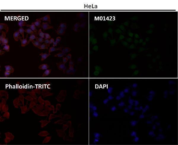
The catalog lists human-, mouse-, and rat-reactive SMAD5 antibodies for IHC, with human and rat tissue images; M01423 also has an IF image (catalog reactivity and image captions).

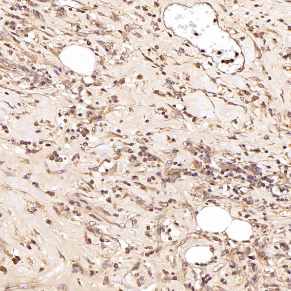
Real IHC data Immunohistochemical analysis of paraffin-embedded Human Hodgkin's lymphoma, using the Antibody at 1:50 dilution.
Anti-Smad5 Rabbit Monoclonal Antibody
Cat # M01423
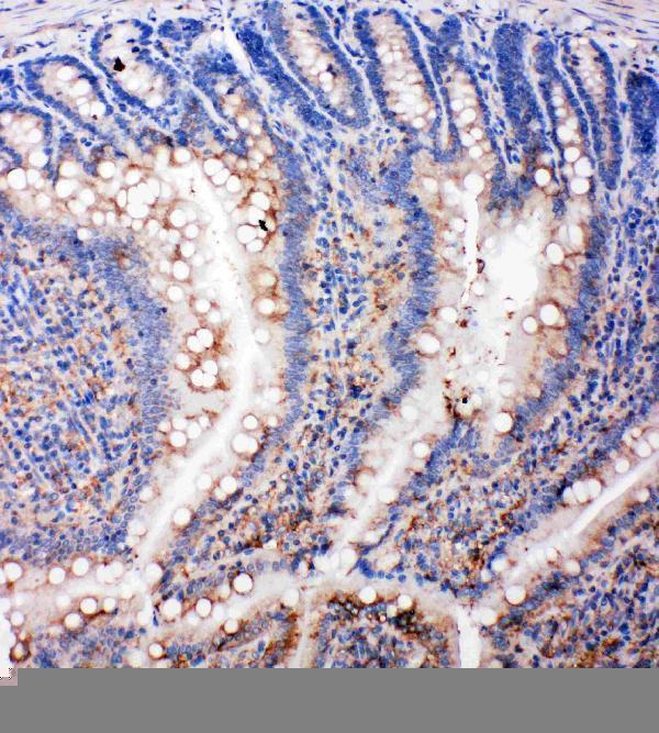
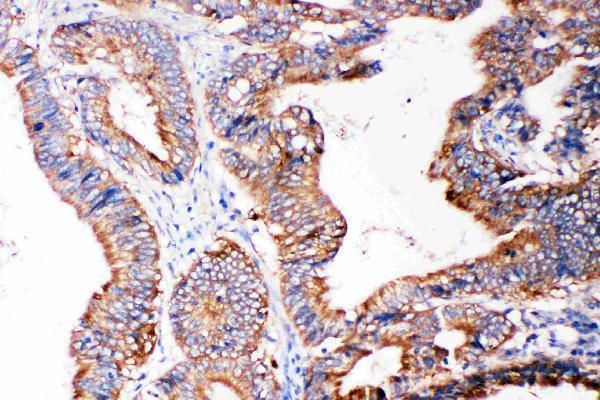
Real IHC data Anti-SMAD5 antibody, PA2115, IHC(P) IHC(P): Rat Intestine Tissue
Anti-SMAD5 Antibody ®
Cat # PA2115
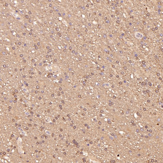
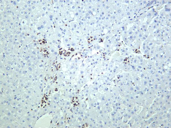
Real IHC data Human liver was stained with anti-Smad5 rabbit antibody
Anti-Smad5 Rabbit Monoclonal Antibody
Cat # M01423-2
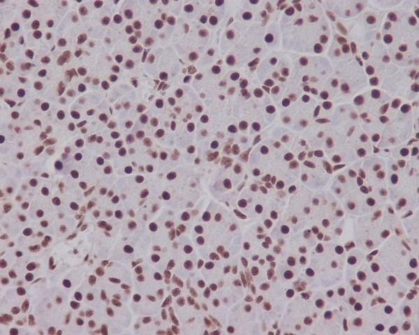
Real IHC data Immunohistochemical analysis of paraffin-embedded human pancreas, using Phospho-Smad5 (S463/465) Antibody.
Anti-Phospho-Smad5 (S463/465) Rabbit Monoclonal Antibody
Cat # P01423-1

M01423 shows paraffin-embedded human Hodgkin’s lymphoma (M01423 image caption), and PA2115 shows rat intestine by IHC(P) (PA2115 image caption). M01423-2 shows human liver (M01423-2 image caption), while P01423-1 shows paraffin-embedded human pancreas stained for phospho-SMAD5 S463/465 (P01423-1 image caption).

Which to pick: For tissue IHC, choose PA2115 for its rat-intestine IHC(P) example (PA2115 image caption), or M01423 for its paraffin-embedded human tissue example (M01423 image caption); the fixative is unreported in both captions. For IF/ICC, M01423 is a rabbit monoclonal listed for both applications and has an IF image (M01423 catalog). All four SKUs list human, mouse, and rat reactivity (catalog reactivity); choose P01423-1 when the target is phospho-SMAD5 S463/465 (P01423-1 title).

Each figure is that product's own IHC / IF validation image from its datasheet.

References

  1. UniProt Consortium. UniProt entry Q99717 (SMAD5_HUMAN, SMAD family member 5).
  2. Human Protein Atlas. SMAD5 tissue IHC expression (reliability: Approved).
  3. Human Protein Atlas. SMAD5 subcellular location (ICC-IF): Mainly localized to the nucleoplasm. In addition localized to the cytosol..
  4. Human Protein Atlas. SMAD5 antibody validation summary (1 antibodies).
  5. MicroRNA-93-5p may participate in the formation of morphine tolerance in bone cancer pain mouse model by targeting Smad5. Oncotarget 2016 — PMC5239538.
  6. MiR-145-5p arrests the cell cycle by modulating SMAD5/cyclin D1 to inhibit gastric cancer progression. Frontiers in cell and developmental biology 2025 — PMC12368585.
  7. Exosomal miRNA-128-3p from mesenchymal stem cells of aged rats regulates osteogenesis and bone fracture healing by targeting Smad5. Journal of nanobiotechnology 2020 — PMC7077029.
  8. Silencing of Long Non-coding RNA SMAD5-AS1 Reverses Epithelial Mesenchymal Transition in Nasopharyngeal Carcinoma via microRNA-195-Dependent Inhibition of SMAD5. Frontiers in oncology 2019 — PMC6923203.
  9. PubMed PMID:8673135 — UniProt-cited evidence.
  10. PubMed PMID:9288787 — UniProt-cited evidence.
  11. PubMed PMID:9264367 — UniProt-cited evidence.