This website uses cookies to ensure you get the best experience on our website.
- Table of Contents
3 Citations
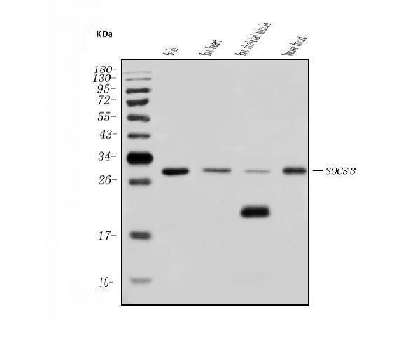
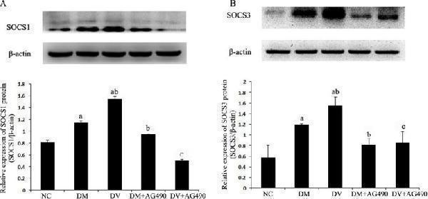
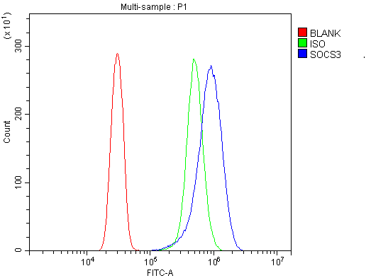
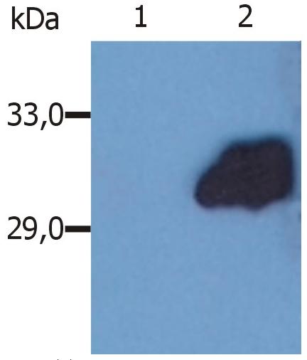
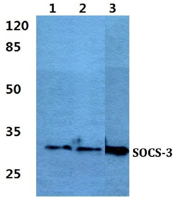
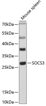
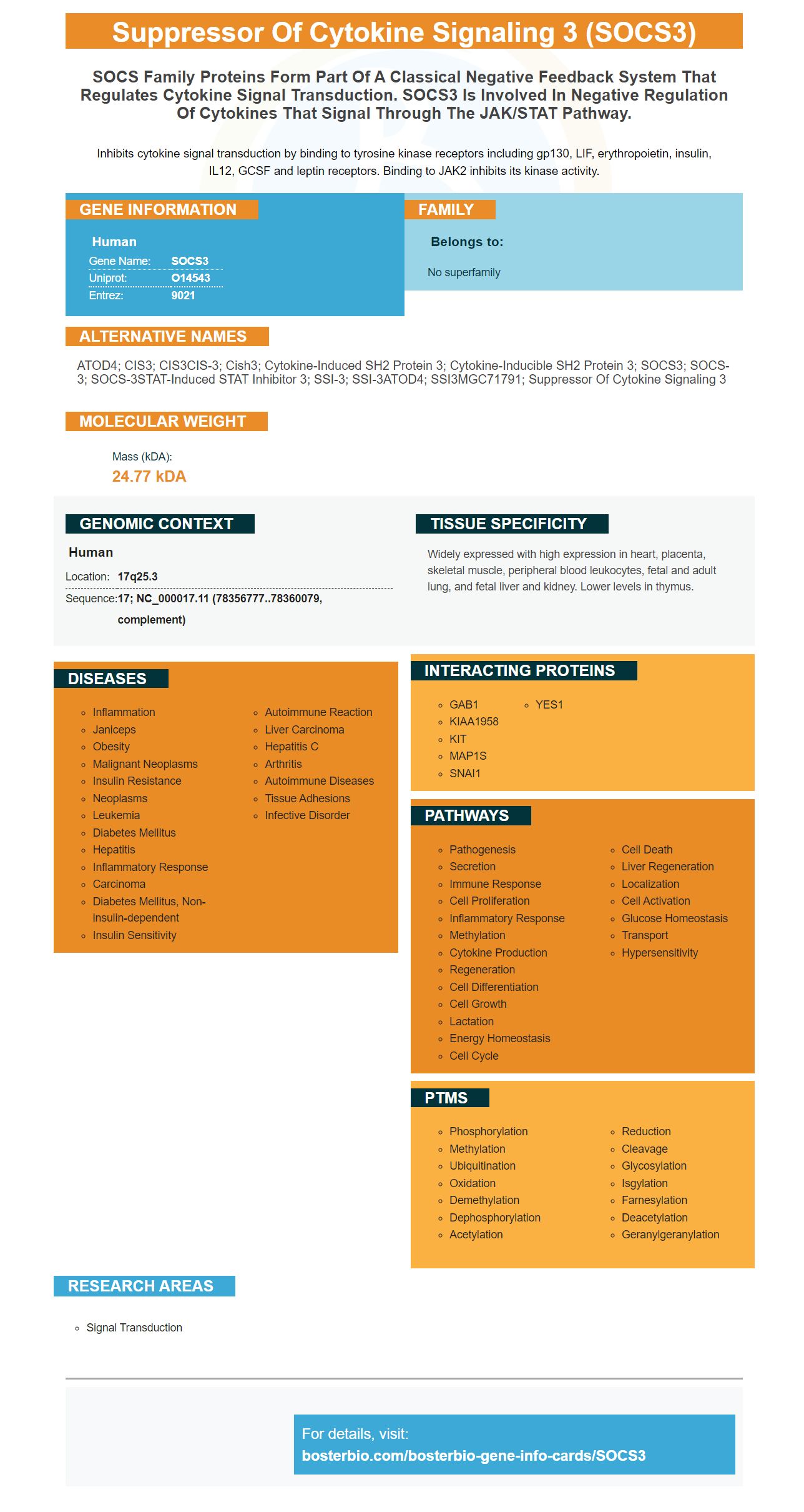
Facts about Suppressor of cytokine signaling 3.

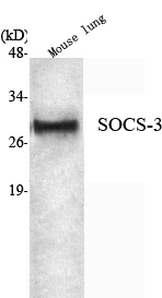
Inhibits cytokine signal transduction by binding to tyrosine kinase receptors including gp130, LIF, erythropoietin, insulin, IL12, GCSF and leptin receptors. Binding to JAK2 inhibits its kinase activity.
| Human | |
|---|---|
| Gene Name: | SOCS3 |
| Uniprot: | O14543 |
| Entrez: | 9021 |

| Belongs to: |
|---|
| No superfamily |

ATOD4; CIS3; CIS3CIS-3; Cish3; cytokine-induced SH2 protein 3; Cytokine-inducible SH2 protein 3; SOCS3; SOCS-3; SOCS-3STAT-induced STAT inhibitor 3; SSI-3; SSI-3ATOD4; SSI3MGC71791; suppressor of cytokine signaling 3
Mass (kDA):
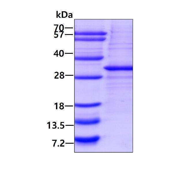
24.77 kDA

| Human | |
|---|---|
| Location: | 17q25.3 |
| Sequence: | 17; NC_000017.11 (78356777..78360079, complement) |
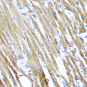
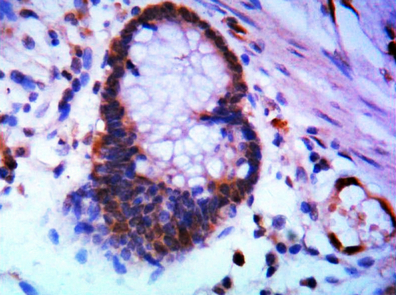
Widely expressed with high expression in heart, placenta, skeletal muscle, peripheral blood leukocytes, fetal and adult lung, and fetal liver and kidney. Lower levels in thymus.


PMID: 9266833 by Minamoto S., et al. Cloning and functional analysis of new members of STAT induced STAT inhibitor (SSI) family: SSI-2 and SSI-3.
PMID: 9344848 by Masuhara M., et al. Cloning and characterization of novel CIS family genes.
*More publications can be found for each product on its corresponding product page