This website uses cookies to ensure you get the best experience on our website.


- Table of Contents
Plan chromogenic SOX2 IHC in paraffin sections using the catalog antibody's documented workflow (datasheet M00105-1). Score nuclear staining by cell type: selected epithelia stain strongly, while brain glia show medium staining (HPA tissue IHC).

Expected localisation, validated protocols, controls and antibodies — the at-a-glance facts below, then the full design guide.
| Expected localisation | Nuclear staining in selected tissue cells (HPA tissue IHC) | |
| Staining pattern | Nuclear signal in respiratory/squamous epithelia and brain glia (HPA tissue IHC) | |
| Antigen retrieval | EDTA pH 8.0 HIER, heat-mediated (datasheet M00105-1) | |
| Positive control | Bronchus+4 more · see all | |
| Negative control | Adipose tissue+4 more · see all |
| Fixation | Keep fixation conditions consistent across sections. (standard IHC practice; not target-specific) | |
| Caveat | Selected cell types stain; whole-tissue scores can mislead (HPA tissue IHC) | |
| Regulation | High in selected epithelia; medium in glia (HPA tissue IHC) | |
| Isoform / epitope | No annotated isoforms; one 1–317 chain (UniProt) |
The catalog antibody uses heat-mediated EDTA pH 8.0 retrieval (datasheet M00105-1). The published SOX2 IHC methods below cover tissue microarrays, vulvar lesions, and frozen pituitary sections (PMC5600089; PMC11640302; PMC6189570).
| Sample | Paraffin-embedded human cervical cancer tissue; fixative not specified (datasheet M00105-1) |
| Fixation | Image fixative and duration unreported (datasheet M00105-1); verify before use. |
| Sectioning | 4–5 µm sections on charged slides (standard) |
| Deparaffinisation | Xylene, graded ethanol series to water (standard) |
| Antigen retrieval | Heat retrieval: EDTA pH 8.0 (datasheet M00105-1); 20 min, 95–100 °C (standard) |
| Peroxidase block | 3% H2O2, 10 min, room temperature (standard) |
| Blocking | 10% goat serum (datasheet M00105-1) |
| Primary antibody | Rabbit monoclonal (clone BIO-19) anti-SOX2, 1:50-1:200 recommended; image 5 μg/ml (datasheet M00105-1) |
| Primary incubation | Overnight at 4 °C (datasheet M00105-1) |
| Detection | HRP-conjugated secondary, DAB chromogen (datasheet M00105-1) |
| Counterstain | Hematoxylin, blue, dehydrate and mount (standard) |
| Expected result | SOX2-positive staining in respiratory epithelial cells of bronchus (HPA tissue IHC: High). HPA tissue profile: Nuclear expression in selected tissues and cell types, including glial cells in brain. No signal in the no-primary control. |
SOX2 staining in paraffin-section IHC should be predominantly nuclear in selected cells, including respiratory and squamous epithelial cells and glia (HPA: tissue IHC). HPA rates the tissue pattern Enhanced, citing consistency with RNA expression (HPA: reliability). UniProt also lists nuclear speckles and cytoplasm, while describing SOX2 as a transcription factor without a transmembrane segment (UniProt P48431: subcellular location, function, topology).
| Strong, discrete nuclear staining in bronchial respiratory epithelium or esophageal squamous epithelium (HPA: High in both cell types). | This matches the reported tissue and compartment pattern (HPA: tissue IHC). Judge positivity in the named cells rather than across the entire section; HPA reports nuclear expression in selected tissues and cell types, with an Enhanced reliability rating (HPA: tissue IHC, reliability). |
| Predominantly cytoplasmic staining, with little or no nuclear staining, in an expected positive epithelium (HPA: nuclear tissue profile). | The compartment disagrees with the observed IHC pattern and warrants an artefact check (HPA: tissue IHC; general IHC interpretation). Cytoplasmic localisation is listed by UniProt, including regulated delocalisation by similarity, so cytoplasmic signal alone cannot establish that an antibody is nonspecific (UniProt P48431: subcellular location). |
| Strong staining of adipocytes or marrow hematopoietic cells (HPA: Not detected in these cell types). | This conflicts with the sampled HPA cell-type patterns (HPA: tissue IHC). Consider antibody cross-reactivity or endogenous chromogenic activity, and inspect a control lacking primary antibody before assigning SOX2 positivity (general IHC practice). HPA's observation does not prove every cell in either tissue is negative (HPA: tissue IHC). |
| Diffuse colour over nuclei, cytoplasm, and tissue-free areas (general IHC interpretation). | A widespread deposit cannot be scored as the selected-cell nuclear pattern reported for SOX2 (HPA: tissue IHC; general IHC interpretation). Check the no-primary control, blocking, washing, and chromogen development to distinguish detection background from interpretable cellular staining (general IHC practice). |
| No nuclear signal in bronchial respiratory or esophageal squamous epithelium (HPA: High in these cell types). | The run has failed its expected-positive check if tissue and cells are identifiable (HPA: tissue IHC; general IHC practice). Review antibody suitability for IHC-P, retrieval, detection, and control performance before calling a study sample negative (general IHC practice). HPA provides a reference pattern, not a guarantee for every specimen (HPA: tissue IHC). |
| Cell-type context (HPA: tissue IHC) | HPA reports High staining in bronchial and nasopharyngeal respiratory epithelium and esophageal and tonsillar squamous epithelium; glial cells in caudate and cerebral cortex, Bergmann glia in cerebellum, and cervical squamous cells are Medium (HPA: tissue IHC). Compare like cell types when selecting controls (general IHC practice). |
| Compartment and regulation (UniProt P48431; HPA: tissue IHC) | HPA's tissue pattern is nuclear, whereas UniProt lists nucleus, nuclear speckles, and cytoplasm and links acetylation state to localisation by similarity (HPA: tissue IHC; UniProt P48431: subcellular location). Score the observed IHC compartment without treating every cytoplasmic pixel as proof of SOX2 (general IHC interpretation). |
| Antibody validation (HPA: antibodies) | HPA lists IHC Enhanced for HPA045725 and CAB079745, Supported for HPA071379, and no IHC status for HPA062851 (HPA: antibodies). Its tissue-level Enhanced rating describes agreement with RNA expression; validation attached to one antibody should not be transferred to another (HPA: reliability, antibodies; general IHC interpretation). |
| Processing and topology (UniProt P48431) | The annotated SOX2 chain spans residues 1–317, with no signal peptide, propeptide, or transmembrane segment and no annotated isoforms (UniProt P48431: processing, topology, isoforms). These annotations do not establish an antibody epitope or antigen-retrieval requirement (UniProt P48431; general IHC interpretation). |
| Situation | Likely cause | Next action |
|---|---|---|
| Expected-positive epithelium is blank (HPA: High in bronchus and esophagus). | Possible retrieval, antibody, or detection failure (general IHC practice); the payload gives no SOX2-specific retrieval condition. | Confirm the expected cells are present, then review the IHC-P antibody instructions, retrieval step, and detection controls (general IHC practice). Do not infer fixation sensitivity from HPA staining levels (HPA: tissue IHC). |
| Nuclear staining is weak where HPA reports High staining (HPA: tissue IHC). | Assay sensitivity or variable specimen performance may limit the visible signal (general IHC practice); a specific cause is unproven. | Compare a concurrent expected-positive section and the same cell type; check the IHC-P antibody and detection steps before changing a scoring threshold (HPA: tissue IHC; general IHC practice). |
| Cytoplasmic colour dominates an expected-positive cell population (HPA: nuclear tissue profile). | Background or compartment misassignment is possible (general IHC interpretation); UniProt also records cytoplasmic SOX2 localisation (UniProt P48431). | Inspect nuclear counterstain alignment and a no-primary control, then report the actual compartment separately from nuclear positivity (general IHC practice). |
| Unexpected cells stain strongly, including HPA-negative adipocytes (HPA: Not detected in adipocytes). | Cross-reactivity or endogenous detection activity is possible (general IHC practice); HPA's reference is cell-type specific (HPA: tissue IHC). | Check a no-primary control and an expected-positive cell population; withhold a SOX2-positive call until the unexpected signal is resolved (general IHC practice). |
| Colour is diffuse across the section (general IHC interpretation). | Nonspecific detection or excess chromogen development may obscure nuclear boundaries (general IHC practice). | Review blocking, washing, and development with a no-primary control; score only cells with interpretable nuclear staining (general IHC practice; HPA: nuclear tissue profile). |
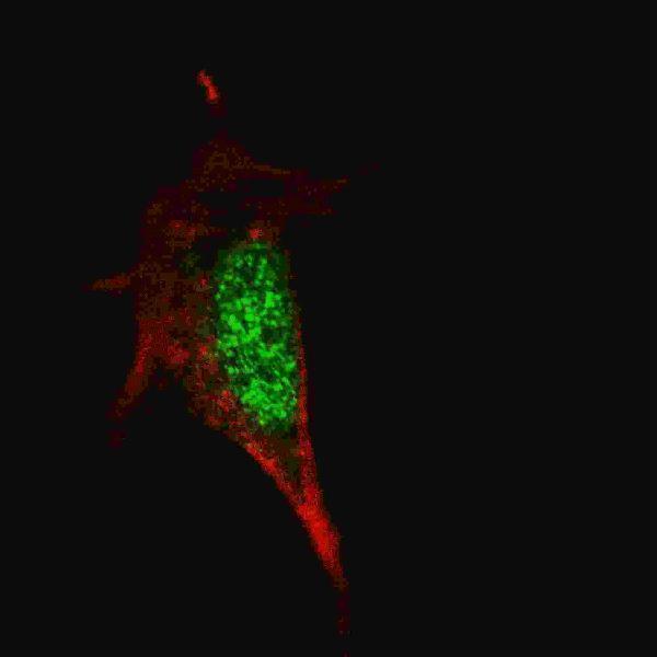
| What pattern should IF/ICC show? (HPA: subcellular ICC-IF) | HPA reports enhanced nucleoplasmic localisation in ICC-IF, while UniProt also lists nuclear speckles and cytoplasm (HPA: subcellular ICC-IF; UniProt P48431). | Look for nucleoplasmic signal with a nuclear counterstain and assess controls (HPA: subcellular ICC-IF; general IF practice). Use the separate IF/ICC guide for assay setup (application scope). |

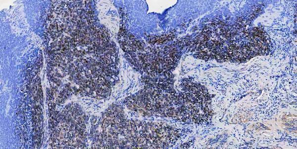
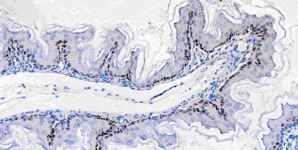


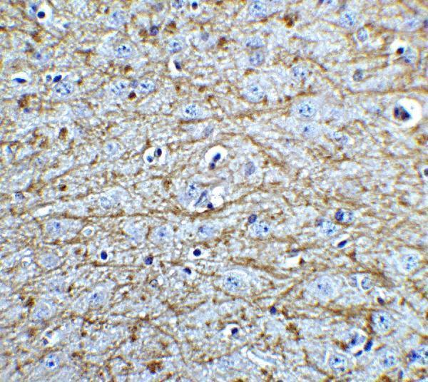


Comprehensive Human Protein Atlas IHC scoring per tissue (reliability: Enhanced — High consistency between antibody staining and RNA expression data.). Rows are taken directly from the HPA tissue chart — click any row's HPA link to view the source.
| Tissue | Cell type | Level | Evidence | Source |
|---|---|---|---|---|
| Bronchus | Respiratory epithelial cells | High | Protein (IHC) | HPA → |
| Esophagus | Squamous epithelial cells | High | Protein (IHC) | HPA → |
| Nasopharynx | Respiratory epithelial cells | High | Protein (IHC) | HPA → |
| Tonsil | Squamous epithelial cells | High | Protein (IHC) | HPA → |
| Caudate | Glial cells | Medium | Protein (IHC) | HPA → |
| Tissue | Cell type | Level | Evidence | Source |
|---|---|---|---|---|
| Adipose tissue | Adipocytes | Not detected | Protein (IHC) | HPA → |
| Adrenal gland | Glandular cells | Not detected | Protein (IHC) | HPA → |
| Appendix | Glandular cells | Not detected | Protein (IHC) | HPA → |
| Bone marrow | Hematopoietic cells | Not detected | Protein (IHC) | HPA → |
| Breast | Adipocytes | Not detected | Protein (IHC) | HPA → |
Troubleshoot SOX2 staining in paraffin sections by checking retrieval, nuclear localisation, tissue context and controls before interpreting chromogenic signal.
Validated anti-SOX2 antibodies have IHC images from human tumors and mouse or rat brain, plus IF images from human tissue, mouse brain and cells (catalog image captions).
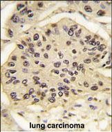
M00105-1 has paraffin-section IHC images from human and mouse tissue and IF images from human paraffin sections; PA2284 has paraffin-section IHC images from human, mouse and rat tissue (catalog image captions). A00105 has a mouse brain IHC image and IF images from 3T3 cells and mouse brain; M00105-3 has an IHC image from formalin-fixed, paraffin-embedded human lung carcinoma and IF images from SY5Y and A549 cells (catalog image captions).
Which to pick: For human tissue IHC with documented fixation, choose mouse monoclonal M00105-3: its IHC image shows paraffin-section, paraffin-embedded lung carcinoma (catalog host, clone and IHC image caption). For IF/ICC, choose rabbit monoclonal M00105-1 when both applications are needed; its IF images use human paraffin sections, while its IHC captions describe paraffin sections without reporting the fixative (catalog applications, clone and image captions). For IHC across species, choose PA2284: its paraffin-section images cover human, mouse and rat tissue, and those captions do not report the fixative (catalog reactivity and IHC image captions). The selected M00105-1 tissue-IHC caption documents paraffin sections, but does not specify the fixative (selected-SKU IHC image M00105-1).