Click image to see more details
-

-
-
-
-
+3
Product Info Summary
| SKU: | M00105-3 |
|---|---|
| Size: | 80 µl |
| Reactive Species: | Human |
| Host: | Mouse |
| Application: | Flow Cytometry, IF, IHC-P, WB |
Customers Who Bought This Also Bought

Product info
Product Name
Anti-SOX2 Antibody
SKU/Catalog Number
M00105-3
Size
80 µl
Description
Boster Bio Anti-SOX2 Antibody (Catalog # M00105-3). Tested in WB, IF, Flow Cytometry, IHC-P application(s). This antibody reacts with Human.
Storage & Handling
Maintain refrigerated at 2-8°C for up to 2 weeks. For long-term storage, store at -20°C in small aliquots to prevent freeze-thaw cycles.
Cite This Product
Anti-SOX2 Antibody (Boster Biological Technology, Pleasanton CA, USA, Catalog # M00105-3)
Host
Mouse
Contents
Purified monoclonal antibody supplied in PBS with 0.09% (W/V) sodium azide.
Clonality
Monoclonal
Clone Number
57CT23.3.4
Isotype
IgG1
Immunogen
SOX2 recombinant protein is used to produce this monoclonal antibody.
Reactive Species
M00105-3 is reactive to SOX2 in Human
Calculated molecular weight
34.3 kDa
Background of SOX2
This intronless gene encodes a member of the SRY-related HMG-box (SOX) family of transcription factors involved in the regulation of embryonic development and in the determination of cell fate. The product of this gene is required for stem-cell maintenance in the central nervous system, and also regulates gene expression in the stomach. Mutations in this gene have been associated with optic nerve hypoplasia and with syndromic microphthalmia, a severe form of structural eye malformation. This gene lies within an intron of another gene called SOX2 overlapping transcript (SOX2OT).
Antibody Validation
Boster validates all antibodies on WB, IHC, ICC, Immunofluorescence, and ELISA with known positive control and negative samples to ensure specificity and high affinity, including thorough antibody incubations.
Application & Images
Applications
M00105-3 is guaranteed for Flow Cytometry, IF, IHC-P, WB Boster Guarantee
Assay Dilutions Recommendation
The recommendations below provide a starting point for assay optimization. The actual working concentration varies and should be decided by the user.
IF: 1:10-1:50
WB: 1:4000
IHC-P: 1:50-1:100
FC: 1:10-1:50
IHC: 1:100
Validation Images & Assay Conditions
Click image to see more details
Fluorescent confocal image of SY5Y cells stained with SOX2 antibody. SY5Y cells were fixed with 4% PFA (20 min), permeabilized with Triton X-100 (0.2%, 30 min). Cells were then incubated with M00105-3 SOX2 primary antibody (1:100, 2 h at room temperature). For secondary antibody, Alexa Fluor® 488 conjugated donkey anti-mouse antibody (green) was used (1:1000, 1h). Note the highly specific localization of the SOX2 mainly to the nucleus.

Click image to see more details
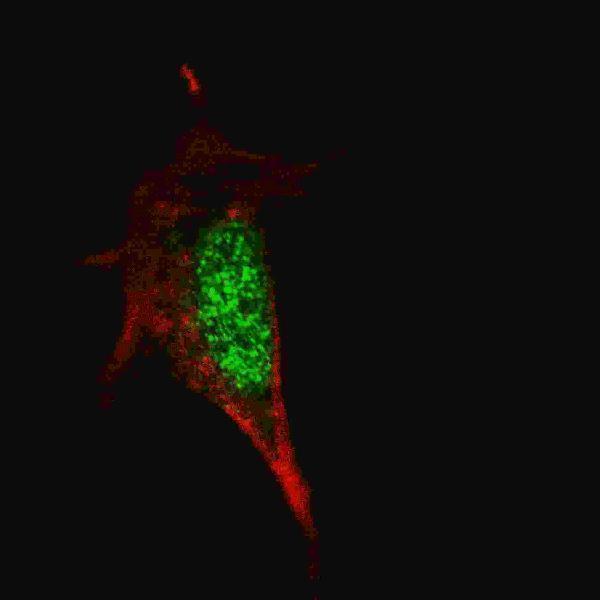
Fluorescent image of A549 cell stained with SOX2 Antibody.A549 cells were fixed with 4% PFA (20 min), permeabilized with Triton X-100 (0.1%, 10 min), then incubated with SOX2 primary antibody (1:25, 1 h at 37℃. For secondary antibody, Alexa Fluor® 488 conjugated donkey anti-mouse antibody (green) was used (1:400, 50 min at 37℃.Cytoplasmic actin was counterstained with Alexa Fluor® 555 (red) conjugated Phalloidin (7units/ml, 1 h at 37℃.SOX2 immunoreactivity is localized to Nucleus significantly.

Click image to see more details
Western blot analysis of SOX2 (arrow) using mouse monoclonal SOX2 antibody. 293 cell lysates (2 g/lane) either nontransfected (Lane 1) or transiently transfected with the SOX2 gene (Lane 2)

Click image to see more details
Western blot analysis of SOX2 Antibody by SOX2 recombinant protein. SOX2(arrow) was detected using the purified Mab.

Click image to see more details
Western blot analysis of lysate from NCCIT cell line, using SOX2 Antibody. M00105-3 was diluted at 1:1000. A goat anti-mouse IgG H&L(HRP) at 1:3000 dilution was used as the secondary antibody. Lysate at 20g.

Click image to see more details
Western blot analysis of lysate from SOX2 protein, using SOX2 Antibody. M00105-3 was diluted at 1:4000. A goat anti-mouse IgG H&L(HRP) at 1:3000 dilution was used as the secondary antibody. Lysate at 20g.

Click image to see more details
Formalin-fixed and paraffin-embedded human lung carcinoma tissue reacted with SOX2 Antibody , which was peroxidase-conjugated to the secondary antibody, followed by DAB staining. This data demonstrates the use of this antibody for immunohistochemistry; clinical relevance has not been evaluated.
Specific Publications For Anti-SOX2 Antibody (M00105-3)
Loading publications
Recommended Resources
Here are featured tools and databases that you might find useful.
- Boster's Pathways Library
- Protein Databases
- Bioscience Research Protocol Resources
- Data Processing & Analysis Software
- Photo Editing Software
- Scientific Literature Resources
- Research Paper Management Tools
- Molecular Biology Software
- Primer Design Tools
- Bioinformatics Tools
- Phylogenetic Tree Analysis
Customer Reviews
Have you used Anti-SOX2 Antibody ?
Share your experimental results or join a short interview to earn up to $1,000 in product credits or other rewards.
0 Reviews For Anti-SOX2 Antibody
Customer Q&As
Have a question?
Find answers in Q&As, reviews.
Can't find your answer?
Submit your question
