Click image to see more details
-

-
-
-
-
+7
Product Info Summary
| SKU: | PA2284 |
|---|---|
| Size: | 100 μg/vial |
| Reactive Species: | Human, Mouse, Rat |
| Host: | Rabbit |
| Application: | IHC, WB |
Customers Who Bought This Also Bought

Product info
Product Name
Anti-Transcription factor SOX-2 SOX2 Antibody Picoband®
SKU/Catalog Number
PA2284
BA3292 is an alternative SKU for this antibody, used in previous lots.
Size
100 μg/vial
Form
Lyophilized
Description
Boster Bio Anti-Transcription factor SOX-2 SOX2 Antibody catalog # PA2284. Tested in IHC, WB applications. This antibody reacts with Human, Mouse, Rat. The brand Picoband indicates this is a premium antibody that guarantees superior quality, high affinity, and strong signals with minimal background in Western blot applications. Only our best-performing antibodies are designated as Picoband, ensuring unmatched performance.
Storage & Handling
Store at -20˚C for one year from date of receipt. After reconstitution, at 4˚C for one month. It can also be aliquotted and stored frozen at -20˚C for six months. Avoid repeated freeze-thaw cycles.
Cite This Product
Anti-Transcription factor SOX-2 SOX2 Antibody Picoband® (Boster Biological Technology, Pleasanton CA, USA, Catalog # PA2284)
Host
Rabbit
Contents
Each vial contains 4 mg Trehalose, 0.9 mg NaCl and 0.2 mg Na2HPO4.
Clonality
Polyclonal
Isotype
Rabbit IgG
Immunogen
A synthetic peptide corresponding to a sequence in the middle region of human SOX2, different from the related mouse and rat sequence by one amino acid.
Cross-reactivity
No cross-reactivity with other proteins
Reactive Species
PA2284 is reactive to SOX2 in Human, Mouse, Rat
Observed Molecular Weight
36 kDa
Calculated molecular weight
34.3 kDa
Background of SOX2
SRY (sex determining region Y)-box 2, also known as SOX2, is a transcription factor that is essential for maintaining self-renewal, or pluripotency of undifferentiated embryonic stem cells. Sox2 is a member of the Sox family of transcription factors, which have been shown to play key roles in many stages of mammalian development. This gene is mapped to 3q26.33. It is found that SOX2 can regulate OCT3/4 expression and maintains ES pluripotency through upstream transcription factors. SOX2 is identified as a lineage-survival oncogene in lung and esophageal squamous cell carcinoma. In addition to those, SOX2 has a critical role in maintenance of embryonic and neural stem cells and holds great promise in research involving induced pluripotency, an emerging and very promising field of regenerative medicine.
Antibody Validation
Boster validates all antibodies on WB, IHC, ICC, Immunofluorescence, and ELISA with known positive control and negative samples to ensure specificity and high affinity, including thorough antibody incubations.
Application & Images
Applications
PA2284 is guaranteed for IHC, WB Boster Guarantee
Recommend Dilution
| Application | Dilution | Species |
|---|---|---|
| Immunohistochemistry (Paraffin-embedded Section) | 2-5μg/ml | Human, Mouse, Rat |
| Western blot | 0.1-0.5μg/ml | Human, Mouse, Rat |
Tested application
Suggested blocking solution with 5% non-fat milk or BSA; (*)Recommended protein loading: 20-40 µg per lane
Use TE buffer pH 9.0 for antigen retrieval; (*) citrate buffer pH 6.0 is an alternative.
Validation Images & Assay Conditions
Click image to see more details
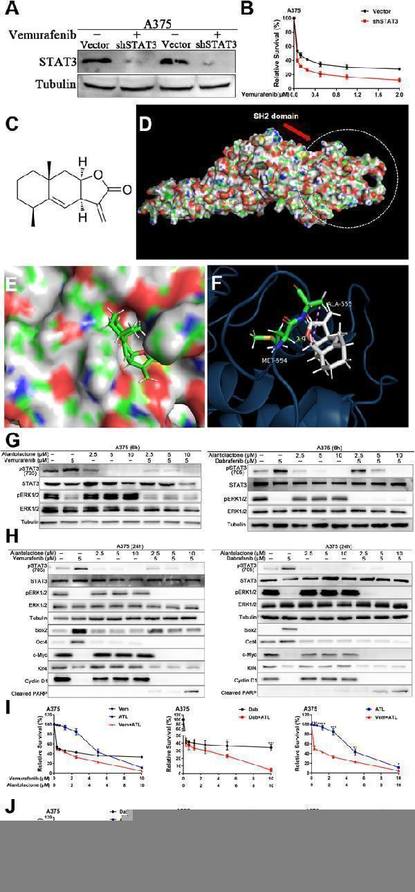
Alantolactone suppressed STAT3 feedback activation induced by BRAFi, downregulating protein expression of Oct4 and Sox2 in A375 cells. ( A ) A375 cells were transfected with shSTAT3 plasmid, and the expression of STAT3 protein was detected 48 h later. ( B ) A375 cells were transfected with shSTAT3 plasmid for 24 h and treated with different concentrations of vemurafenib. After another 72 h, cell viability was detected by CCK8 assays. ( C ) Chemical structure of alantolactone. ( D ) Predicted model of alantolactone binding to STAT3β SH2, as shown by computational modeling. Protein structure information was obtained from Protein Data Bank (PDB) entry 6NJS. ( E ) Binding model of alantolactone to the SH2 domain. The molecular surface of the STAT3β SH2 domain is electrostatically colored with blue and red representing potentially positively- and negatively-charged regions, respectively. ( F ) Predicted interactions between the amino acid residues of the SH2 domain and alantolactone. Oxygen atoms of alantolactone are shown in red. Alantolactone forms carbon hydrogen bond with MET554, and there is a alkyl bond between alantolactone and ALA555. (G and H) A375 cells were treated with alantolactone and BRAFi single or combination for 6 ( G ) or 24 ( H ) hours, phospho-STAT3 (705), STAT3, phospho-ERK1/2, ERK1/2, SOX2, Oct4, c-Myc, Klf4, cyclin D1 and cleaved PARP levels were analyzed by western blotting, and tubulin served as a loading control. (I and J) A375 cells were treated with different concentrations of alantolactone and vemurafenib ( I ) or alantolactone and dabrafenib ( J ) for 3 days. Cell viability was determined by CCK8 assays. The two images on the left in Figures G and H are split from the image on the right. Data are mean ± SD. * p < 0.05; ** p < 0.01; *** p < 0.001; Student’s t -test
Index in PubMed under a CC BY license. PMID: 38822350

Click image to see more details
The combination of alantolactone and MAPKi simultaneously inhibited the STAT3 and BRAF/MEK/ERK pathways, regulating the expression level of downstream effectors in A375 cells. ( A ) A375 cells were treated with vemurafenib, cobimetinib and alantolactone alone or in combination for 6 h. The gene expression levels of c-Myc, Klf4, Sox2 and Oct4 were detected by real-time fluorescent quantitative PCR, and β-actin served as a loading control. ( B ) A375 cells were treated with vemurafenib, cobimetinib and alantolactone for 24 h. Phospho-STAT3 (705), STAT3, phospho-ERK1/2, ERK1/2, Sox2, Oct4, c-Myc, Klf4, cyclin D1 and cleaved PARP levels were analyzed by western blotting, and tubulin served as a loading control. (C and D) A375 cells were treated with different concentrations of cobimetinib and alantolactone ( C ) or trametinib and alantolactone ( D ) for 72 h. Cell viability was determined by CCK8 assays. The two images on the left in Figures C and D are split from the image on the right. (E and F) A375 cells were treated with different concentrations of vemurafenib, cobimetinib, and alantolactone ( E ) or dabrafenib, trametinib and alantolactone ( F ) for 72 h. Cell viability was determined by CCK8 assays. The two images on the left in Figures C and D are split from the image on the right. ( G ) A375 cells were treated with vemurafenib (5 µM), cobimetinib (1 µM), and alantolactone (6 µM) alone or in combination for 24 h. Flow cytometry analysis of cell death (Annexin V/PI labelling) in A375 cells. The histogram on the right shows the proportion of dead cells. Data, means ± SDs. * p < 0.05; ** p < 0.01; *** p < 0.001; Student’s t test
Index in PubMed under a CC BY license. PMID: 38822350

Click image to see more details
Combined treatment with alantolactone and MAPK pathway inhibitors showed highly selective cytotoxic effects on A375 cells, but had no obvious cytotoxicity on renal tubular epithelial cells. ( A ) HK2 and A375 cells were treated with vemurafenib and alantolactone or vemurafenib, cobimetinib and alantolactone for 24 h. Phospho-STAT3 (705), STAT3, phospho-ERK1/2, ERK1/2, Sox2, c-Myc and cleaved PARP levels were analyzed by western blotting, and tubulin served as a loading control. ( B ) HK2 and A375 cells were treated with different concentrations of alantolactone, vemurafenib and cobimetinib for 72 h. Cell viability was determined by CCK8 assays. Data are the mean ± SD. * p < 0.05; ** p < 0.01; *** p < 0.001; Student’s t test
Index in PubMed under a CC BY license. PMID: 38822350

Click image to see more details
Alantolactone could synergistically enhance the cytotoxic effects with MAPKi in A375 xenografts of nude mice. ( A ) A375 xenografts were administrated by alantolactone (ATL; 20 mg/kg administered intraperitoneally once daily) and vemurafenib (Vem; 25 mg/kg administered intraperitoneally once daily) + cobimetinib (Cobi; 1 mg/kg administered intraperitoneally once daily) individually or in combination. During the treatment period, measure and record the tumor volume every other day. ( B ) After 12 days of treatment, tumor grafts were removed and weighed. ( C ) Photographs of xenograft A375 tumors treated with single or combination drugs. ( D ) During the drug treatment, the animals were weighed every other day. ( E ) Haematoxylin-eosin staining of heart, liver and kidney tissue sections (magnification: ×100). ( F ) Representative images of immunohistochemical staining of p-STAT3(705), p-ERK1/2, c-Myc, Klf4, Sox2, Oct4 and Ki67 in tumor tissues. Data are the mean ± SD. * p < 0.05; ** p < 0.01; *** p < 0.001; Student’s t test
Index in PubMed under a CC BY license. PMID: 38822350

Click image to see more details
Alantolactone sensitized intrinsic-resistant A2058 cells to MAPKi targeted therapy by inhibiting STAT3 activation. ( A ) A2058 cells were treated with vemurafenib and alantolactone or vemurafenib, cobimetinib and alantolactone for 24 h. Phospho-STAT3 (705), STAT3, phospho-ERK1/2, ERK1/2, Sox2, Oct4, c-Myc, Klf4, cyclin D1 and cleaved PARP levels were analyzed by western blotting, and tubulin served as a loading control. (B-D) A2058 cells were treated with different concentrations of vemurafenib and alantolactone ( B ), cobimetinib and alantolactone ( C ), or vemurafenib, cobimetinib and alantolactone ( D ) for 72 h. Cell viability was determined by CCK8 assays. Data are the mean ± SD. * p < 0.05; ** p < 0.01; *** p < 0.001; Student’s t test
Index in PubMed under a CC BY license. PMID: 38822350

Click image to see more details
The combination of alantolactone and MAPKi could inhibit the proliferation of MAPKi-resistant A375R cells. ( A ) A375R cells were treated with vemurafenib and alantolactone or vemurafenib, cobimetinib and alantolactone for 24 h. Phospho-STAT3 (705), STAT3, phospho-ERK1/2, ERK1/2, Sox2, Oct4, c-Myc, Klf4, cyclin D1 and cleaved PARP levels were analyzed by western blotting, and tubulin served as a loading control. (B-D) A375R cells were treated with different concentrations of vemurafenib and alantolactone ( B ), cobimetinib and alantolactone ( C ), or vemurafenib, cobimetinib and alantolactone ( D ) for 72 h. Cell viability was determined by CCK8 assays. Data are the mean ± SD. * p < 0.05; ** p < 0.01; *** p < 0.001; Student’s t test
Index in PubMed under a CC BY license. PMID: 38822350

Click image to see more details
Alantolactone could synergistically enhance the cytotoxic effects with MAPKi in A375 xenografts of nude mice. ( A ) Tumor growth curves of A375R xenograft models treated with alantolactone (ATL; 20 mg/kg administered intraperitoneally once daily) and vemurafenib (Vem; 25 mg/kg administered intraperitoneally once daily) + cobimetinib (Cobi; 1 mg/kg administered intraperitoneally once daily) alone or in combination. ( B ) After 12 days of treatment, tumor grafts were removed and weighed. ( C ) Photographs of xenograft A375R tumors treated with single or combination drugs. ( D ) During the drug treatment, the animals were weighed every other day. ( E ) Representative images of immunohistochemical staining of p-STAT3(705), p-ERK1/2, c-Myc, Klf4, Sox2, Oct4 and Ki67 in tumor tissues. Data are the mean ± SD. * p < 0.05; ** p < 0.01; *** p < 0.001; Student’s t test
Index in PubMed under a CC BY license. PMID: 38822350

Click image to see more details
Western blot analysis of SOX2 using anti-SOX2 antibody (PA2284).
Electrophoresis was performed on a 5-20% SDS-PAGE gel at 70V (Stacking gel) / 90V (Resolving gel) for 2-3 hours. The sample well of each lane was loaded with 30 ug of sample under reducing conditions.
Lane 1: human U251 whole cell lysates,
Lane 2: human 293T whole cell lysates.
After electrophoresis, proteins were transferred to a nitrocellulose membrane at 150 mA for 50-90 minutes. Blocked the membrane with 5% non-fat milk/TBS for 1.5 hour at RT. The membrane was incubated with rabbit anti-SOX2 antigen affinity purified polyclonal antibody (Catalog # PA2284) at 0.5 μg/mL overnight at 4°C, then washed with TBS-0.1%Tween 3 times with 5 minutes each and probed with a goat anti-rabbit IgG-HRP secondary antibody at a dilution of 1:5000 for 1.5 hour at RT. The signal is developed using an Enhanced Chemiluminescent detection (ECL) kit (Catalog # EK1002) with Tanon 5200 system. A specific band was detected for SOX2 at approximately 36 kDa. The expected band size for SOX2 is at 34 kDa.

Click image to see more details
IHC analysis of SOX2 using anti-SOX2 antibody (PA2284).
SOX2 was detected in a paraffin-embedded section of human laryngeal squamous cell carcinoma tissue. Heat mediated antigen retrieval was performed in EDTA buffer (pH 8.0, epitope retrieval solution). The tissue section was blocked with 10% goat serum. The tissue section was then incubated with 2 μg/ml rabbit anti-SOX2 Antibody (PA2284) overnight at 4°C. Peroxidase Conjugated Goat Anti-rabbit IgG was used as secondary antibody and incubated for 30 minutes at 37°C. The tissue section was developed using HRP Conjugated Rabbit IgG Super Vision Assay Kit (Catalog # SV0002) with DAB as the chromogen.

Click image to see more details
IHC analysis of SOX2 using anti-SOX2 antibody (PA2284).
SOX2 was detected in a paraffin-embedded section of mouse brain tissue. Heat mediated antigen retrieval was performed in EDTA buffer (pH 8.0, epitope retrieval solution). The tissue section was blocked with 10% goat serum. The tissue section was then incubated with 2 μg/ml rabbit anti-SOX2 Antibody (PA2284) overnight at 4°C. Peroxidase Conjugated Goat Anti-rabbit IgG was used as secondary antibody and incubated for 30 minutes at 37°C. The tissue section was developed using HRP Conjugated Rabbit IgG Super Vision Assay Kit (Catalog # SV0002) with DAB as the chromogen.

Click image to see more details
IHC analysis of SOX2 using anti-SOX2 antibody (PA2284).
SOX2 was detected in a paraffin-embedded section of rat brain tissue. Heat mediated antigen retrieval was performed in EDTA buffer (pH 8.0, epitope retrieval solution). The tissue section was blocked with 10% goat serum. The tissue section was then incubated with 2 μg/ml rabbit anti-SOX2 Antibody (PA2284) overnight at 4°C. Peroxidase Conjugated Goat Anti-rabbit IgG was used as secondary antibody and incubated for 30 minutes at 37°C. The tissue section was developed using HRP Conjugated Rabbit IgG Super Vision Assay Kit (Catalog # SV0002) with DAB as the chromogen.
Specific Publications For Anti-Transcription factor SOX-2 SOX2 Antibody Picoband® (PA2284)
Loading publications
Recommended Resources
Here are featured tools and databases that you might find useful.
- Boster's Pathways Library
- Protein Databases
- Bioscience Research Protocol Resources
- Data Processing & Analysis Software
- Photo Editing Software
- Scientific Literature Resources
- Research Paper Management Tools
- Molecular Biology Software
- Primer Design Tools
- Bioinformatics Tools
- Phylogenetic Tree Analysis
Customer Reviews
Have you used Anti-Transcription factor SOX-2 SOX2 Antibody Picoband®?
Share your experimental results or join a short interview to earn up to $1,000 in product credits or other rewards.
0 Reviews For Anti-Transcription factor SOX-2 SOX2 Antibody Picoband®
Customer Q&As
Have a question?
Find answers in Q&As, reviews.
Can't find your answer?
Submit your question
17 Customer Q&As for Anti-Transcription factor SOX-2 SOX2 Antibody Picoband®
Question
We have seen staining in rat retina. What should we do? Is anti-SOX2 antibody supposed to stain retina positively?
Verified Customer
Verified customer
Asked: 2020-01-20
Answer
According to literature retina does express SOX2. According to Uniprot.org, SOX2 is expressed in cerebral cortex, fetal brain, retina, lung, among other tissues. Regarding which tissues have SOX2 expression, here are a few articles citing expression in various tissues:
Fetal brain, Pubmed ID: 7849401
Lung, Pubmed ID: 15489334
Boster Scientific Support
Answered: 2020-01-20
Question
Can you help my question with product PA2284, anti-SOX2 antibody. I was wondering if it would be possible to conjugate this antibody with biotin. I would need it to be without BSA or sodium azide. I am planning on using a buffer exchange of sodium azide with PBS only. Would there be problems for me to conjugate the antibody and store it in -20 degrees in small aliquots?
Verified Customer
Verified customer
Asked: 2020-01-14
Answer
We do not recommend storing this antibody with PBS buffer only in -20 degrees. If you want to store it in -20 degrees it is best to add some cryoprotectant like glycerol. If you want carrier free PA2284 anti-SOX2 antibody, we can provide it to you in a special formula with trehalose and/or glycerol. These molecules will not interfere with conjugation chemistry and provide a good level of protection for the antibody from degradation. Please be sure to specify this in your purchase order.
Boster Scientific Support
Answered: 2020-01-14
Question
I would like using your anti-SOX2 antibody for neuronal stem cell population maintenance studies. Has this antibody been tested with western blotting on brain tissue? We would like to see some validation images before ordering.
Verified Customer
Verified customer
Asked: 2020-01-06
Answer
Thank you for your inquiry. This PA2284 anti-SOX2 antibody is tested on rat lung tissue, tissue lysate, mouse brain, brain tissue, cancer tissue. It is guaranteed to work for IHC, WB in human, mouse, rat. Our Boster guarantee will cover your intended experiment even if the sample type has not been be directly tested.
Boster Scientific Support
Answered: 2020-01-06
Question
Our team were well pleased with the WB result of your anti-SOX2 antibody. However we have been able to see positive staining in retina nucleus. using this antibody. Is that expected? Could you tell me where is SOX2 supposed to be expressed?
H. Anderson
Verified customer
Asked: 2019-11-18
Answer
According to literature, retina does express SOX2. Generally SOX2 expresses in nucleus. Regarding which tissues have SOX2 expression, here are a few articles citing expression in various tissues:
Fetal brain, Pubmed ID: 7849401
Lung, Pubmed ID: 15489334
Boster Scientific Support
Answered: 2019-11-18
Question
Does anti-SOX2 antibody PA2284 work on pig IHC with retina?
Verified Customer
Verified customer
Asked: 2019-08-20
Answer
Our lab technicians have not tested anti-SOX2 antibody PA2284 on pig. You can run a BLAST between pig and the immunogen sequence of anti-SOX2 antibody PA2284 to see if they may cross-react. If the sequence homology is close, then you can perform a pilot test. Keep in mind that since we have not validated pig samples, this use of the antibody is not covered by our guarantee. However we have an innovator award program that if you test this antibody and show it works in pig retina in IHC, you can get your next antibody for free.
Boster Scientific Support
Answered: 2019-08-20
Question
Would anti-SOX2 antibody PA2284 work for IHC with fetal brain?
Verified Customer
Verified customer
Asked: 2019-07-09
Answer
According to the expression profile of fetal brain, SOX2 is highly expressed in fetal brain. So, it is likely that anti-SOX2 antibody PA2284 will work for IHC with fetal brain.
Boster Scientific Support
Answered: 2019-07-09
Question
Is this PA2284 anti-SOX2 antibody reactive to the isotypes of SOX2?
Verified Customer
Verified customer
Asked: 2019-06-03
Answer
The immunogen of PA2284 anti-SOX2 antibody is A synthetic peptide corresponding to a sequence in the middle region of human SOX2(171-183aa YSMMQDQLGYPQH), different from the related mouse and rat sequence by one amino acid. Could you tell me which isotype you are interested in so I can help see if the immunogen is part of this isotype?
Boster Scientific Support
Answered: 2019-06-03
Question
I see that the anti-SOX2 antibody PA2284 works with IHC, what is the protocol used to produce the result images on the product page?
A. Johnson
Verified customer
Asked: 2019-02-15
Answer
You can find protocols for IHC on the "support/technical resources" section of our navigation menu. If you have any further questions, please send an email to support@bosterbio.com
Boster Scientific Support
Answered: 2019-02-15
Question
Would PA2284 anti-SOX2 antibody work on parafin embedded sections? If so, which fixation method do you recommend we use (PFA, paraformaldehyde, other)?
Verified Customer
Verified customer
Asked: 2019-01-17
Answer
You can see on the product datasheet, PA2284 anti-SOX2 antibody as been validated on IHC. It is best to use PFA for fixation because it has better tissue penetration ability. PFA needs to be prepared fresh before use. Long term stored PFA turns into formalin, as the PFA molecules congregate and become formalin.
Boster Scientific Support
Answered: 2019-01-17
Question
Is a blocking peptide available for product anti-SOX2 antibody (PA2284)?
Verified Customer
Verified customer
Asked: 2019-01-07
Answer
We do provide the blocking peptide for product anti-SOX2 antibody (PA2284). If you would like to place an order for it please contact support@bosterbio.com and make a special request.
Boster Scientific Support
Answered: 2019-01-07
Question
I was wanting to use your anti-SOX2 antibody for IHC for human fetal brain on frozen tissues, but I want to know if it has been validated for this particular application. Has this antibody been validated and is this antibody a good choice for human fetal brain identification?
J. Anderson
Verified customer
Asked: 2018-10-10
Answer
You can see on the product datasheet, PA2284 anti-SOX2 antibody has been validated for IHC, WB on human, mouse, rat tissues. We have an innovator award program that if you test this antibody and show it works in human fetal brain in IHC-frozen, you can get your next antibody for free.
Boster Scientific Support
Answered: 2018-10-10
Question
See below the WB image, lot number and protocol we used for fetal brain using anti-SOX2 antibody PA2284. Please let me know if you require anything else.
Verified Customer
Verified customer
Asked: 2018-09-24
Answer
Thank you very much for the data. Our lab team are working to resolve this as quickly as possible, and we appreciate your patience and understanding! You have provided everything we needed. Please let me know if there is anything you need in the meantime.
Boster Scientific Support
Answered: 2018-09-24
Question
We ordered your anti-SOX2 antibody for IHC on lung in a previous project. I am using rat, and I plan to use the antibody for WB next. My lab would like examining lung as well as cerebral cortex in our next experiment. Could you please give me some suggestion on which antibody would work the best for WB?
Verified Customer
Verified customer
Asked: 2018-04-11
Answer
I have checked the website and datasheets of our anti-SOX2 antibody and it appears that PA2284 has been validated on rat in both IHC and WB. Thus PA2284 should work for your application. Our Boster satisfaction guarantee will cover this product for WB in rat even if the specific tissue type has not been validated. We do have a comprehensive range of products for WB detection and you can check out our website bosterbio.com to find out more information about them.
Boster Scientific Support
Answered: 2018-04-11
Question
I am looking for to test anti-SOX2 antibody PA2284 on human fetal brain for research purposes, then I may be interested in using anti-SOX2 antibody PA2284 for diagnostic purposes as well. Is the antibody suitable for diagnostic purposes?
V. Singh
Verified customer
Asked: 2017-11-09
Answer
The products we sell, including anti-SOX2 antibody PA2284, are only intended for research use. They would not be suitable for use in diagnostic work. If you have the means to develop a product into diagnostic use, and are interested in collaborating with us and develop our product into an IVD product, please contact us for more discussions.
Boster Scientific Support
Answered: 2017-11-09
Question
We appreciate helping with my inquiry over the phone. Here are the WB image, lot number and protocol we used for fetal brain using anti-SOX2 antibody PA2284. Let me know if you need anything else.
Verified Customer
Verified customer
Asked: 2017-06-21
Answer
Thank you for the data. You have provided everything we needed. Our lab team are working to resolve your inquiry as quickly as possible, and we appreciate your patience and understanding! Please let me know if there is anything you need in the meantime.
Boster Scientific Support
Answered: 2017-06-21
Question
We are currently using anti-SOX2 antibody PA2284 for rat tissue, and we are satisfied with the IHC results. The species of reactivity given in the datasheet says human, mouse, rat. Is it possible that the antibody can work on monkey tissues as well?
B. Jones
Verified customer
Asked: 2016-09-21
Answer
The anti-SOX2 antibody (PA2284) has not been validated for cross reactivity specifically with monkey tissues, though there is a good chance of cross reactivity. We have an innovator award program that if you test this antibody and show it works in monkey you can get your next antibody for free. Please contact me if I can help you with anything.
Boster Scientific Support
Answered: 2016-09-21
Question
Is there a BSA free version of anti-SOX2 antibody PA2284 available?
B. Singh
Verified customer
Asked: 2016-04-06
Answer
Thanks for your recent telephone inquiry. I can confirm that some lots of this anti-SOX2 antibody PA2284 are BSA free. For now, these lots are available and we can make a BSA free formula for you free of charge. It will take 3 extra days to prepare. If you require this antibody BSA free again in future, please do not hesitate to contact me and I will be pleased to check which lots we have in stock that are BSA free.
Boster Scientific Support
Answered: 2016-04-06
