Click image to see more details
-

-
-
-
-
+8
Product Info Summary
| SKU: | M00105-1 |
|---|---|
| Size: | 100 μl |
| Reactive Species: | Human, Mouse, Rat |
| Host: | Rabbit |
| Application: | IF, IHC, ICC, WB |
Customers Who Bought This Also Bought

Product info
Product Name
Anti-SOX2 Rabbit Monoclonal Antibody
SKU/Catalog Number
M00105-1
BM4147 is an alternative SKU for this antibody, used in previous lots.
Size
100 μl
Form
Liquid
Description
Boster Bio Anti-SOX2 Rabbit Monoclonal Antibody catalog # M00105-1. Tested in WB, IHC, ICC/IF applications. This antibody reacts with Human, Mouse, Rat.
Storage & Handling
Store at -20°C for one year. For short term storage and frequent use, store at 4°C for up to one month. Avoid repeated freeze-thaw cycles.
Cite This Product
Anti-SOX2 Rabbit Monoclonal Antibody (Boster Biological Technology, Pleasanton CA, USA, Catalog # M00105-1)
Host
Rabbit
Contents
Rabbit IgG in stabilizing components, phosphate buffered saline, pH 7.4, 150mM NaCl, 0.02% sodium azide and 50% glycerol.
*This antibody is supplied in a stabilized formulation.
Compatibility with conjugation reactions depends on the chemistry of the conjugation method used.
For conjugation methods that are not compatible with the stabilizing components present in this formulation, a carrier-free antibody format is required.
Clonality
Monoclonal
Clone Number
BIO-19
Isotype
Rabbit IgG
Immunogen
A synthesized peptide derived from human SOX2
Reactive Species
M00105-1 is reactive to SOX2 in Human, Mouse, Rat
Observed Molecular Weight
34 kDa
Calculated molecular weight
34.3 kDa
Antibody Validation
Boster validates all antibodies on WB, IHC, ICC, Immunofluorescence, and ELISA with known positive control and negative samples to ensure specificity and high affinity, including thorough antibody incubations.
Application & Images
Applications
M00105-1 is guaranteed for IF, IHC, ICC, WB Boster Guarantee
Assay Dilutions Recommendation
The recommendations below provide a starting point for assay optimization. The actual working concentration varies and should be decided by the user.
WB 1:1000-2000
IHC 1:50-200
ICC/IF 1:50-200
Positive Control
IHC: human cervix carcinoma tissue, human cervical cancer tissue, human tonsilitis tissue, mouse kidney tissue, mouse stomach tissue
IF: human cervical cancer tissue, human testis cancer tissue
Validation Images & Assay Conditions
Click image to see more details
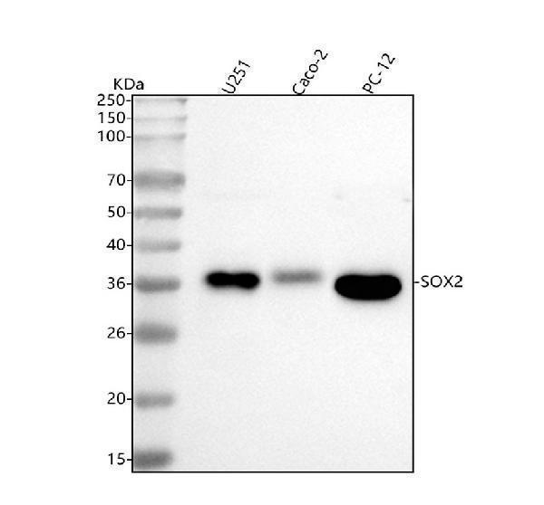
Western blot analysis of SOX2 using anti-SOX2 antibody (M00105-1).
Electrophoresis was performed on a 5-20% SDS-PAGE gel at 70V (Stacking gel) / 90V (Resolving gel) for 2-3 hours. The sample well of each lane was loaded with 30 ug of sample under reducing conditions.
Lane 1: human U251 whole cell lysates,
Lane 2: human CACO-2 whole cell lysates,
Lane 3: rat PC-12 whole cell lysates.
After electrophoresis, proteins were transferred to a nitrocellulose membrane at 150 mA for 50-90 minutes. Blocked the membrane with 5% non-fat milk/TBS for 1.5 hour at RT. The membrane was incubated with rabbit anti-SOX2 antigen affinity purified monoclonal antibody (Catalog # M00105-1) at 1:1000 overnight at 4°C, then washed with TBS-0.1%Tween 3 times with 5 minutes each and probed with a goat anti-rabbit IgG-HRP secondary antibody at a dilution of 1:500 for 1.5 hour at RT. The signal is developed using an Enhanced Chemiluminescent detection (ECL) kit (Catalog # EK1002) with Tanon 5200 system. A specific band was detected for SOX2 at approximately 34 kDa. The expected band size for SOX2 is at 34 kDa.

Click image to see more details
IHC analysis of FLOT1 using anti-FLOT1 antibody (M00105-1).
FLOT1 was detected in a paraffin-embedded section of human lung cancer tissue. Heat mediated antigen retrieval was performed in EDTA buffer (pH 8.0, epitope retrieval solution). The tissue section was blocked with 10% goat serum. The tissue section was then incubated with 1:50 rabbit anti-FLOT1 Antibody (M00105-1) overnight at 4°C. Peroxidase Conjugated Goat Anti-rabbit IgG was used as secondary antibody and incubated for 30 minutes at 37°C. The tissue section was developed using HRP Conjugated Rabbit IgG Super Vision Assay Kit (Catalog # SV0002) with DAB as the chromogen.

Click image to see more details
Identification of NSCs. (A) NSCs were cultured in serum-free medium for 3 days (Scale bar: 200/100 μm). (B)Representative images of NSCs labelled with Nestin(red) and SOX2(green). The cell nuclei are stained blue with DAPI (Scale bar: 20/50 μm). (C) Images of NSCs-derived differentiated cells. TUBB3 (purple), GFAP (red), and Olig2 (green) are positively expressed, indicating the presence of neurons, astrocytes, and oligodendrocytes, respectively (Scale bar: 100 μm).
Index in Materials Today Advances under a CC BY license. DOI: 10.1016/j.mtadv.2025.100639

Click image to see more details
IHC analysis of SOX2 using anti-SOX2 antibody (M00105-1).
SOX2 was detected in a paraffin-embedded section of human cervical cancer tissue. Heat mediated antigen retrieval was performed in EDTA buffer (pH 8.0, epitope retrieval solution). The tissue section was blocked with 10% goat serum. The tissue section was then incubated with 5 μg/ml rabbit anti-SOX2 Antibody (M00105-1) overnight at 4°C. HRP Conjugated Goat Anti-rabbit IgG was used as secondary antibody and incubated for 30 minutes at 37°C. The tissue section was developed using HRP Conjugated Rabbit IgG Super Vision Assay Kit (Catalog # SV0002) with DAB as the chromogen.

Click image to see more details
IHC analysis of SOX2 using anti-SOX2 antibody (M00105-1).
SOX2 was detected in a paraffin-embedded section of human tonsilitis tissue. Heat mediated antigen retrieval was performed in EDTA buffer (pH 8.0, epitope retrieval solution). The tissue section was blocked with 10% goat serum. The tissue section was then incubated with 5 μg/ml rabbit anti-SOX2 Antibody (M00105-1) overnight at 4°C. HRP Conjugated Goat Anti-rabbit IgG was used as secondary antibody and incubated for 30 minutes at 37°C. The tissue section was developed using HRP Conjugated Rabbit IgG Super Vision Assay Kit (Catalog # SV0002) with DAB as the chromogen.

Click image to see more details
IHC analysis of SOX2 using anti-SOX2 antibody (M00105-1).
SOX2 was detected in a paraffin-embedded section of mouse kidney tissue. Heat mediated antigen retrieval was performed in EDTA buffer (pH 8.0, epitope retrieval solution). The tissue section was blocked with 10% goat serum. The tissue section was then incubated with 5 μg/ml rabbit anti-SOX2 Antibody (M00105-1) overnight at 4°C. HRP Conjugated Goat Anti-rabbit IgG was used as secondary antibody and incubated for 30 minutes at 37°C. The tissue section was developed using HRP Conjugated Rabbit IgG Super Vision Assay Kit (Catalog # SV0002) with DAB as the chromogen.

Click image to see more details
IHC analysis of SOX2 using anti-SOX2 antibody (M00105-1).
SOX2 was detected in a paraffin-embedded section of mouse stomach tissue. Heat mediated antigen retrieval was performed in EDTA buffer (pH 8.0, epitope retrieval solution). The tissue section was blocked with 10% goat serum. The tissue section was then incubated with 5 μg/ml rabbit anti-SOX2 Antibody (M00105-1) overnight at 4°C. HRP Conjugated Goat Anti-rabbit IgG was used as secondary antibody and incubated for 30 minutes at 37°C. The tissue section was developed using HRP Conjugated Rabbit IgG Super Vision Assay Kit (Catalog # SV0002) with DAB as the chromogen.

Click image to see more details
IHC analysis of FLOT1 using anti-FLOT1 antibody (M00105-1).
FLOT1 was detected in a paraffin-embedded section of mouse brain tissue. Heat mediated antigen retrieval was performed in EDTA buffer (pH 8.0, epitope retrieval solution). The tissue section was blocked with 10% goat serum. The tissue section was then incubated with 1:50 rabbit anti-FLOT1 Antibody (M00105-1) overnight at 4°C. Peroxidase Conjugated Goat Anti-rabbit IgG was used as secondary antibody and incubated for 30 minutes at 37°C. The tissue section was developed using HRP Conjugated Rabbit IgG Super Vision Assay Kit (Catalog # SV0002) with DAB as the chromogen.

Click image to see more details
IHC analysis of FLOT1 using anti-FLOT1 antibody (M00105-1).
FLOT1 was detected in a paraffin-embedded section of rat brain tissue. Heat mediated antigen retrieval was performed in EDTA buffer (pH 8.0, epitope retrieval solution). The tissue section was blocked with 10% goat serum. The tissue section was then incubated with 1:50 rabbit anti-FLOT1 Antibody (M00105-1) overnight at 4°C. Peroxidase Conjugated Goat Anti-rabbit IgG was used as secondary antibody and incubated for 30 minutes at 37°C. The tissue section was developed using HRP Conjugated Rabbit IgG Super Vision Assay Kit (Catalog # SV0002) with DAB as the chromogen.

Click image to see more details
IF analysis of SOX2 using anti-SOX2 antibody (M00105-1).
SOX2 was detected in a paraffin-embedded section of human cervical cancer tissue. Heat mediated antigen retrieval was performed in EDTA buffer (pH 8.0, epitope retrieval solution). The tissue section was blocked with 10% goat serum. The tissue section was then incubated with 25 μg/mL rabbit anti-SOX2 Antibody (M00105-1) overnight at 4°C. DyLight®594 Conjugated Goat Anti-Rabbit IgG (BA1142) was used as secondary antibody at 1:100 dilution and incubated for 30 minutes at 37°C. The section was counterstained with DAPI. Visualize using a fluorescence microscope and filter sets appropriate for the label used.

Click image to see more details
IF analysis of SOX2 using anti-SOX2 antibody (M00105-1).
SOX2 was detected in a paraffin-embedded section of human testis cancer tissue. Heat mediated antigen retrieval was performed in EDTA buffer (pH 8.0, epitope retrieval solution). The tissue section was blocked with 10% goat serum. The tissue section was then incubated with 25 μg/mL rabbit anti-SOX2 Antibody (M00105-1) overnight at 4°C. DyLight®594 Conjugated Goat Anti-Rabbit IgG (BA1142) was used as secondary antibody at 1:100 dilution and incubated for 30 minutes at 37°C. The section was counterstained with DAPI. Visualize using a fluorescence microscope and filter sets appropriate for the label used.

Click image to see more details
Specific Publications For Anti-SOX2 Rabbit Monoclonal Antibody (M00105-1)
Loading publications
Recommended Resources
Here are featured tools and databases that you might find useful.
- Boster's Pathways Library
- Protein Databases
- Bioscience Research Protocol Resources
- Data Processing & Analysis Software
- Photo Editing Software
- Scientific Literature Resources
- Research Paper Management Tools
- Molecular Biology Software
- Primer Design Tools
- Bioinformatics Tools
- Phylogenetic Tree Analysis
Customer Reviews
Have you used Anti-SOX2 Rabbit Monoclonal Antibody?
Share your experimental results or join a short interview to earn up to $1,000 in product credits or other rewards.
0 Reviews For Anti-SOX2 Rabbit Monoclonal Antibody
Customer Q&As
Have a question?
Find answers in Q&As, reviews.
Can't find your answer?
Submit your question
4 Customer Q&As for Anti-SOX2 Rabbit Monoclonal Antibody
Question
We have seen staining in human fetal brain. Any tips? Is anti-SOX2 Rabbit Monoclonal antibody supposed to stain fetal brain positively?
Verified Customer
Verified customer
Asked: 2019-09-13
Answer
Based on literature fetal brain does express SOX2. Based on Uniprot.org, SOX2 is expressed in cerebral cortex, fetal brain, retina, lung, among other tissues. Regarding which tissues have SOX2 expression, here are a few articles citing expression in various tissues:
Fetal brain, Pubmed ID: 7849401
Lung, Pubmed ID: 15489334
Boster Scientific Support
Answered: 2019-09-13
Question
My boss were satisfied with the WB result of your anti-SOX2 Rabbit Monoclonal antibody. However we have seen positive staining in fetal brain nucleus. using this antibody. Is that expected? Could you tell me where is SOX2 supposed to be expressed?
Verified Customer
Verified customer
Asked: 2019-07-29
Answer
From what I have seen in literature, fetal brain does express SOX2. Generally SOX2 expresses in nucleus. Regarding which tissues have SOX2 expression, here are a few articles citing expression in various tissues:
Fetal brain, Pubmed ID: 7849401
Lung, Pubmed ID: 15489334
Boster Scientific Support
Answered: 2019-07-29
Question
We bought anti-SOX2 Rabbit Monoclonal antibody for IHC on retina in a previous experiment. I am using mouse, and We intend to use the antibody for IF next. you antibody examining retina as well as lung in our next experiment. Do you have any suggestion on which antibody would work the best for IF?
Verified Customer
Verified customer
Asked: 2019-07-22
Answer
I viewed the website and datasheets of our anti-SOX2 Rabbit Monoclonal antibody and it seems that M00105-1 has been tested on mouse in both IHC and IF. Thus M00105-1 should work for your application. Our Boster satisfaction guarantee will cover this product for IF in mouse even if the specific tissue type has not been validated. We do have a comprehensive range of products for IF detection and you can check out our website bosterbio.com to find out more information about them.
Boster Scientific Support
Answered: 2019-07-22
Question
We are currently using anti-SOX2 Rabbit Monoclonal antibody M00105-1 for human tissue, and we are happy with the IF results. The species of reactivity given in the datasheet says human, mouse. Is it true that the antibody can work on feline tissues as well?
Verified Customer
Verified customer
Asked: 2019-02-15
Answer
The anti-SOX2 Rabbit Monoclonal antibody (M00105-1) has not been validated for cross reactivity specifically with feline tissues, though there is a good chance of cross reactivity. We have an innovator award program that if you test this antibody and show it works in feline you can get your next antibody for free. Please contact me if I can help you with anything.
Boster Scientific Support
Answered: 2019-02-15
