This website uses cookies to ensure you get the best experience on our website.
- Table of Contents
30 Citations 10 Q&As
21 Citations 7 Q&As
12 Citations 10 Q&As
23 Citations 6 Q&As
3 Citations 1 Q&As
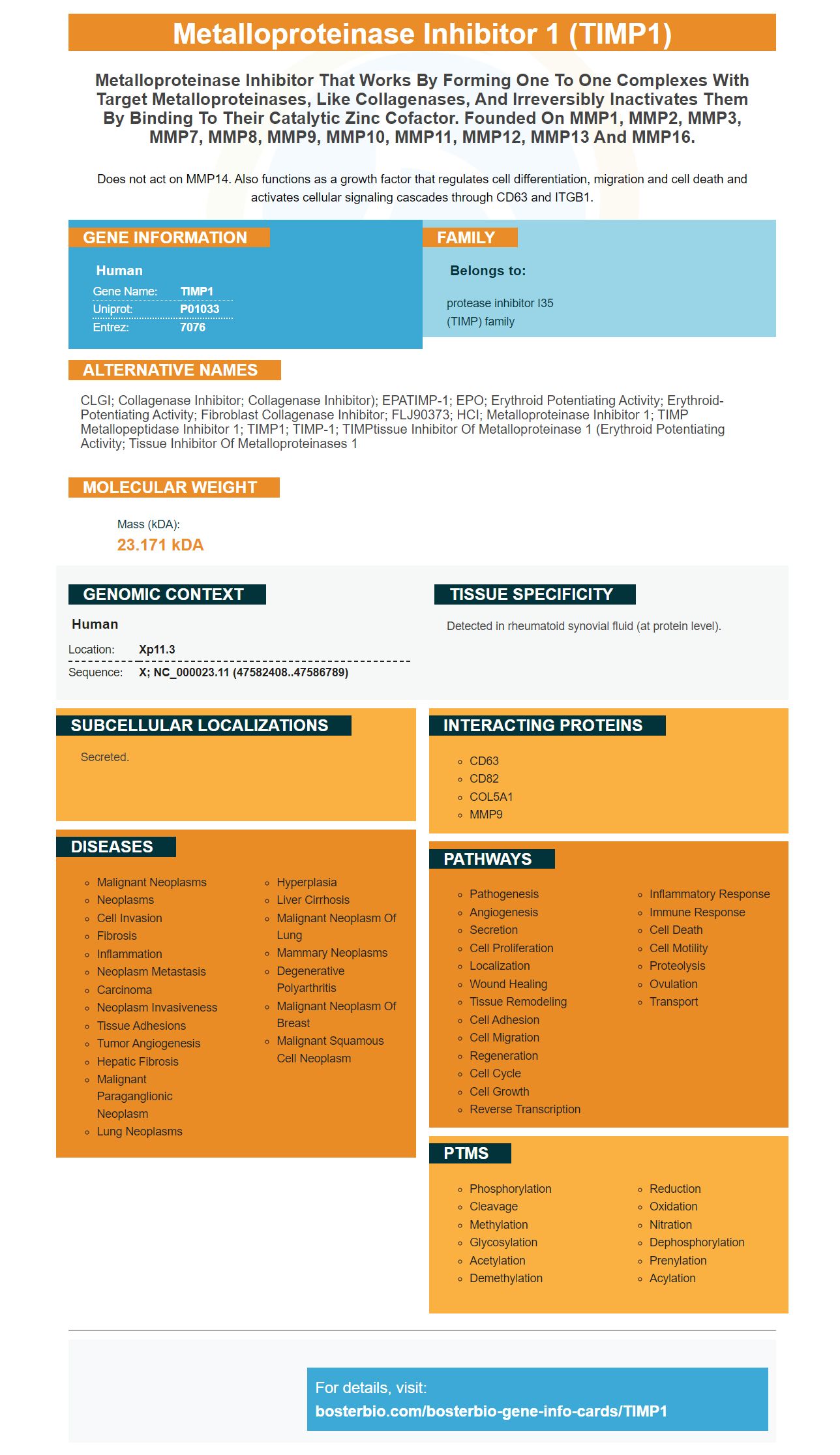
Facts about Metalloproteinase inhibitor 1.
Does not act on MMP14. Also functions as a growth factor that regulates cell differentiation, migration and cell death and activates cellular signaling cascades through CD63 and ITGB1.
Human | |
---|---|
Gene Name: | TIMP1 |
Uniprot: | P01033 |
Entrez: | 7076 |
Belongs to: |
---|
protease inhibitor I35 (TIMP) family |
CLGI; Collagenase inhibitor; collagenase inhibitor); EPATIMP-1; EPO; erythroid potentiating activity; Erythroid-potentiating activity; Fibroblast collagenase inhibitor; FLJ90373; HCI; metalloproteinase inhibitor 1; TIMP metallopeptidase inhibitor 1; TIMP1; TIMP-1; TIMPtissue inhibitor of metalloproteinase 1 (erythroid potentiating activity; Tissue inhibitor of metalloproteinases 1
Mass (kDA):
23.171 kDA
Human | |
---|---|
Location: | Xp11.3 |
Sequence: | X; NC_000023.11 (47582408..47586789) |
Detected in rheumatoid synovial fluid (at protein level).
Secreted.
You may be wondering how to maximize your BosterBio experiment. Here are some suggestions and tricks to aid you in maximizing your results. You can reduce the risk that your experiment will fail by implementing proper controls. Boster Bio's troubleshooting manual can assist you in identifying the most prevalent sources of error. This guide will help you identify the most common problems.
TIMP1 is a tumor marker that stimulates the cycle of cells in leukemic blasts from AML patients. It has been shown as a diagnostic tool to determine the presence of leukemia. In this study, the presence of TIMP-1 in the blasts from leukemic patients AML was measured by ELISA. The TIMP-1 levels in both AML samples and patient serum were compared to those of healthy controls.
TIMP1 was recently identified as a potential tumor marker. It was only recently discovered that TIMP1 is present in body fluids and blood. However its significance in biology has not been established. The results of this study provide a novel prognosis prediction tool , and suggest TIMP1 expression may be a novel therapeutic target. Researchers from the Broad Institute of MIT as along with the Cancer Genome Atlas carried out the study.
In addition to being a tumor marker, TIMP-1 is a tumor growth regulator. It also affects the clonogenic potential of CD34+ cells from healthy donors. Furthermore, it is involved in the CXCL12-mediated migration process, which is important in AML and BM stromal cells. In addition, TIMP-1 is involved in the retention of LSCs in the bone marrow.
The study was conducted with leukemia cells from 14 patients. The cells were stained with the pAkt (Thr308) antibody. The procedure was repeated with the same cells . The results it was found that TIMP-1 significantly promoted the survival of leukemic blasts. TIMP-1 also inhibited the apoptosis process, according to the study. This suggests that TIMP-1 could be used to detect leukemia-causing cells.
The TIMP1 marker serves a variety of functions. One of these functions is to stop the growth of colon cancer cells. It has been proven that it prevents the growth of colony in patients suffering from colon cancer. TIMP1 expression is also essential for colon cell proliferativeness, motility, and metastasis. Moreover, the TIMP1-KD cells showed less subcutaneous metastasis than the scramble cells.
TIMP1 is a secretory protein that are found in blood. It is not understood how it influences colon cancer development. This marker was previously used to identify metastatic disease. The mechanism of action remains unknown, therefore further studies are required to know the role played by TIMP1 in colon cancer. The TIMP1 marker could be a valuable tool for patients with colon cancer in the near future.
TIMP1 expression was shown to be associated with the progress of colon cancer during an initial study conducted by the Chinese Academy of Sciences. The study also showed that high levels of TIMP1 were associated with a shorter time to cure and overall survival. However, these results don't necessarily mean high levels of TIMP1 are a bad indicator. They suggest that high levels of TIMP1 expression is an early indicator for colon cancer progression.
Researchers from Boster Bio and the Broad Institute at MIT have identified a gene that has an abnormal expression is associated with a poor prognosis. TIMP1 expression has been associated with TNM stage, disease-free survival and the extent of liver metastasis within a variety of tumors. The gene is secretory, and can be detected in blood. TIMP1 can also be detected in the blood of patients with familial pancreatic and gastric cancers.
Boster Bio's TIMP1 marker does not only regulate cell proliferation but also inhibits cyclinB1 or cyclin D1 expressions in TNBC cells. The latter are required for the transition of cells from G1 to M phase. Furthermore the knockdown of TIMP-1 reduced the level of cyclin D1 in MDA MB-468 and MCF-10A cells, whereas TIMP-1's increased levels of regulation triggered cell growth in both types of cells.
In addition to demonstrating its efficacy in a variety of cancer cell varieties, TIMP-1 was also evaluated in human TNBC cells. TIMP-1 was targeted by shRNAs. Three shRNAs were employed and knockdown efficacy was determined using ELISA and real-time PCR. Further Boster Bio's TIMP1 marker blocks cyclinB1 expression in human TNBC cells.
In addition to being a powerful tumor suppressant, TIMP-1 has anti-apoptotic as well as matrix metalloproteinase activity. Numerous studies have revealed elevated levels of TIMP1 in tumors. Breast cancer can be classified into different molecular subtypes including triple-negative. The triple-negative subtype is linked to the worst clinical outcome.
The Boster Bio Anti-Cyclin B1 antibody has been confirmed for use in Flow Cytometry. The antibody recognizes Cyclin B1 in both mice and humans , and is extremely specific. It also blocks PIK3CG, and PDK1 which are vital to understand the regulation of the cell cycle. Inhibiting cyclin B1 protein expression in cancer cells is essential for proper tumorigenesis.
The use of sal is believed to induce apoptosis in a variety of cancer cells. Sal is able to induce apoptosis via caspase-dependent and independent pathways. This study looked at the activation of caspases in MCF-7 cells to discover the mechanism that artesunate works. Caspases are the most important regulator proteins that play a role in the process of apoptosis. They initiate an extrinsic and intrinsic apoptosis.
Sal treatment increased the number of Hs578T cells' pre-G1 phase. It also increased apoptosis at an increased rate than Sal alone. The Hs578T cells co-treated with Sal increased side-by-side, while cells treated with Sal alone did not grow. The degree of cellular separation increased. This is in line with the role of Sal in increasing cell separation.
The Sal compound enhanced DNA breakage and phosphorylated p53 and H2AX, and induced DNA foci by inhibiting p53. It also reduced p21 protein levels and increased the activity of the proteasome. The results may be helpful for combination-chemotherapy treatments. These compounds can also increase cytochrome C, which is essential for apoptosis.
Oxygen deprivation during slaughter of animals triggers caspases and triggers anapoptosis. The cells that lack oxygen undergo apoptosis by activating the intrinsic mitochondrial pathway. Caspase-3 is most commonly used as the mediator of this cleavage. This protease cuts proteins in the cytoskeletal system and other molecules. Caspase-9 is activated during apoptosis.
Recent studies have proven that TIMP1 boosts leukemic blast survival and clonogenicity, and decreased sensitivity to chemotherapy. These findings are in line with previous research on other hematological malignancies such as children with ALL. The expression of TIMP-1 within the leukemia cell line suggests that it may play a significant role in the microenvironment that leukemia cells reside. Additionally, these findings suggest that there exists a therapeutic concentration optimal for studies of functional.
Recent studies have suggested that TIMP1 could play a part in colon cancer progression. While the exact mechanism for this effect isn't fully understood, TIMP1 was found to block the BAD-mediated phosphorylation process. In addition, TIMP1 has anti-apoptotic activity. It reduces the sensitivity colon cancer cells to chemotherapy. This is an important result that will be further explored in the future research.
The Boster Bio TIMP1 gene is expressed in many types of cancer. Its aberrant expression has been associated with poor prognosis, TNM stage and the rate of recurrence of cancer. Additionally that the TIMP1 gene also affects liver metastasis, a key indicator of prognosis. Furthermore, TIMP1 expression is found in blood samples, and is a crucial marker in finding tumors and evaluating their response to chemotherapy.
It is also possible that TIMP-1 is an alternative therapeutic target for conventional cancer drugs. This hypothesis can be explored more deeply using the TIMP-1 cell model. Future studies using TIMP-1 cell models are expected to provide valuable data that will help optimize conventional anticancer drugs. The authors would like to thank Bristol-Myers Squibb, Pfizer, and Faulding for their support for this study.
PMID: 3903517 by Docherty A.J.P., et al. Sequence of human tissue inhibitor of metalloproteinases and its identity to erythroid-potentiating activity.
PMID: 3839290 by Gasson J.C., et al. Molecular characterization and expression of the gene encoding human erythroid-potentiating activity.
*Showing only the more recent 20. More publications can be found for each product on its corresponding product page