Click image to see more details
Product Info Summary
| SKU: | DZ07988 |
|---|---|
| Size: | 200 μl/vial |
| Reactive Species: | Zebrafish |
| Host: | Rabbit |
| Application: | IF, IHC |
Customers Who Bought This Also Bought

Product info
Product Name
Anti-Zebrafish cdhr1a Antibody
SKU/Catalog Number
DZ07988
Size
200 μl/vial
Form
Liquid
Description
Boster Bio Polyclonal Anti-cdhr1a Antibody catalog # DZ07988. This antibody reacts with Zebrafish.
Storage & Handling
Store at -20˚C for one year from date of receipt. After reconstitution, at 4˚C for one month. It can also be aliquotted and stored frozen at -20˚C for six months. Avoid repeated freeze-thaw cycles.
Cite This Product
Anti-Zebrafish cdhr1a Antibody (Boster Biological Technology, Pleasanton CA, USA, Catalog # DZ07988)
Host
Rabbit
Contents
Each vial contains 50% glycerol, 0.9% NaCl, 0.2% Na2HPO4, 0.02% NaN3.
Clonality
Polyclonal
Isotype
Rabbit IgG
Reactive Species
DZ07988 is reactive to cdhr1 in Zebrafish
Application & Images
Applications
DZ07988 is guaranteed for IF, IHC Boster Guarantee
Recommend Dilution
| Application | Dilution | Species |
|---|---|---|
| Immunohistochemistry(Paraffin-embedded Section) | 2-5μg/ml | |
| Immunofluorescence | 5 μg/ml |
Validation Images & Assay Conditions
Click image to see more details
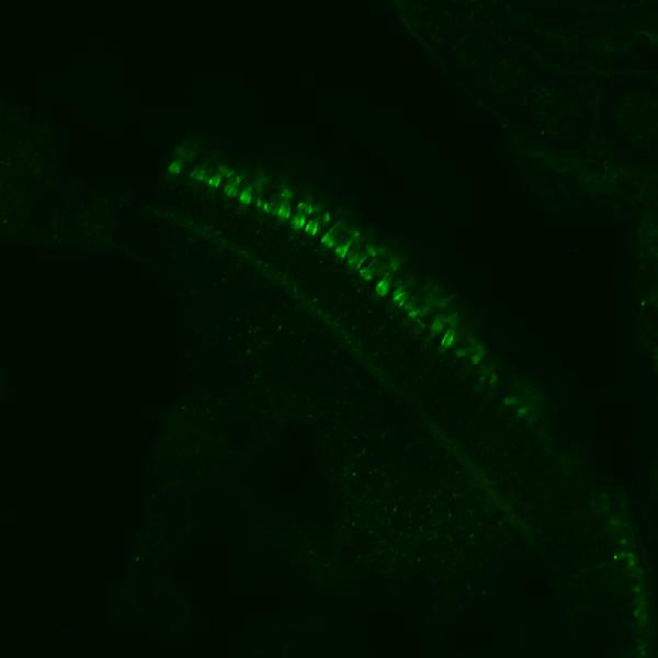
IF analysis of Cdhr1a using anti-Cdhr1a antibody (DZ07988).
Cdhr1a was detected in paraffin-embedded section of five-day-old zebrafish embryos tissue. Heat mediated antigen retrieval was performed in EDTA buffer (pH8.0, epitope retrieval solution). The tissue section was blocked with 10% goat serum. The tissue section was then incubated with 5μg/mL rabbit anti-Cdhr1a Antibody (DZ07988) overnight at 4°C. Biotin conjugated goat anti-rabbit IgG (BA1003) was used as secondary antibody and incubated for 30 minutes at 37°C. The tissue section was developed using DyLight®488 Conjugated Avidin (BA1128). Visualize using a fluorescence microscope and filter sets appropriate for the label used.

Click image to see more details
Evolutionarily conserved localization of pcdh15 and cdhr1 in photoreceptors predicts interactions linking outer segments and calyceal processes.
A-A”) Confocal microscopy of wildtype 5dpf retinal cryosections probed with cdhr1a antibody (green), Peanut germ agglutinin (PNA - magenta), Wheat germ agglutinin (WG - teal) and pcdh15b (red). White boxes indicate the location of the inset enlargement. White arrowheads highlight the linear localization of cdhr1a along cone OSs (A) and pcdh15b outlining the calyceal process (A’). Merge of all three signals highlights the proximity between cdhr1a and pcdh15b (A”). Scale bar = 10μm. B) Confocal microscopy of wildtype 5dpf retinal cryosections probed with cdhr1a antibody (green), pcdh15b (red) and WGA (teal). White boxes indicate the location of the inset enlargement. White arrowheads highlight the linear localization of cdhr1a along rod OSs (B) and pcdh15b outlining the calyceal process (B’). Merge of all three signals highlights the proximity between cdhr1a and pcdh15b (B”). Scale bar = 10μm. C) Structured illumination microscopy (SIM) of wildtype zebrafish retinal cryosections probed with cdhr1a (green) and pcdh15b (red). White box represents the inset enlargement. White arrowheads highlight the juxtaposition of the cdhr1a and pcdh15b signals. Scale bar = 10μm. D) Diagrammatic model of the connection between the OS discs and CPs in both rod and cone cells mediated by the interaction between OS bound cdhr1a and CP bound pcdh15b. E-L) Structured illumination microscopy (SIM) of wildtype xenopus (E), mallard duck (F), mouse (G), rat (H), spiny mouse (I), gerbil (J), macaque (K) and human (L) retinal sections probed for cdhr1 (green) and pcdh15 (red). White boxes represent the inset enlargements. White arrowheads highlight the juxtaposition of the cdhr1a and pchd15b signals in each species. The model figure was prepared using Biorender. Scale bar = 10μm.
Index in eLife under a CC BY license. DOI: 10.7554/eLife.102258.1

Click image to see more details
IHC analysis of Cdhr1a using anti-Cdhr1a antibody (DZ07988).
Cdhr1a was detected in paraffin-embedded section of zebrafish kidney tissue. Heat mediated antigen retrieval was performed in EDTA buffer (pH8.0, epitope retrieval solution). The tissue section was blocked with 10% goat serum. The tissue section was then incubated with 2μg/ml rabbit anti-Cdhr1a Antibody (DZ07988) overnight at 4°C. Peroxidase Conjugated Goat Anti-rabbit IgG was used as secondary antibody and incubated for 30 minutes at 37°C. The tissue section was developed using HRP Conjugated Rabbit IgG Super Vision Assay Kit (Catalog # SV0002) with DAB as the chromogen.
Specific Publications For Anti-Zebrafish cdhr1a Antibody (DZ07988)
Loading publications
Recommended Resources
Here are featured tools and databases that you might find useful.
- Boster's Pathways Library
- Protein Databases
- Bioscience Research Protocol Resources
- Data Processing & Analysis Software
- Photo Editing Software
- Scientific Literature Resources
- Research Paper Management Tools
- Molecular Biology Software
- Primer Design Tools
- Bioinformatics Tools
- Phylogenetic Tree Analysis
Customer Reviews
Have you used Anti-Zebrafish cdhr1a Antibody?
Share your experimental results or join a short interview to earn up to $1,000 in product credits or other rewards.
1 Reviews For Anti-Zebrafish cdhr1a Antibody
Good Zebrafish CDHR1a Antibody from Boster Bio--Jakub Famulski, University of Kentucky, Biology, Principal Investigator
Excellent

Source: Biocompare.com
| SKU | DZ07988 |
|---|---|
| Application | Immunofluorescence |
| Sample | Zebrafish embryos, 5 days old |
| Detection | Confocal Microscopy |
"The antibody clearly labelled the outer segments of photoreceptor cells. The antibody is clean and precise. A zebrafish specific antibody for cdhr1a that WORKS!"
Jakub Famulski
Verified customer
Submitted 2019-12-06
Customer Q&As
Have a question?
Find answers in Q&As, reviews.
Can't find your answer?
Submit your question
