Click image to see more details
-

-
-
-
-
+1
Product Info Summary
| SKU: | M00096-7 |
|---|---|
| Size: | 100 μl/vial |
| Reactive Species: | Human, Mouse, Rat |
| Host: | Rabbit |
| Application: | Flow Cytometry, IP, IF, IHC, ICC, WB |
Customers Who Bought This Also Bought

Product info
Product Name
Anti-PDGFRB Antibody (Monoclonal, 33P28)
SKU/Catalog Number
M00096-7
Size
100 μl/vial
Form
Liquid
Description
Boster Bio Anti-PDGFRB Antibody (Monoclonal, 33P28) catalog # M00096-7. Tested in WB, IHC, IF, ICC/IF, IP, Flow Cytometry applications. This antibody reacts with Human, Mouse, Rat.
Storage & Handling
Store at -20°C for one year. For short term storage and frequent use, store at 4°C for up to one month. Avoid repeated freeze-thaw cycles.
Cite This Product
Anti-PDGFRB Antibody (Monoclonal, 33P28) (Boster Biological Technology, Pleasanton CA, USA, Catalog # M00096-7)
Host
Rabbit
Contents
Rabbit IgG in stabilizing components, phosphate buffered saline, pH 7.4, 150mM NaCl, 0.02% sodium azide and 50% glycerol.
This antibody is supplied in a stabilized formulation.
Compatibility with conjugation reactions depends on the chemistry of the conjugation method used.
For conjugation methods that are not compatible with the stabilizing components present in this formulation, a carrier-free antibody format is required.
Clonality
Monoclonal
Clone Number
33P28
Immunogen
Recombinant protein within Human PDGFR beta aa 963-1104.
Reactive Species
M00096-7 is reactive to Pdgfrb in Human, Mouse, Rat
Observed Molecular Weight
180 kDa
Calculated molecular weight
122.8 kDa
Background of Pdgfrb
Beta-type platelet-derived growth factor receptor is a protein that in humans is encoded by the PDGFRB gene. The protein encoded by this gene is a cell surface tyrosine kinase receptor for members of the platelet-derived growth factor family. These growth factors are mitogens for cells of mesenchymal origin. The identity of the growth factor bound to a receptor monomer determines whether the functional receptor is a homodimer (PDGFB or PDGFD) or a heterodimer (PDGFA and PDGFB). This gene is essential for normal development of the cardiovascular system and aids in rearrangement of the actin cytoskeleton. This gene is flanked on chromosome 5 by the genes for granulocyte-macrophage colony-stimulating factor and macrophage-colony stimulating factor receptor; all three genes may be implicated in the 5-q syndrome. A translocation between chromosomes 5 and 12, that fuses this gene to that of the ETV6 gene, results in chronic myeloproliferative disorder with eosinophilia.
Antibody Validation
Boster validates all antibodies on WB, IHC, ICC, Immunofluorescence, and ELISA with known positive control and negative samples to ensure specificity and high affinity, including thorough antibody incubations.
Application & Images
Applications
M00096-7 is guaranteed for Flow Cytometry, IP, IF, IHC, ICC, WB Boster Guarantee
Assay Dilutions Recommendation
The recommendations below provide a starting point for assay optimization. The actual working concentration varies and should be decided by the user.
Western blot, 1:500-2000
Immunohistochemistry, 1:50-200
Immunofluorescence, 1:50-200
Immunocytochemistry/Immunofluorescence, 1:50-200
ImmunoPrecipitation, 1:50
Flow Cytometry (Fixed), 1:50-200
Positive Control
WB: human SH-SY5Y whole cell, rat L6 whole cell, rat C6 whole cell, mouse C2C12 whole cell, mouse NIH/3T3 whole cell
IHC: human melanoma tissue, human breast cancer tissue, mouse kidney tissue, rat kidney tissue
Validation Images & Assay Conditions
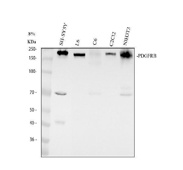
Click image to see more details
Western blot analysis of PDGFRB using anti-PDGFRB antibody (M00096-7).
Electrophoresis was performed on a 8% SDS-PAGE gel at 80V (Stacking gel) / 120V (Resolving gel) for 2 hours. The sample well of each lane was loaded with 30 ug of sample under reducing conditions.
Lane 1: human SH-SY5Y whole cell lysates,
Lane 2: rat L6 whole cell lysates,
Lane 3: rat C6 whole cell lysates,
Lane 4: mouse C2C12 whole cell lysates,
Lane 5: mouse NIH/3T3 whole cell lysates.
After electrophoresis, proteins were transferred to a nitrocellulose membrane at 150 mA for 50-90 minutes. Blocked the membrane with 5% non-fat milk/TBS for 1.5 hour at RT. The membrane was incubated with rabbit anti-PDGFRB antigen affinity purified monoclonal antibody (M00096-7) at 1:1000 overnight at 4°C, then washed with TBS-0.1%Tween 3 times with 5 minutes each and probed with a goat anti-rabbit IgG-HRP secondary antibody at a dilution of 1:5000 for 1.5 hour at RT. The signal is developed using an ECL Plus Western Blotting Substrate (Catalog # AR1196-200) with Tanon 5200 system. A specific band was detected for PDGFRB at approximately 180 kDa. The expected band size for PDGFRB is at 123 kDa.

Click image to see more details
IHC analysis of PDGFRB using anti-PDGFRB antibody (M00096-7).
PDGFRB was detected in a paraffin-embedded section of human melanoma tissue. Heat mediated antigen retrieval was performed in EDTA buffer (pH 8.0, epitope retrieval solution). The tissue section was blocked with 10% goat serum. The tissue section was then incubated with 1:200 rabbit anti-PDGFRB Antibody (M00096-7) overnight at 4°C. Peroxidase Conjugated Goat Anti-rabbit IgG was used as secondary antibody and incubated for 30 minutes at 37°C. The tissue section was developed using HRP Conjugated Rabbit IgG Super Vision Assay Kit (Catalog # SV0002) with DAB as the chromogen.

Click image to see more details
IHC analysis of PDGFRB using anti-PDGFRB antibody (M00096-7).
PDGFRB was detected in a paraffin-embedded section of human breast cancer tissue. Heat mediated antigen retrieval was performed in EDTA buffer (pH 8.0, epitope retrieval solution). The tissue section was blocked with 10% goat serum. The tissue section was then incubated with 1:200 rabbit anti-PDGFRB Antibody (M00096-7) overnight at 4°C. Peroxidase Conjugated Goat Anti-rabbit IgG was used as secondary antibody and incubated for 30 minutes at 37°C. The tissue section was developed using HRP Conjugated Rabbit IgG Super Vision Assay Kit (Catalog # SV0002) with DAB as the chromogen.

Click image to see more details
IHC analysis of PDGFRB using anti-PDGFRB antibody (M00096-7).
PDGFRB was detected in a paraffin-embedded section of mouse kidney tissue. Heat mediated antigen retrieval was performed in EDTA buffer (pH 8.0, epitope retrieval solution). The tissue section was blocked with 10% goat serum. The tissue section was then incubated with 1:200 rabbit anti-PDGFRB Antibody (M00096-7) overnight at 4°C. Peroxidase Conjugated Goat Anti-rabbit IgG was used as secondary antibody and incubated for 30 minutes at 37°C. The tissue section was developed using HRP Conjugated Rabbit IgG Super Vision Assay Kit (Catalog # SV0002) with DAB as the chromogen.

Click image to see more details
IHC analysis of PDGFRB using anti-PDGFRB antibody (M00096-7).
PDGFRB was detected in a paraffin-embedded section of rat kidney tissue. Heat mediated antigen retrieval was performed in EDTA buffer (pH 8.0, epitope retrieval solution). The tissue section was blocked with 10% goat serum. The tissue section was then incubated with 1:200 rabbit anti-PDGFRB Antibody (M00096-7) overnight at 4°C. Peroxidase Conjugated Goat Anti-rabbit IgG was used as secondary antibody and incubated for 30 minutes at 37°C. The tissue section was developed using HRP Conjugated Rabbit IgG Super Vision Assay Kit (Catalog # SV0002) with DAB as the chromogen.
Specific Publications For Anti-PDGFRB Antibody (Monoclonal, 33P28) (M00096-7)
Loading publications
Recommended Resources
Here are featured tools and databases that you might find useful.
- Boster's Pathways Library
- Protein Databases
- Bioscience Research Protocol Resources
- Data Processing & Analysis Software
- Photo Editing Software
- Scientific Literature Resources
- Research Paper Management Tools
- Molecular Biology Software
- Primer Design Tools
- Bioinformatics Tools
- Phylogenetic Tree Analysis
Customer Reviews
Have you used Anti-PDGFRB Antibody (Monoclonal, 33P28)?
Share your experimental results or join a short interview to earn up to $1,000 in product credits or other rewards.
0 Reviews For Anti-PDGFRB Antibody (Monoclonal, 33P28)
Customer Q&As
Have a question?
Find answers in Q&As, reviews.
Can't find your answer?
Submit your question
