Click image to see more details
-

-
-
-
-
+5
Product Info Summary
| SKU: | M03989 |
|---|---|
| Size: | 0.1 mg |
| Reactive Species: | Human, Mouse, Pig, Turkey, Yeast, Arabidopsis, Eisenia, Paramecium, Nicotiana |
| Host: | Mouse |
| Application: | Flow Cytometry, IP, IHC-P, ICC, WB |
Customers Who Bought This Also Bought

Product info
Product Name
Anti-alpha-Tubulin Purified TUBA1A Monoclonal Antibody
SKU/Catalog Number
M03989
Size
0.1 mg
Form
Liquid
Description
Boster Bio Anti-alpha-Tubulin Purified TUBA1A Monoclonal Antibody (Catalog# M03989). Tested in WB, IHC-P, ICC, IP, Flow Cytometry application(s). This antibody reacts with Mouse, Pig, Human, Turkey, Eisenia, Paramecium, Nicotiana, Yeast, Arabidopsis.
Storage & Handling
Store at 2-8°C. Do not freeze.
Cite This Product
Anti-alpha-Tubulin Purified TUBA1A Monoclonal Antibody (Boster Biological Technology, Pleasanton CA, USA, Catalog # M03989)
Host
Mouse
Contents
Phosphate buffered saline (PBS), pH 7.4, 15 mM sodium azide
Clonality
Monoclonal
Clone Number
TU-01
Isotype
Mouse IgG1
Immunogen
Fraction of tubulin purified from porcine brain by two cycles of polymerization - depolymerization. The antibody TU-01 recognizes a defined epitope (aa 65-97) on N-terminal structural domain of alpha-tubulin.
Cross-reactivity
This antibody does not cross-react with Thy-1.1 alloantigen.
Reactive Species
M03989 is reactive to TUBA1 in Human, Mouse, Pig, Turkey, Yeast, Arabidopsis, Eisenia, Paramecium, Nicotiana
Observed Molecular Weight
42 kDa
Calculated molecular weight
50.1 kDa
Background of TUBA1
The microtubules are intracellular dynamic polymers made up of evolutionarily conserved polymorphic alpha/beta-tubulin heterodimers and a large number of microtubule-associated proteins (MAPs). The microtubules consist of 13 protofilaments and have an outer diameter 25 nm. Microtubules have their intrinsic polarity; highly dynamic plus ends and less dynamic minus ends. Microtubules are required for vital processes in eukaryotic cells including mitosis, meiosis, maintenance of cell shape and intracellular transport. Microtubules are also necessary for movement of cells by means of flagella and cilia. In mammalian tissue culture cells microtubules have their minus ends anchored in microtubule organizing centers (MTOCs). The GTP (guanosintriphosphate) molecule is an essential for tubulin heterodimer to associate with other heterodimers to form microtubule. In vivo, microtubule dynamics vary considerably. Microtubule polymerization is reversible and a populations of microtubules in cells are on their minus ends either growing or shortening –; this phenomenon is called dynamic instability of microtubules. On a practical level, microtubules can easily be stabilized by the addition of non-hydrolysable analogues of GTP (eg. GMPPCP) or more commonly by anti-cancer drugs such as Taxol. Taxol stabilizes microtubules at room temperature for many hours. Using limited proteolysis by enzymes both tubulin subunits can be divided into N-terminal and C-terminal structural domains. The alpha-tubulin (relative molecular weight around 50 kDa) is globular protein that exists in cells as part of soluble alpha/beta-tubulin dimer or it is polymerized into microtubules. In different species it is coded by multiple tubulin genes that form tubulin classes (in human 6 genes). Expressed tubulin genes are named tubulin isotypes. Some of the tubulin isotypes are expressed ubiquitously, while some have more restricted tissue expression. Alpha-tubulin is also subject of numerous post-translational modifications. Tubulin isotypes and their posttranslational modifications are responsible for multiple tubulin charge variants - tubulin isoforms. Heterogeneity of alpha-tubulin is concentrated in C-terminal structural domain.
Antibody Validation
Boster validates all antibodies on WB, IHC, ICC, Immunofluorescence, and ELISA with known positive control and negative samples to ensure specificity and high affinity, including thorough antibody incubations.
Application & Images
Applications
M03989 is guaranteed for Flow Cytometry, IP, IHC-P, ICC, WB Boster Guarantee
Recommend Dilution
| Application | Dilution | Species |
|---|---|---|
| Flow cytometry: 1-4 μg/ml | intracellular staining. | |
| Western blotting: 1-2 μg/ml | reducing conditions. |
Validation Images & Assay Conditions
Click image to see more details
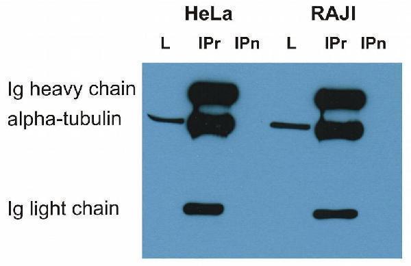
Immunoprecipitation of alpha-tubulin from HeLa and RAJI cell lysate by antibody TU-16 and its detection by antibody TU-01. IgM heavy chain (76-92 kDa) and IgM light chain (25-30 kDa) indicated. Mr of alpha tubulin is around 50 kDa.
L = lysate
IPr = immunoprecipitate (reducing conditions)
IPn = immunoprecipitate (non-reducing conditions)

Click image to see more details
Separation of HeLa cells stained using anti-alpha-Tubulin (TU-01) purified antibody (concentration in sample 3 µg/ml, GAM APC, red-filled) from HeLa cells unstained by primary antibody (GAM APC, black-dashed) in flow cytometry analysis (intracellular staining).

Click image to see more details
Immunocytochemistry staining of 3T3 mouse embryonal fibroblast cell line using anti-alpha-tubulin (TU-01; green) and anti-Vimentin (VI-01; red). Nucleus is stained with DAPI (blue).

Click image to see more details
Immunocytochemistry staining of HeLa human cervix carcinoma cell line using anti-alpha-tubulin (TU-01; red). Nucleus is stained with DAPI (blue).

Click image to see more details
Immunocytochemistry staining of 3T3 mouse embryonal fibroblast cell line using anti-alpha-tubulin (TU-01; green). Nucleus is stained with DAPI (blue).

Click image to see more details
Immunohistochemistry staining of human heart (paraffin sections) using anti-alpha-tubulin (TU-01). Commercially tested by LifeSpan BioSciences.

Click image to see more details
Western blotting analysis of human alpha-tubulin using mouse monoclonal antibody TU-01 on lysates of various cell lines under reducing and non-reducing conditions. Nitrocellulose membrane was probed with 2 µg/ml of mouse anti-alpha-tubulin monoclonal antibody followed by IRDye800-conjugated anti-mouse secondary antibody. A specific band was detected for alpha-tubulin at approximately 54 kDa.

Click image to see more details
Use of anti-alpha-tubulin antibody TU-01 as a loading control (A) in an Western blotting experiment revealing the staining pattern of various cell lysates by a newly developed monoclonal antibody (B).

Click image to see more details
Anti-alpha-Tubulin Purified (TU-01) works in WB application under reducing conditions.
Western blotting analysis was performed on whole cell extracts (RIPA lysis buffer) of JAR, JEG3, HTr-8/SVneo, and HeLa cell lines mixed and heated (100°C, 5 min) with reducing (2-mercaptoethanol) or non-reducing SDS-loading buffer. Samples were resolved using 10% SDS-PAGE gel.
Nitrocellulose membrane blot was probed simultaneously with mouse IgG1 monoclonal antibody TU-01 (1 µg/ml) and mouse IgM monoclonal antibody VI-01 detecting vimentin. Subclass-specific secondary antibodies IRDye 800CW Goat-anti-Mouse IgG (green) and IRDye 680RD Goat-anti-Mouse IgM (red) were used for multiplex fluorescent Western blot detection.
Alpha-tubulin was detected at ~50 kDa and vimentin at ~55 kDa.
Specific Publications For Anti-alpha-Tubulin Purified TUBA1A Monoclonal Antibody (M03989)
Loading publications
Recommended Resources
Here are featured tools and databases that you might find useful.
- Boster's Pathways Library
- Protein Databases
- Bioscience Research Protocol Resources
- Data Processing & Analysis Software
- Photo Editing Software
- Scientific Literature Resources
- Research Paper Management Tools
- Molecular Biology Software
- Primer Design Tools
- Bioinformatics Tools
- Phylogenetic Tree Analysis
Customer Reviews
Have you used Anti-alpha-Tubulin Purified TUBA1A Monoclonal Antibody?
Share your experimental results or join a short interview to earn up to $1,000 in product credits or other rewards.
0 Reviews For Anti-alpha-Tubulin Purified TUBA1A Monoclonal Antibody
Customer Q&As
Have a question?
Find answers in Q&As, reviews.
Can't find your answer?
Submit your question
