Click image to see more details
Product Info Summary
| SKU: | M00133-2 |
|---|---|
| Size: | 100 μl/vial |
| Reactive Species: | Human, Mouse |
| Host: | Rabbit |
| Application: | Flow Cytometry, IF, IHC, ICC, WB |
Customers Who Bought This Also Bought

Product info
Product Name
Anti-ABL1 Antibody (Monoclonal, 33A80)
SKU/Catalog Number
M00133-2
Size
100 μl/vial
Form
Liquid
Description
Boster Bio Anti-ABL1 Antibody (Monoclonal, 33A80) catalog # M00133-2. Tested in WB, IHC, ICC/IF, Flow Cytometry applications. This antibody reacts with Human, Mouse.
Storage & Handling
Store at -20°C for one year. For short term storage and frequent use, store at 4°C for up to one month. Avoid repeated freeze-thaw cycles.
Cite This Product
Anti-ABL1 Antibody (Monoclonal, 33A80) (Boster Biological Technology, Pleasanton CA, USA, Catalog # M00133-2)
Host
Rabbit
Contents
Rabbit IgG in stabilizing components, phosphate buffered saline, pH 7.4, 150mM NaCl, 0.02% sodium azide and 50% glycerol.
This antibody is supplied in a stabilized formulation.
Compatibility with conjugation reactions depends on the chemistry of the conjugation method used.
For conjugation methods that are not compatible with the stabilizing components present in this formulation, a carrier-free antibody format is required.
Clonality
Monoclonal
Clone Number
33A80
Immunogen
Recombinant protein within human ABL1 aa 603-998.
Reactive Species
M00133-2 is reactive to ABL1 in Human, Mouse
Calculated molecular weight
122.9 kDa
Background of ABL1
c Abl is also called as ABL1. This gene is a protooncogene that encodes a protein tyrosine kinase involved in a variety of cellular processes, including cell division, adhesion, differentiation, and response to stress. The activity of the protein is negatively regulated by its SH3 domain, whereby deletion of the region encoding this domain results in an oncogene. The ubiquitously expressed protein has DNA-binding activity that is regulated by CDC2-mediated phosphorylation, suggesting a cell cycle function. This gene has been found fused to a variety of translocation partner genes in various leukemias, most notably the t(9;22) translocation that results in a fusion with the 5' end of the breakpoint cluster region gene. Alternative splicing of this gene results in two transcript variants, which contain alternative first exons that are spliced to the remaining common exons.
Antibody Validation
Boster validates all antibodies on WB, IHC, ICC, Immunofluorescence, and ELISA with known positive control and negative samples to ensure specificity and high affinity, including thorough antibody incubations.
Application & Images
Applications
M00133-2 is guaranteed for Flow Cytometry, IF, IHC, ICC, WB Boster Guarantee
Recommend Dilution
| Application | Dilution | Species |
|---|---|---|
| Western blot | 1:500-2000 | |
| Immunohistochemistry | 1:50-200 | |
| Immunocytochemistry/Immunofluorescence | 1:50-200 | |
| Flow Cytometry (Fixed) | 1:50-200 |
Tested application
Use TE buffer pH 9.0 for antigen retrieval; (*) citrate buffer pH 6.0 is an alternative.
Validation Images & Assay Conditions
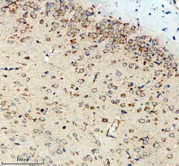
Click image to see more details
IHC analysis of ABL1 using anti-ABL1 antibody (M00133-2).
ABL1 was detected in a paraffin-embedded section of mouse brain tissue. Heat mediated antigen retrieval was performed in EDTA buffer (pH 8.0, epitope retrieval solution). The tissue section was blocked with 10% goat serum. The tissue section was then incubated with 1:50 rabbit anti-ABL1 Antibody (M00133-2) overnight at 4°C. Peroxidase Conjugated Goat Anti-rabbit IgG was used as secondary antibody and incubated for 30 minutes at 37°C. The tissue section was developed using HRP Conjugated Rabbit IgG Super Vision Assay Kit (Catalog # SV0002) with DAB as the chromogen.

Click image to see more details
IHC analysis of ABL1 using anti-ABL1 antibody (M00133-2).
ABL1 was detected in a paraffin-embedded section of rat brain tissue. Heat mediated antigen retrieval was performed in EDTA buffer (pH 8.0, epitope retrieval solution). The tissue section was blocked with 10% goat serum. The tissue section was then incubated with 1:50 rabbit anti-ABL1 Antibody (M00133-2) overnight at 4°C. Peroxidase Conjugated Goat Anti-rabbit IgG was used as secondary antibody and incubated for 30 minutes at 37°C. The tissue section was developed using HRP Conjugated Rabbit IgG Super Vision Assay Kit (Catalog # SV0002) with DAB as the chromogen.
Specific Publications For Anti-ABL1 Antibody (Monoclonal, 33A80) (M00133-2)
Loading publications
Recommended Resources
Here are featured tools and databases that you might find useful.
- Boster's Pathways Library
- Protein Databases
- Bioscience Research Protocol Resources
- Data Processing & Analysis Software
- Photo Editing Software
- Scientific Literature Resources
- Research Paper Management Tools
- Molecular Biology Software
- Primer Design Tools
- Bioinformatics Tools
- Phylogenetic Tree Analysis
Customer Reviews
Have you used Anti-ABL1 Antibody (Monoclonal, 33A80)?
Share your experimental results or join a short interview to earn up to $1,000 in product credits or other rewards.
0 Reviews For Anti-ABL1 Antibody (Monoclonal, 33A80)
Customer Q&As
Have a question?
Find answers in Q&As, reviews.
Can't find your answer?
Submit your question
