Click image to see more details
-

-
-
-
-
+1
Product Info Summary
| SKU: | PB9752 |
|---|---|
| Size: | 100 μg/vial |
| Reactive Species: | Human, Mouse, Rat |
| Host: | Rabbit |
| Application: | WB |
Customers Who Bought This Also Bought

Product info
Product Name
Anti-alpha 1d Adrenergic Receptor/ADRA1A Antibody Picoband®
SKU/Catalog Number
PB9752
PB0798 is an alternative SKU for this antibody, used in previous lots.
Size
100 μg/vial
Form
Lyophilized
Description
Boster Bio Anti-alpha 1d Adrenergic Receptor/ADRA1A Antibody Picoband® catalog # PB9752. Tested in WB applications. This antibody reacts with Human, Mouse, Rat. The brand Picoband indicates this is a premium antibody that guarantees superior quality, high affinity, and strong signals with minimal background in Western blot applications. Only our best-performing antibodies are designated as Picoband, ensuring unmatched performance.
Storage & Handling
Store at -20˚C for one year from date of receipt. After reconstitution, at 4˚C for one month. It can also be aliquotted and stored frozen at -20˚C for six months. Avoid repeated freeze-thaw cycles.
Cite This Product
Anti-alpha 1d Adrenergic Receptor/ADRA1A Antibody Picoband® (Boster Biological Technology, Pleasanton CA, USA, Catalog # PB9752)
Host
Rabbit
Contents
Each vial contains antibody formulated with stabilizing components, 0.9 mg NaCl, 0.2 mg Na2HPO4, and 0.05 mg NaN3.
*This antibody is supplied in a stabilized formulation.
Compatibility with conjugation reactions depends on the chemistry of the conjugation method used.
For conjugation methods that are not compatible with the stabilizing components present in this formulation, a carrier-free antibody format is required.
Clonality
Polyclonal
Isotype
Rabbit IgG
Immunogen
A synthetic peptide corresponding to a sequence at the C-terminus of human ADRA1A, different from the related mouse and rat sequences by four amino acids.
Cross-reactivity
No cross-reactivity with other proteins.
Reactive Species
PB9752 is reactive to ADRA1A in Human, Mouse, Rat
Observed Molecular Weight
51 kDa
Calculated molecular weight
51.5 kDa
Background of ADRA1A
ADRA1A, also known as alpha-1A adrenergic receptor, is an alpha-1 adrenergic receptor, and also denotes the human gene encoding it. This gene is mapped to 8p21.2. Alpha-1-adrenergic receptors are G protein-coupled transmembrane receptors that mediate actions in the sympathetic nervous system through the binding of the catecholamines, epinephrine and norepinephrine. It has been found that ADRA1A transcripts in heart, brain, liver, and prostate. ADRA1A is the predominant ADRA1 subtype in liver and heart, and it can mediate the contraction of prostate smooth muscle.
Antibody Validation
Boster validates all antibodies on WB, IHC, ICC, Immunofluorescence, and ELISA with known positive control and negative samples to ensure specificity and high affinity, including thorough antibody incubations.
Application & Images
Applications
PB9752 is guaranteed for WB Boster Guarantee
Assay Dilutions Recommendation
The recommendations below provide a starting point for assay optimization. The actual working concentration varies and should be decided by the user.
Western blot, 0.1-0.5μg/ml, Human, Mouse, Rat
Positive Control
WB: rat brain tissue, rat heart tissue, mouse heart tissue, mouse liver tissue
IHC: human liver cancer tissue, rat brain tissue, rat brain tissue
FCM: A431 cell
Validation Images & Assay Conditions
Click image to see more details
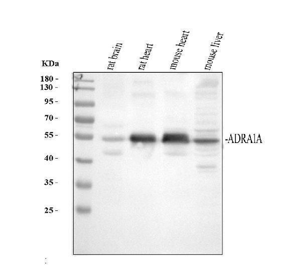
Western blot analysis of ADRA1A using anti-ADRA1A antibody (PB9752).
Electrophoresis was performed on a 10 SDS-PAGE gel at 80V (Stacking gel) / 120V (Resolving gel) for 2 hours. The sample well of each lane was loaded with 30 ug of sample under reducing conditions.
Lane 1: rat brain tissue lysates,
Lane 2: rat heart tissue lysates,
Lane 3: mouse heart tissue lysates,
Lane 4: mouse liver tissue lysates.
After electrophoresis, proteins were transferred to a nitrocellulose membrane at 150 mA for 50-90 minutes. Blocked the membrane with 5% non-fat milk/TBS for 1.5 hour at RT. The membrane was incubated with rabbit anti-ADRA1A antigen affinity purified polyclonal antibody (Catalog # PB9752) at 0.5 μg/mL overnight at 4°C, then washed with TBS-0.1%Tween 3 times with 5 minutes each and probed with a goat anti-rabbit IgG-HRP secondary antibody at a dilution of 1:5000 for 1.5 hour at RT. The signal is developed using an ECL Plus Western Blotting Substrate (Catalog # AR1196-200) with Tanon 5200 system. A specific band was detected for ADRA1A at approximately 51 kDa. The expected band size for ADRA1A is at 51 kDa.

Click image to see more details
IHC analysis of ADRA1A using anti-ADRA1A antibody (PB9752).
ADRA1A was detected in paraffin-embedded section of human liver cancer tissues. Heat mediated antigen retrieval was performed in citrate buffer (pH6, epitope retrieval solution) for 20 mins. The tissue section was blocked with 10% goat serum. The tissue section was then incubated with 1μg/ml rabbit anti-ADRA1A Antibody (PB9752) overnight at 4°C. Biotinylated goat anti-rabbit IgG was used as secondary antibody and incubated for 30 minutes at 37°C. The tissue section was developed using Strepavidin-Biotin-Complex (SABC)(Catalog # SA1022) with DAB as the chromogen.

Click image to see more details
IHC analysis of ADRA1A using anti-ADRA1A antibody (PB9752).
ADRA1A was detected in paraffin-embedded section of rat brain tissue tissues. Heat mediated antigen retrieval was performed in citrate buffer (pH6, epitope retrieval solution) for 20 mins. The tissue section was blocked with 10% goat serum. The tissue section was then incubated with 1μg/ml rabbit anti-ADRA1A Antibody (PB9752) overnight at 4°C. Biotinylated goat anti-rabbit IgG was used as secondary antibody and incubated for 30 minutes at 37°C. The tissue section was developed using Strepavidin-Biotin-Complex (SABC)(Catalog # SA1022) with DAB as the chromogen.

Click image to see more details
Flow Cytometry analysis of A431 cells using anti-ADRA1A antibody (PB9752).
Overlay histogram showing A431 cells stained with PB9752 (Blue line).The cells were blocked with 10% normal goat serum. And then incubated with rabbit anti-ADRA1A Antibody (PB9752,1μg/1x106 cells) for 30 min at 20°C. DyLight®488 conjugated goat anti-rabbit IgG (BA1127, 5-10μg/1x106 cells) was used as secondary antibody for 30 minutes at 20°C. Isotype control antibody (Green line) was rabbit IgG (1μg/1x106) used under the same conditions. Unlabelled sample (Red line) was also used as a control.
Specific Publications For Anti-alpha 1d Adrenergic Receptor/ADRA1A Antibody Picoband® (PB9752)
Loading publications
Recommended Resources
Here are featured tools and databases that you might find useful.
- Boster's Pathways Library
- Protein Databases
- Bioscience Research Protocol Resources
- Data Processing & Analysis Software
- Photo Editing Software
- Scientific Literature Resources
- Research Paper Management Tools
- Molecular Biology Software
- Primer Design Tools
- Bioinformatics Tools
- Phylogenetic Tree Analysis
Customer Reviews
Have you used Anti-alpha 1d Adrenergic Receptor/ADRA1A Antibody Picoband®?
Share your experimental results or join a short interview to earn up to $1,000 in product credits or other rewards.
0 Reviews For Anti-alpha 1d Adrenergic Receptor/ADRA1A Antibody Picoband®
Customer Q&As
Have a question?
Find answers in Q&As, reviews.
Can't find your answer?
Submit your question
3 Customer Q&As for Anti-alpha 1d Adrenergic Receptor/ADRA1A Antibody Picoband®
Question
I would like to ask if it is possible to do a dual staining experiment using your product PB9752 Anti-CD41/Integrin alpha 2b/ITGA2B Antibody Picoband antibody in mouse and rat tissue with staining of the endothelial layer.
Verified Customer
Verified customer
Asked: 2020-03-31
Answer
We only have a data on goat anti-VEGF receptor 1 that has been tested successfully with dual staining of both mouse and rat sample. However, we think that this antibody PB9752 Anti-CD41/Integrin alpha 2b/ITGA2B Antibody Picoband that you want to use will work as well.
Boster Scientific Support
Answered: 2020-03-31
Question
We are currently using anti-alpha 1d Adrenergic Receptor/ADRA1A antibody PB9752 for mouse tissue, and we are happy with the WB results. The species of reactivity given in the datasheet says human, mouse, rat. Is it true that the antibody can work on canine tissues as well?
Verified Customer
Verified customer
Asked: 2019-10-10
Answer
The anti-alpha 1d Adrenergic Receptor/ADRA1A antibody (PB9752) has not been validated for cross reactivity specifically with canine tissues, but there is a good chance of cross reactivity. We have an innovator award program that if you test this antibody and show it works in canine you can get your next antibody for free. Please contact me if I can help you with anything.
Boster Scientific Support
Answered: 2019-10-10
Question
Hi: I would like to know the concentration of this PB9752 antibody.
Verified Customer
Verified customer
Asked: 2018-08-21
Answer
The concentration of this PB9752 antibody is 0.5mg/ml
Boster Scientific Support
Answered: 2018-08-21
