Click image to see more details
-

-
-
-
-
+7
Product Info Summary
| SKU: | PB10037 |
|---|---|
| Size: | 100 μg/vial |
| Reactive Species: | Human, Monkey, Mouse, Rat |
| Host: | Rabbit |
| Application: | Flow Cytometry, IF, IHC, ICC, WB |
Customers Who Bought This Also Bought

Product info
Product Name
Anti-ALDH1B1 Antibody Picoband®
SKU/Catalog Number
PB10037
PB1092 is an alternative SKU for this antibody, used in previous lots.
Size
100 μg/vial
Form
Lyophilized
Description
Boster Bio Anti-ALDH1B1 Antibody Picoband® catalog # PB10037. Tested in Flow Cytometry, IF, IHC, ICC, WB applications. This antibody reacts with Human, Monkey, Mouse, Rat. The brand Picoband indicates this is a premium antibody that guarantees superior quality, high affinity, and strong signals with minimal background in Western blot applications. Only our best-performing antibodies are designated as Picoband, ensuring unmatched performance. ALDH1B1 (mitochondrial aldehyde dehydrogenase X) is described as an aldehyde dehydrogenase family enzyme and as the second enzyme in the major oxidative pathway of alcohol metabolism, with locus variation noted as potentially affecting alcohol-related problems. Assay context: validated for Flow Cytometry, IF, IHC, ICC, and WB; observed MW ~55 kDa. Often contextualized with other metabolic/oxidoreductase markers such as AKR1D1 and AKR7A2 when profiling redox and small-molecule metabolism in tissue/cell systems; recombinant antigens are commonly used as assay controls/standards (see recombinant protein expression for methodological context). "
Storage & Handling
Store at -20˚C for one year from date of receipt. After reconstitution, at 4˚C for one month. It can also be aliquotted and stored frozen at -20˚C for six months. Avoid repeated freeze-thaw cycles.
Cite This Product
Anti-ALDH1B1 Antibody Picoband® (Boster Biological Technology, Pleasanton CA, USA, Catalog # PB10037)
Host
Rabbit
Contents
Each vial contains 4 mg Trehalose, 0.9 mg NaCl and 0.2 mg Na2HPO4.
Clonality
Polyclonal
Isotype
Rabbit IgG
Immunogen
A synthetic peptide corresponding to a sequence at the N-terminus of human ALDH1B1, different from the related mouse sequence by one amino acid, and from the related rat sequence by two amino acids.
Cross-reactivity
No cross-reactivity with other proteins.
Reactive Species
PB10037 is reactive to ALDH1B1 in Human, Monkey, Mouse, Rat
Observed Molecular Weight
55 kDa
Calculated molecular weight
57.2 kDa
Background of ALDH1B1
Aldehyde dehydrogenase X, mitochondrial is an enzyme that in humans is encoded by the ALDH1B1 gene. This protein belongs to the aldehyde dehydrogenases family of proteins. Aldehyde dehydrogenase is the second enzyme of the major oxidative pathway of alcohol metabolism. This gene does not contain introns in the coding sequence. The variation of this locus may affect the development of alcohol-related problems.
Antibody Validation
Boster validates all antibodies on WB, IHC, ICC, Immunofluorescence, and ELISA with known positive control and negative samples to ensure specificity and high affinity, including thorough antibody incubations.
Application & Images
Applications
PB10037 is guaranteed for Flow Cytometry, IF, IHC, ICC, WB Boster Guarantee
Recommend Dilution
| Application | Dilution | Species |
|---|---|---|
| Western blot | 0.1-0.5μg/ml | Human, Monkey, Mouse, Rat |
| Immunohistochemistry (Paraffin-embedded Section) | 2-5μg/ml | Human, Mouse, Rat |
| Immunocytochemistry/Immunofluorescence | 5μg/ml | Human |
| Flow Cytometry (Fixed) | 1-3μg/1x106 cells | Human |
Tested application
Suggested blocking solution with 5% non-fat milk or BSA; (*)Recommended protein loading: 20-40 µg per lane
Use TE buffer pH 9.0 for antigen retrieval; (*) citrate buffer pH 6.0 is an alternative.
Validation Images & Assay Conditions
Click image to see more details
IHC analysis of ALDH1B1 using anti-ALDH1B1 antibody (PB10037).
ALDH1B1 was detected in a paraffin-embedded section of human colon cancer tissue. Heat mediated antigen retrieval was performed in EDTA buffer (pH 8.0, epitope retrieval solution). The tissue section was blocked with 10% goat serum. The tissue section was then incubated with 2 μg/ml rabbit anti-ALDH1B1 Antibody (PB10037) overnight at 4°C. Peroxidase Conjugated Goat Anti-rabbit IgG was used as secondary antibody and incubated for 30 minutes at 37°C. The tissue section was developed using HRP Conjugated Rabbit IgG Super Vision Assay Kit (Catalog # SV0002) with DAB as the chromogen.

Click image to see more details
Western blot analysis of ALDH1B1 using anti-ALDH1B1 antibody (PB10037).
Electrophoresis was performed on a 5-20% SDS-PAGE gel at 70V (Stacking gel) / 90V (Resolving gel) for 2-3 hours. The sample well of each lane was loaded with 30 ug of sample under reducing conditions.
Lane 1: human HepG2 whole cell lysates,
Lane 2: human K562 whole cell lysates,
Lane 3: human A431 whole cell lysates,
Lane 4: human A549 whole cell lysates,
Lane 5: human U20S whole cell lysates,
Lane 6: human Hela whole cell lysates,
Lane 7: monkey COS-7 whole cell lysates.
red to a nitrocellulose membrane at 150 mA for 50-90 minutes. Blocked the membrane with 5% non-fat milk/TBS for 1.5 hour at RT. The membrane was incubated with rabbit anti-ALDH1B1 antigen affinity purified polyclonal antibody (Catalog # PB10037) at 0.5 μg/mL overnight at 4°C, then washed with TBS-0.1%Tween 3 times with 5 minutes each and probed with a goat anti-rabbit IgG-HRP secondary antibody at a dilution of 1:5000 for 1.5 hour at RT. The signal is developed using an Enhanced Chemiluminescent detection (ECL) kit (Catalog # EK1002) with Tanon 5200 system. A specific band was detected for ALDH1B1 at approximately 55 kDa. The expected band size for ALDH1B1 is at 57 kDa.

Click image to see more details
Western blot analysis of ALDH1B1 using anti-ALDH1B1 antibody (PB10037).
Electrophoresis was performed on a 5-20% SDS-PAGE gel at 70V (Stacking gel) / 90V (Resolving gel) for 2-3 hours. The sample well of each lane was loaded with 30 ug of sample under reducing conditions.
Lane 1: rat liver tissue lysates,
Lane 2: rat testis tissue lysates,
Lane 3: rat RH35 whole cell lysates,
Lane 4: mouse brain tissue lysates,
Lane 5: mouse liver tissue lysates.
red to a nitrocellulose membrane at 150 mA for 50-90 minutes. Blocked the membrane with 5% non-fat milk/TBS for 1.5 hour at RT. The membrane was incubated with rabbit anti-ALDH1B1 antigen affinity purified polyclonal antibody (Catalog # PB10037) at 0.5 μg/mL overnight at 4°C, then washed with TBS-0.1%Tween 3 times with 5 minutes each and probed with a goat anti-rabbit IgG-HRP secondary antibody at a dilution of 1:5000 for 1.5 hour at RT. The signal is developed using an Enhanced Chemiluminescent detection (ECL) kit (Catalog # EK1002) with Tanon 5200 system. A specific band was detected for ALDH1B1 at approximately 55 kDa. The expected band size for ALDH1B1 is at 57 kDa.

Click image to see more details
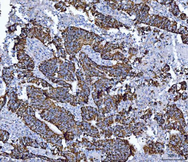
IHC analysis of ALDH1B1 using anti-ALDH1B1 antibody (PB10037).
ALDH1B1 was detected in a paraffin-embedded section of human colonic adenocarcinoma tissue. Heat mediated antigen retrieval was performed in EDTA buffer (pH 8.0, epitope retrieval solution). The tissue section was blocked with 10% goat serum. The tissue section was then incubated with 2 μg/ml rabbit anti-ALDH1B1 Antibody (PB10037) overnight at 4°C. Peroxidase Conjugated Goat Anti-rabbit IgG was used as secondary antibody and incubated for 30 minutes at 37°C. The tissue section was developed using HRP Conjugated Rabbit IgG Super Vision Assay Kit (Catalog # SV0002) with DAB as the chromogen.

Click image to see more details
IHC analysis of ALDH1B1 using anti-ALDH1B1 antibody (PB10037).
ALDH1B1 was detected in a paraffin-embedded section of human endometrial adenocarcinoma tissue. Heat mediated antigen retrieval was performed in EDTA buffer (pH 8.0, epitope retrieval solution). The tissue section was blocked with 10% goat serum. The tissue section was then incubated with 2 μg/ml rabbit anti-ALDH1B1 Antibody (PB10037) overnight at 4°C. Peroxidase Conjugated Goat Anti-rabbit IgG was used as secondary antibody and incubated for 30 minutes at 37°C. The tissue section was developed using HRP Conjugated Rabbit IgG Super Vision Assay Kit (Catalog # SV0002) with DAB as the chromogen.

Click image to see more details
IHC analysis of ALDH1B1 using anti-ALDH1B1 antibody (PB10037).
ALDH1B1 was detected in a paraffin-embedded section of human hepatocellular carcinoma tissue. Heat mediated antigen retrieval was performed in EDTA buffer (pH 8.0, epitope retrieval solution). The tissue section was blocked with 10% goat serum. The tissue section was then incubated with 2 μg/ml rabbit anti-ALDH1B1 Antibody (PB10037) overnight at 4°C. Peroxidase Conjugated Goat Anti-rabbit IgG was used as secondary antibody and incubated for 30 minutes at 37°C. The tissue section was developed using HRP Conjugated Rabbit IgG Super Vision Assay Kit (Catalog # SV0002) with DAB as the chromogen.

Click image to see more details
Flow Cytometry analysis of HEL cells using anti-ALDH1B1 antibody (PB10037).
Overlay histogram showing HEL cells stained with PB10037 (Blue line). To facilitate intracellular staining, cells were fixed with 4% paraformaldehyde and permeabilized with permeabilization buffer. The cells were blocked with 10% normal goat serum. And then incubated with rabbit anti-ALDH1B1 Antibody (PB10037, 1 μg/1x106 cells) for 30 min at 20°C. DyLight®488 conjugated goat anti-rabbit IgG (BA1127, 5-10 μg/1x106 cells) was used as secondary antibody for 30 minutes at 20°C. Isotype control antibody (Green line) was rabbit IgG (1 μg/1x106) used under the same conditions. Unlabelled sample without incubation with primary antibody and secondary antibody (Red line) was used as a blank control.

Click image to see more details
IHC analysis of ALDH1B1 using anti-ALDH1B1 antibody (PB10037).
ALDH1B1 was detected in a paraffin-embedded section of human laryngeal squamous cell carcinoma tissue. Heat mediated antigen retrieval was performed in EDTA buffer (pH 8.0, epitope retrieval solution). The tissue section was blocked with 10% goat serum. The tissue section was then incubated with 2 μg/ml rabbit anti-ALDH1B1 Antibody (PB10037) overnight at 4°C. Peroxidase Conjugated Goat Anti-rabbit IgG was used as secondary antibody and incubated for 30 minutes at 37°C. The tissue section was developed using HRP Conjugated Rabbit IgG Super Vision Assay Kit (Catalog # SV0002) with DAB as the chromogen.

Click image to see more details
IHC analysis of ALDH1B1 using anti-ALDH1B1 antibody (PB10037).
ALDH1B1 was detected in a paraffin-embedded section of mouse colon tissue. Heat mediated antigen retrieval was performed in EDTA buffer (pH 8.0, epitope retrieval solution). The tissue section was blocked with 10% goat serum. The tissue section was then incubated with 2 μg/ml rabbit anti-ALDH1B1 Antibody (PB10037) overnight at 4°C. Peroxidase Conjugated Goat Anti-rabbit IgG was used as secondary antibody and incubated for 30 minutes at 37°C. The tissue section was developed using HRP Conjugated Rabbit IgG Super Vision Assay Kit (Catalog # SV0002) with DAB as the chromogen.

Click image to see more details
IHC analysis of ALDH1B1 using anti-ALDH1B1 antibody (PB10037).
ALDH1B1 was detected in a paraffin-embedded section of rat colon tissue. Heat mediated antigen retrieval was performed in EDTA buffer (pH 8.0, epitope retrieval solution). The tissue section was blocked with 10% goat serum. The tissue section was then incubated with 2 μg/ml rabbit anti-ALDH1B1 Antibody (PB10037) overnight at 4°C. Peroxidase Conjugated Goat Anti-rabbit IgG was used as secondary antibody and incubated for 30 minutes at 37°C. The tissue section was developed using HRP Conjugated Rabbit IgG Super Vision Assay Kit (Catalog # SV0002) with DAB as the chromogen.

Click image to see more details
IF analysis of ALDH1B1 using anti-ALDH1B1 antibody (PB10037).
ALDH1B1 was detected in an immunocytochemical section of A431 cells. Enzyme antigen retrieval was performed using IHC enzyme antigen retrieval reagent (AR0022) for 15 mins. The cells were blocked with 10% goat serum. And then incubated with 5 μg/mL rabbit anti-ALDH1B1 Antibody (PB10037) overnight at 4°C. DyLight®488 Conjugated Goat Anti-Rabbit IgG (BA1127) was used as secondary antibody at 1:100 dilution and incubated for 30 minutes at 37°C. The section was counterstained with DAPI. Visualize using a fluorescence microscope and filter sets appropriate for the label used.
Specific Publications For Anti-ALDH1B1 Antibody Picoband® (PB10037)
Loading publications
Recommended Resources
Here are featured tools and databases that you might find useful.
- Boster's Pathways Library
- Protein Databases
- Bioscience Research Protocol Resources
- Data Processing & Analysis Software
- Photo Editing Software
- Scientific Literature Resources
- Research Paper Management Tools
- Molecular Biology Software
- Primer Design Tools
- Bioinformatics Tools
- Phylogenetic Tree Analysis
Customer Reviews
Have you used Anti-ALDH1B1 Antibody Picoband®?
Share your experimental results or join a short interview to earn up to $1,000 in product credits or other rewards.
0 Reviews For Anti-ALDH1B1 Antibody Picoband®
Customer Q&As
Have a question?
Find answers in Q&As, reviews.
Can't find your answer?
Submit your question
6 Customer Q&As for Anti-ALDH1B1 Antibody Picoband®
Question
Will anti-ALDH1B1 antibody PB10037 work for WB with eye?
Verified Customer
Verified customer
Asked: 2020-04-14
Answer
According to the expression profile of eye, ALDH1B1 is highly expressed in eye. So, it is likely that anti-ALDH1B1 antibody PB10037 will work for WB with eye.
Boster Scientific Support
Answered: 2020-04-14
Question
We are currently using anti-ALDH1B1 antibody PB10037 for rat tissue, and we are happy with the WB results. The species of reactivity given in the datasheet says human, mouse, rat. Is it possible that the antibody can work on horse tissues as well?
Verified Customer
Verified customer
Asked: 2019-11-22
Answer
The anti-ALDH1B1 antibody (PB10037) has not been tested for cross reactivity specifically with horse tissues, though there is a good chance of cross reactivity. We have an innovator award program that if you test this antibody and show it works in horse you can get your next antibody for free. Please contact me if I can help you with anything.
Boster Scientific Support
Answered: 2019-11-22
Question
Would anti-ALDH1B1 antibody PB10037 work on dog WB with eye?
Verified Customer
Verified customer
Asked: 2019-10-17
Answer
Our lab technicians have not validated anti-ALDH1B1 antibody PB10037 on dog. You can run a BLAST between dog and the immunogen sequence of anti-ALDH1B1 antibody PB10037 to see if they may cross-react. If the sequence homology is close, then you can perform a pilot test. Keep in mind that since we have not validated dog samples, this use of the antibody is not covered by our guarantee. However we have an innovator award program that if you test this antibody and show it works in dog eye in WB, you can get your next antibody for free.
Boster Scientific Support
Answered: 2019-10-17
Question
Do you have a BSA free version of anti-ALDH1B1 antibody PB10037 available?
Verified Customer
Verified customer
Asked: 2019-06-13
Answer
We appreciate your recent telephone inquiry. I can confirm that some lots of this anti-ALDH1B1 antibody PB10037 are BSA free. For now, these lots are available and we can make a BSA free formula for you free of charge. It will take 3 extra days to prepare. If you require this antibody BSA free again in future, please do not hesitate to contact me and I will be pleased to check which lots we have in stock that are BSA free.
Boster Scientific Support
Answered: 2019-06-13
Question
I am interested in to test anti-ALDH1B1 antibody PB10037 on mouse eye for research purposes, then I may be interested in using anti-ALDH1B1 antibody PB10037 for diagnostic purposes as well. Is the antibody suitable for diagnostic purposes?
Verified Customer
Verified customer
Asked: 2019-05-21
Answer
The products we sell, including anti-ALDH1B1 antibody PB10037, are only intended for research use. They would not be suitable for use in diagnostic work. If you have the means to develop a product into diagnostic use, and are interested in collaborating with us and develop our product into an IVD product, please contact us for more discussions.
Boster Scientific Support
Answered: 2019-05-21
Question
Is a blocking peptide available for product anti-ALDH1B1 antibody (PB10037)?
Verified Customer
Verified customer
Asked: 2019-05-13
Answer
We do provide the blocking peptide for product anti-ALDH1B1 antibody (PB10037). If you would like to place an order for it please contact support@bosterbio.com and make a special request.
Boster Scientific Support
Answered: 2019-05-13
