Click image to see more details
Product Info Summary
| SKU: | M05278 |
|---|---|
| Size: | 100 μg/vial |
| Reactive Species: | Human, Mouse, Rat |
| Host: | Mouse |
| Application: | Flow Cytometry, IHC, WB |
Customers Who Bought This Also Bought

Product info
Product Name
Anti-AKR1D1 Antibody Picoband® (monoclonal, 6I4)
SKU/Catalog Number
M05278
Size
100 μg/vial
Form
Lyophilized
Description
Boster Bio Anti-AKR1D1 Antibody Picoband® (monoclonal, 6I4) catalog # M05278. Tested in Flow Cytometry, IHC, WB applications. This antibody reacts with Human, Mouse, Rat. The brand Picoband indicates this is a premium antibody that guarantees superior quality, high affinity, and strong signals with minimal background in Western blot applications. Only our best-performing antibodies are designated as Picoband, ensuring unmatched performance. AKR1D1 (3-oxo-5β-steroid 4-dehydrogenase) is a key enzyme in steroid/bile acid metabolism, catalyzing 5β-reduction steps important for bile acid biosynthesis (canonical). Assay context: monoclonal (6I4) antibody validated for Flow Cytometry, IHC, and Western blot across Human/Mouse/Rat; metabolic profiling workflows often contextualize AKR1D1 with mitochondrial oxidation markers such as ACADM/MCAD (putative, systems-level).
Storage & Handling
Store at -20˚C for one year from date of receipt. After reconstitution, at 4˚C for one month. It can also be aliquotted and stored frozen at -20˚C for six months. Avoid repeated freeze-thaw cycles.
Cite This Product
Anti-AKR1D1 Antibody Picoband® (monoclonal, 6I4) (Boster Biological Technology, Pleasanton CA, USA, Catalog # M05278)
Host
Mouse
Contents
Each vial contains 4mg Trehalose, 0.9mg NaCl, 0.2mg Na2HPO4, 0.05mg NaN3.
Clonality
Monoclonal
Clone Number
6I4
Isotype
Mouse IgG2b
Immunogen
A synthetic peptide corresponding to a sequence at the C-terminus of human AKR1D1, which shares 90.9% and 93.9% amino acid (aa) sequence identity with mouse and rat AKR1D1, respectively.
Cross-reactivity
No cross-reactivity with other proteins.
Reactive Species
M05278 is reactive to AKR1D1 in Human, Mouse, Rat
Observed Molecular Weight
37 kDa
Calculated molecular weight
37.4 kDa
Background of AKR1D1
Human delta (4)-3-oxosteroid 5-beta-reductase (steroid 5-beta-reductase) catalyzes 5-beta-reduction of bile acid intermediates and steroid hormones carrying a delta (4)-3-one structure. This gene is mapped to 7q33. The enzyme encoded by this gene is responsible for the catalysis of the 5-beta-reduction of bile acid intermediates and steroid hormones carrying a delta (4)-3-one structure. Deficiency of this enzyme may contribute to hepatic dysfunction. Three transcript variants encoding different isoforms have been found for this gene. Other variants may be present, but their full-length natures have not been determined yet.
Antibody Validation
Boster validates all antibodies on WB, IHC, ICC, Immunofluorescence, and ELISA with known positive control and negative samples to ensure specificity and high affinity, including thorough antibody incubations.
Application & Images
Applications
M05278 is guaranteed for Flow Cytometry, IHC, WB Boster Guarantee
Recommend Dilution
| Application | Dilution | Species |
|---|---|---|
| Western blot | 0.1-0.5μg/ml | Human, Mouse, Rat |
| Immunohistochemistry (Paraffin-embedded Section) | 0.5-1μg/ml | Human |
| Flow Cytometry (Fixed) | 1-3μg/1x106 cells | Human |
Tested application
Suggested blocking solution with 5% non-fat milk or BSA; (*)Recommended protein loading: 20-40 µg per lane
Use TE buffer pH 9.0 for antigen retrieval; (*) citrate buffer pH 6.0 is an alternative.
Validation Images & Assay Conditions
Click image to see more details
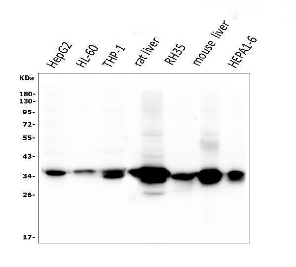
Western blot analysis of AKR1D1 using anti-AKR1D1 antibody (M05278).
Electrophoresis was performed on a 5-20% SDS-PAGE gel at 70V (Stacking gel) / 90V (Resolving gel) for 2-3 hours. The sample well of each lane was loaded with 50ug of sample under reducing conditions.
Lane 1: human HepG2 whole cell lysates;
Lane 2: human HL-60 whole cell lysates;
Lane 3: human THP-1 whole cell lysates;
Lane 4: rat liver tissue lysates;
Lane 5: rat RH35 whole cell lysates;
Lane 6: mouse liver tissue lysates;
Lane 7: mouse HEPA1-6 whole cell lysates;
After Electrophoresis, proteins were transferred to a Nitrocellulose membrane at 150mA for 50-90 minutes. Blocked the membrane with 5% Non-fat Milk/ TBS for 1.5 hour at RT. The membrane was incubated with mouse anti-AKR1D1 antigen affinity purified monoclonal antibody (Catalog # M05278) at 0.5 μg/mL overnight at 4°C, then washed with TBS-0.1%Tween 3 times with 5 minutes each and probed with a goat anti-mouse IgG-HRP secondary antibody at a dilution of 1:10000 for 1.5 hour at RT. The signal is developed using an Enhanced Chemiluminescent detection (ECL) kit (Catalog # EK1001) with Tanon 5200 system. A specific band was detected for AKR1D1 at approximately 37KD. The expected band size for AKR1D1 is at 37KD.

Click image to see more details
IHC analysis of AKR1D1 using anti-AKR1D1 antibody (M05278).
AKR1D1 was detected in paraffin-embedded section of human liver cancer tissue. Heat mediated antigen retrieval was performed in EDTA buffer (pH8.0, epitope retrieval solution). The tissue section was blocked with 10% goat serum. The tissue section was then incubated with 1μg/ml mouse anti-AKR1D1 Antibody (M05278) overnight at 4°C. Biotinylated goat anti-mouse IgG was used as secondary antibody and incubated for 30 minutes at 37°C. The tissue section was developed using Strepavidin-Biotin-Complex (SABC) (Catalog # SA1021) with DAB as the chromogen.

Click image to see more details
Flow Cytometry analysis of HepG2 cells using anti-AKR1D1 antibody (M05278).
Overlay histogram showing HepG2 cells stained with M04586-2 (Blue line). To facilitate intracellular staining, cells were fixed with 4% paraformaldehyde and permeabilized with permeabilization buffer. The cells were blocked with 10% normal goat serum. And then incubated with mouse anti-AKR1D1 Antibody (M05278,1μg/1x106 cells) for 30 min at 20°C. DyLight®488 conjugated goat anti-mouse IgG (BA1126, 5-10μg/1x106 cells) was used as secondary antibody for 30 minutes at 20°C. Isotype control antibody (Green line) was mouse IgG (1μg/1x106) used under the same conditions. Unlabelled sample without incubation with primary antibody and secondary antibody (Red line) was used as a blank control.
Specific Publications For Anti-AKR1D1 Antibody Picoband® (monoclonal, 6I4) (M05278)
Loading publications
Recommended Resources
Here are featured tools and databases that you might find useful.
- Boster's Pathways Library
- Protein Databases
- Bioscience Research Protocol Resources
- Data Processing & Analysis Software
- Photo Editing Software
- Scientific Literature Resources
- Research Paper Management Tools
- Molecular Biology Software
- Primer Design Tools
- Bioinformatics Tools
- Phylogenetic Tree Analysis
Customer Reviews
Have you used Anti-AKR1D1 Antibody Picoband® (monoclonal, 6I4)?
Share your experimental results or join a short interview to earn up to $1,000 in product credits or other rewards.
0 Reviews For Anti-AKR1D1 Antibody Picoband® (monoclonal, 6I4)
Customer Q&As
Have a question?
Find answers in Q&As, reviews.
Can't find your answer?
Submit your question
