Click image to see more details
-

-
-
-
-
+1
Product Info Summary
| SKU: | A03799 |
|---|---|
| Size: | 0.1 mg |
| Reactive Species: | Human, Mouse, Rat |
| Host: | Rabbit |
| Application: | ELISA, IF, IHC-P, ICC, WB |
Customers Who Bought This Also Bought

Product info
Product Name
Anti-Ambra1 Antibody
SKU/Catalog Number
A03799
Size
0.1 mg
Form
Liquid
Description
Boster Bio Anti-Ambra1 Antibody (Catalog # A03799). Tested in ELISA, ICC/IF, IHC-P, WB applications. This antibody reacts with Human, Mouse, Rat.
Storage & Handling
Ambra1 antibody can be stored at 4°C for three months and -20°C, stable for up to one year. Avoid repeated freeze-thaw cycles. Antibodies should not be exposed to prolonged high temperatures.
Cite This Product
Anti-Ambra1 Antibody (Boster Biological Technology, Pleasanton CA, USA, Catalog # A03799)
Host
Rabbit
Contents
Ambra1 Antibody is supplied in PBS containing 0.02% sodium azide.
Clonality
Polyclonal
Isotype
IgG
Immunogen
Ambra1 antibody was raised against a 15 amino acid synthetic peptide from near the carboxy terminus of human Ambra1. The immunogen is located within the last 50 amino acids of Ambra1.
Reactive Species
A03799 is reactive to AMBRA1 in Human, Mouse, Rat
Observed Molecular Weight
143 kDa
Calculated molecular weight
142.5 kDa
Background of AMBRA1
Autophagy, the process of bulk degradation of cellular proteins through an autophagosomic-lysosomal pathway is important for normal growth control and may be defective in tumor cells. It is involved in the preservation of cellular nutrients under starvation conditions as well as the normal turnover of cytosolic components. Beclin-1, a principal regulator of autophagosome formation, is in turn regulated by Ambra1. Ambra1 associates with Beclin-1 through a region near its center as determined by yeast two-hybrid assay. Null mutations in this gene in mice resulted in embryonic lethality with severe neural tube defects associated with autophagy impairment, accumulation of ubiquitinated proteins, unbalanced cell proliferation and excessive apoptotic death. Furthermore, down-regulation of Ambra1 in cultured cells though RNA interference decreased the level of rapamycin- and nutrient starvation-induced autophagy. Multiple isoforms of Ambra1 are known to exist.
Antibody Validation
Boster validates all antibodies on WB, IHC, ICC, Immunofluorescence, and ELISA with known positive control and negative samples to ensure specificity and high affinity, including thorough antibody incubations.
Application & Images
Applications
A03799 is guaranteed for ELISA, IF, IHC-P, ICC, WB Boster Guarantee
Assay Dilutions Recommendation
The recommendations below provide a starting point for assay optimization. The actual working concentration varies and should be decided by the user.
WB: 1-2 μg/mL; IHC: 1-5 μg/mL; ICC/IF: 20 μg/mL.
Antibody validated: Western Blot in human and mouse samples; Immunocytochemistry and Immunofluorescence in human samples. All other applications and species not yet tested.
Validation Images & Assay Conditions
Click image to see more details
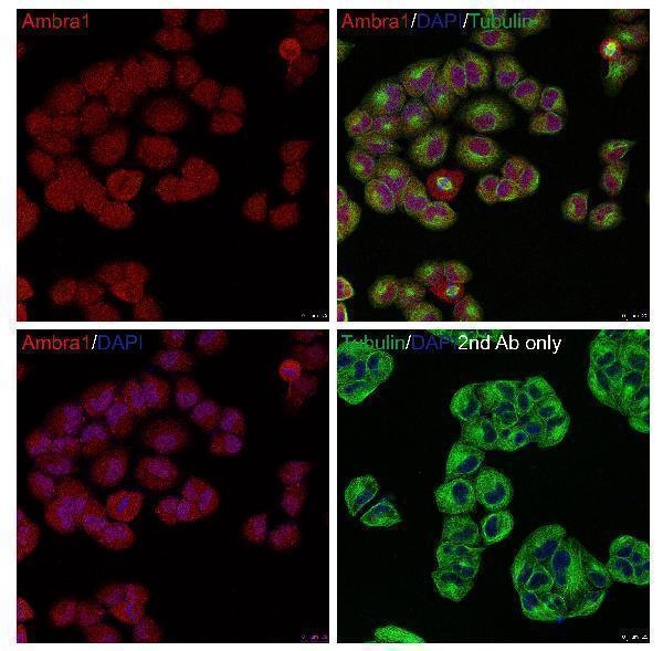
Immunoflouorescence Validation of AMBRA1 in HeLa Cells
Immunofluorescent analysis of PFA-fixed HeLa cells labeling AMBRA1 with A03799 at 20 μg/mL, followed by goat anti-rabbit IgG secondary antibody at 1/1000 dilution (red) and DAPI staining (blue). Alpha tubulin was stained with anti-alpha tubulin antibody following by goat anti-mouse IgG secondary antibody (green). Images were captured with confocal microscopy.

Click image to see more details
WB Validation in Human Cell Lines
Loading: 15 μg of lysate per lane
Antibodies: AMBRA1 A03799, 1 μg/mL , 1 h incubation at RT in 5% NFDM/TBST.
Secondary: Goat Anti-Rabbit IgG HRP conjugate at 1:10000 dilution.

Click image to see more details
Western Blot Validation in 3T3/NIH Cells
Loading: 15 μg of lysate
Antibodies: AMBRA1 A03799, 1 μg/mL, 1h incubation at RT in 5% NFDM/TBST.
Secondary: Goat anti-rabbit IgG HRP conjugate at 1:10000 dilution.

Click image to see more details
Immunohistochemistry Validation of AMBRA1 in Rat Colon Tissue
Immunohistochemical analysis of paraffin-embedded rat colon tissue using anti-AMBRA1 antibody (A03799) at 2 μg/ml. Tissue was fixed with formaldehyde and blocked with 10% serum for 1 h at RT; antigen retrieval was by heat mediation with a citrate buffer (pH6). Samples were incubated with primary antibody overnight at 4 ˚C. A goat anti-rabbit IgG H&L (HRP) at 1/250 was used as secondary. Counter stained with Hematoxylin.

Click image to see more details
Immunofluorescence Validation of AMBRA1 in K562 Cells
Immunofluorescent analysis of 4% paraformaldehyde-fixed K562 Cells labeling AMBRA1 with A03799 at 20 μg/mL, followed by goat anti-rabbit IgG secondary antibody at 1/500 dilution (red) DAPI staining (blue).
Specific Publications For Anti-Ambra1 Antibody (A03799)
Loading publications
Recommended Resources
Here are featured tools and databases that you might find useful.
- Boster's Pathways Library
- Protein Databases
- Bioscience Research Protocol Resources
- Data Processing & Analysis Software
- Photo Editing Software
- Scientific Literature Resources
- Research Paper Management Tools
- Molecular Biology Software
- Primer Design Tools
- Bioinformatics Tools
- Phylogenetic Tree Analysis
Customer Reviews
Have you used Anti-Ambra1 Antibody?
Share your experimental results or join a short interview to earn up to $1,000 in product credits or other rewards.
0 Reviews For Anti-Ambra1 Antibody
Customer Q&As
Have a question?
Find answers in Q&As, reviews.
Can't find your answer?
Submit your question
