Click image to see more details
-

-
-
-
-
+1
Product Info Summary
| SKU: | PB9420 |
|---|---|
| Size: | 100 μg/vial |
| Reactive Species: | Human, Mouse, Rat |
| Host: | Rabbit |
| Application: | Flow Cytometry, IP, IF, IHC, ICC, WB |
Customers Who Bought This Also Bought

Product info
Product Name
Anti-Annexin A3/ANXA3 Antibody Picoband®
SKU/Catalog Number
PB9420
Size
100 μg/vial
Form
Lyophilized
Description
Boster Bio Anti-Annexin A3/ANXA3 Antibody Picoband® catalog # PB9420. Tested in Flow Cytometry, IP, ICC/IF, IHC, WB applications. This antibody reacts with Human, Mouse, Rat. The brand Picoband indicates this is a premium antibody that guarantees superior quality, high affinity, and strong signals with minimal background in Western blot applications. Only our best-performing antibodies are designated as Picoband, ensuring unmatched performance.
Storage & Handling
Store at -20˚C for one year from date of receipt. After reconstitution, at 4˚C for one month. It can also be aliquotted and stored frozen at -20˚C for six months. Avoid repeated freeze-thaw cycles.
Cite This Product
Anti-Annexin A3/ANXA3 Antibody Picoband® (Boster Biological Technology, Pleasanton CA, USA, Catalog # PB9420)
Host
Rabbit
Contents
Each vial contains 4 mg Trehalose, 0.9 mg NaCl and 0.2 mg Na2HPO4.
Clonality
Polyclonal
Isotype
Rabbit IgG
Immunogen
A synthetic peptide corresponding to a sequence in the middle region of human Annexin A3, different from the related mouse sequence by one amino acid, and from the related rat sequence by three amino acids.
Cross-reactivity
No cross-reactivity with other proteins
Reactive Species
PB9420 is reactive to ANXA3 in Human, Mouse, Rat
Observed Molecular Weight
36 kDa
Calculated molecular weight
36.4 kDa
Background of ANXA3
Annexin A3 is a protein that in humans is encoded by the Annexin A3 gene. The Annexin A3 gene contains 13 exons and spans 58 kb of genomic DNA. The Annexin A3 gene is mapped to 4q21. It is abnormally expressed in fetuses of both IVF and ICSI, which may contribute to the increase risk of birth defects in these ART. This gene encodes a member of the annexin family. Members of this calcium-dependent phospholipid-binding protein family play a role in the regulation of cellular growth and in signal transduction pathways. This protein functions in the inhibition of phospholipase A2 and cleavage of inositol 1,2-cyclic phosphate to form inositol 1-phosphate. This protein may also play a role in anti-coagulation.
Antibody Validation
Boster validates all antibodies on WB, IHC, ICC, Immunofluorescence, and ELISA with known positive control and negative samples to ensure specificity and high affinity, including thorough antibody incubations.
Application & Images
Applications
PB9420 is guaranteed for Flow Cytometry, IP, IF, IHC, ICC, WB Boster Guarantee
Recommend Dilution
| Application | Dilution | Species |
|---|---|---|
| Western blot | 0.1-0.5μg/ml | Human, Mouse, Rat |
| Immunohistochemistry (Paraffin-embedded Section) | 2-5μg/ml | Human |
| Immunocytochemistry/Immunofluorescence | 5μg/ml | Human |
| Immunoprecipitation | 0.5-2 μg/ml | Human |
| Flow Cytometry (Fixed) | 1-3μg/1x106 cells | Human |
Tested application
Suggested blocking solution with 5% non-fat milk or BSA; (*)Recommended protein loading: 20-40 µg per lane
Use TE buffer pH 9.0 for antigen retrieval; (*) citrate buffer pH 6.0 is an alternative.
Validation Images & Assay Conditions
Click image to see more details
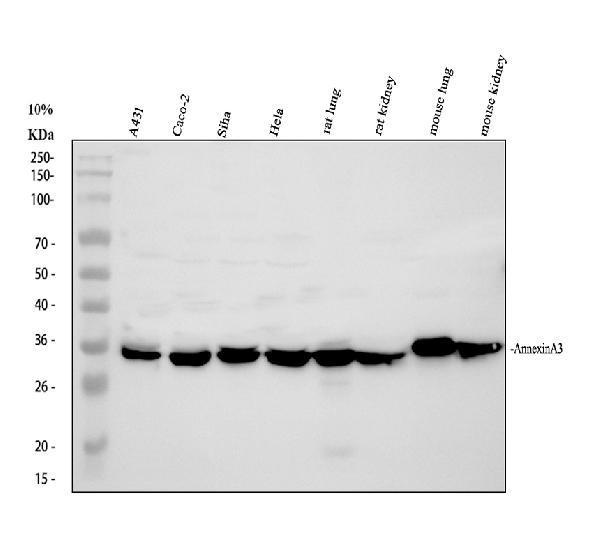
Western blot analysis of ANXA3 using anti-ANXA3 antibody (PB9420).
Electrophoresis was performed on a 10% SDS-PAGE gel at 80V (Stacking gel) / 120V (Resolving gel) for 2 hours. The sample well of each lane was loaded with 30 ug of sample under reducing conditions.
Lane 1: human A431 whole cell lysates,
Lane 2: human Caco-2 whole cell lysates,
Lane 3: human SiHa whole cell lysates,
Lane 4: human Hela whole cell lysates,
Lane 5: rat lung tissue lysates,
Lane 6: rat kidney tissue lysates,
Lane 7: mouse lung tissue lysates,
Lane 8: mouse kidney tissue lysates.
After electrophoresis, proteins were transferred to a nitrocellulose membrane at 150 mA for 50-90 minutes. Blocked the membrane with 5% non-fat milk/TBS for 1.5 hour at RT. The membrane was incubated with rabbit anti-ANXA3 antigen affinity purified polyclonal antibody (PB9420) at 0.5 μg/mL overnight at 4°C, then washed with TBS-0.1%Tween 3 times with 5 minutes each and probed with a goat anti-rabbit IgG-HRP secondary antibody (Catalog # BA1054) at a dilution of 1:5000 for 1.5 hour at RT. The signal is developed using an ECL Plus Western Blotting Substrate (Catalog # AR1196-200) with Tanon 5200 system. A specific band was detected for ANXA3 at approximately 36 kDa. The expected band size for ANXA3 is at 36 kDa.

Click image to see more details
IHC analysis of ANXA3 using anti-ANXA3 antibody (PB9420).
ANXA3 was detected in a paraffin-embedded section of human gastric carcinoma tissue. Heat mediated antigen retrieval was performed in EDTA buffer (pH 8.0, epitope retrieval solution). The tissue section was blocked with 10% goat serum. The tissue section was then incubated with 2 μg/ml rabbit anti-ANXA3 Antibody (PB9420) overnight at 4°C. Peroxidase Conjugated Goat Anti-rabbit IgG was used as secondary antibody and incubated for 30 minutes at 37°C. The tissue section was developed using HRP Conjugated Rabbit IgG Super Vision Assay Kit (Catalog # SV0002) with DAB as the chromogen.

Click image to see more details
IF analysis of ANXA3 using antiANXA3 antibody (PB9420).
ANXA3 was detected in an immunocytochemical section of Hela cells. Enzyme antigen retrieval was performed using IHC enzyme antigen retrieval reagent (AR0022) for 15 mins. The cells were blocked with 10% goat serum. And then incubated with 5 μg/mL rabbit anti-ANXA3 Antibody (PB9420) overnight at 4°C. Fluoro488 Conjugated Goat Anti-Rabbit IgG (BA1127) was used as secondary antibody at 1:500 dilution and incubated for 30 minutes at 37°C. The section was counterstained with DAPI. Visualize using a fluorescence microscope and filter sets appropriate for the label used.

Click image to see more details
Immunoprecipitating ANXA3 in A431 whole cell lysate.
Western blot analysis of ANXA3 using anti-ANXA3 antibody (PB9420);
Lane 1: A431 whole cell lysates (30ug);
Lane 2: Rabbit control IgG instead of anti-ANXA3 antibody in A431 whole cell lysate;
Lane 3: anti-ANXA3 antibody (2μg) + A431 whole cell lysate (500μg).
After electrophoresis, proteins were transferred to a membrane. Then the membrane was incubated with rabbit anti-ANXA3 antigen affinity purified polyclonal antibody (PB9420) at a dilution of 0.5 μg/mL and probed with a goat anti-rabbit IgG-HRP secondary antibody (Catalog # BA1054). The signal is developed using ECL Plus Western Blotting Substrate (Catalog # AR1196-200). A specific band was detected for ANXA3 at approximately 36 kDa. The expected band size for ANXA3 is at 36 kDa.

Click image to see more details
Flow Cytometry analysis of Hela cells using anti-ANXA3 antibody (PB9420).
Overlay histogram showing Hela cells stained with PB9420 (Blue line). The cells were fixed with 4% paraformaldehyde and blocked with 10% normal goat serum. And then incubated with rabbit anti-ANXA3 Antibody (PB9420, 1 μg/1x106 cells) for 30 min at 20°C. Fluoro488 conjugated goat anti-rabbit IgG (BA1127, 5-10 μg/1x106 cells) was used as secondary antibody for 30 minutes at 20°C. Isotype control antibody (Green line) was rabbit IgG (1 μg/1x106) used under the same conditions. Unlabelled sample without incubation with primary antibody and secondary antibody (Red line) was used as a blank control.
Specific Publications For Anti-Annexin A3/ANXA3 Antibody Picoband® (PB9420)
Loading publications
Recommended Resources
Here are featured tools and databases that you might find useful.
- Boster's Pathways Library
- Protein Databases
- Bioscience Research Protocol Resources
- Data Processing & Analysis Software
- Photo Editing Software
- Scientific Literature Resources
- Research Paper Management Tools
- Molecular Biology Software
- Primer Design Tools
- Bioinformatics Tools
- Phylogenetic Tree Analysis
Customer Reviews
Have you used Anti-Annexin A3/ANXA3 Antibody Picoband®?
Share your experimental results or join a short interview to earn up to $1,000 in product credits or other rewards.
0 Reviews For Anti-Annexin A3/ANXA3 Antibody Picoband®
Customer Q&As
Have a question?
Find answers in Q&As, reviews.
Can't find your answer?
Submit your question
1 Customer Q&As for Anti-Annexin A3/ANXA3 Antibody Picoband®
Question
We are currently using anti-Annexin A3/ANXA3 antibody PB9420 for rat tissue, and we are well pleased with the WB results. The species of reactivity given in the datasheet says human, mouse, rat. Is it likely that the antibody can work on primate tissues as well?
D. Krishna
Verified customer
Asked: 2014-01-31
Answer
The anti-Annexin A3/ANXA3 antibody (PB9420) has not been tested for cross reactivity specifically with primate tissues, though there is a good chance of cross reactivity. We have an innovator award program that if you test this antibody and show it works in primate you can get your next antibody for free. Please contact me if I can help you with anything.
Boster Scientific Support
Answered: 2014-01-31
