Click image to see more details
Product Info Summary
| SKU: | P03471 |
|---|---|
| Size: | 100ug |
| Reactive Species: | Human |
| Host: | Rabbit |
| Application: | ELISA, IHC, WB |
Customers Who Bought This Also Bought

Product info
Product Name
Anti-Phospho-APC1 S355 ANAPC1 Antibody
SKU/Catalog Number
P03471
Size
100ug
Form
Liquid (sterile filtered)
Description
Boster Bio Anti-Phospho-APC1 S355 ANAPC1 Antibody (Catalog # P03471). Tested in ELISA, IHC, WB applications. This antibody reacts with Human.
Storage & Handling
Store vial at -20°C prior to opening. Aliquot contents and freeze at -20°C or below for extended storage. Avoid cycles of freezing and thawing. Centrifuge product if not completely clear after standing at room temperature. This product is stable for several weeks at 4°C as an undiluted liquid. Dilute only prior to immediate use. Expiration date is one (1) year from date of opening. (Ship on dry ice.)
Cite This Product
Anti-Phospho-APC1 S355 ANAPC1 Antibody (Boster Biological Technology, Pleasanton CA, USA, Catalog # P03471)
Host
Rabbit
Contents
0.02 M Potassium Phosphate, 0.15 M Sodium Chloride, pH 7.2, 0.01% (w/v) Sodium Azide
Clonality
Polyclonal
Isotype
IgG
Immunogen
This affinity purified antibody was prepared from whole rabbit serum produced by repeated immunizations with a synthetic peptide corresponding to an internal region surrounding pS377 of Human Apc1 protein.
Cross-reactivity
No cross reactivity with other proteins.
Reactive Species
P03471 is reactive to ANAPC1 in Human
Observed Molecular Weight
68 kDa
Calculated molecular weight
216.5 kDa
Background of ANAPC1
APC1 (also known as Anaphase promoting complex subunit 1, Cyclosome subunit 1, Protein Tsg24, Mitotic checkpoint regulator and ANAPC1) is 1 of at least 11 subunits of the anaphase-promoting complex (APC), which functions at the metaphase-to-anaphase transition of the cell cycle and is regulated by spindle checkpoint proteins. The APC is an E3 ubiquitin ligase that targets cell cycle regulatory proteins for degradation by the proteasome, thereby allowing progression through the cell cycle.
Antibody Validation
Boster validates all antibodies on WB, IHC, ICC, Immunofluorescence, and ELISA with known positive control and negative samples to ensure specificity and high affinity, including thorough antibody incubations.
Application & Images
Applications
P03471 is guaranteed for ELISA, IHC, WB Boster Guarantee
Assay Dilutions Recommendation
The recommendations below provide a starting point for assay optimization. The actual working concentration varies and should be decided by the user.
ELISA: 1:2,000 - 1:10,000
IHC: 5.0 µg/ml
WB: 1:200 - 1:1,000
This affinity purified antibody has been tested for use in ELISA, immunohistochemistry and western blot. Specific conditions for reactivity should be optimized by the end user. Expect a band ~ 215 kDa in size corresponding to APC1 by western blotting in the appropriate cell lysate or extract.
Validation Images & Assay Conditions
Click image to see more details
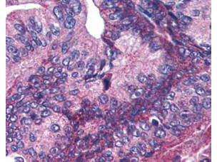
Boster's affinity purified anti-APC1 pS377 antibody was used at 5.0µg/ml to detect signal in a variety of tissues including multi-human, multi-brain and multi-cancer slides. This image shows moderate positive cytoplasmic and occasional nuclear staining of pancreatic carcinoma cells at 60X. Tissue was formalin-fixed and paraffin embedded. The image shows localization of the antibody as the precipitated red signal, with a hematoxylin purple nuclear counterstain. Personal Communication, Tina Roush, LifeSpanBiosciences, Seattle, WA.

Click image to see more details
Western blot using Boster's Affinity Purified anti-APC1 pS377 antibody shows detection of a band ~215 kDa corresponding to phosphorylated human APC1 (arrowhead). Lane 1 shows lysate from asynchronous cells. Lane 2 shows lysate from cells treated with nocodazole. While some phosphorylated APC1 is present in untreated cell, the amount of phosphorylated protein is increased in cell preparations arrested in mitosis. Each lane contains approximately 30 ug of HeLa whole cell lysates, separated by 4-8% SDS-PAGE followed by transfer to nitrocellulose. After blocking the membrane was probed with the primary antibody diluted to 1:1,000 overnight at 4°C followed by washes and reaction with a 1:10,000 dilution of IRDye800 conjugated Gt-a-Rabbit IgG [H&L] MX (611-132-122) for 45 min at room temperature. IRDye800 fluorescence image was captured using the Odyssey® Infrared Imaging System developed by LI-COR. IRDye is a trademark of LI-COR, Inc. Other detection systems will yield similar results.
Specific Publications For Anti-Phospho-APC1 S355 ANAPC1 Antibody (P03471)
Loading publications
Recommended Resources
Here are featured tools and databases that you might find useful.
- Boster's Pathways Library
- Protein Databases
- Bioscience Research Protocol Resources
- Data Processing & Analysis Software
- Photo Editing Software
- Scientific Literature Resources
- Research Paper Management Tools
- Molecular Biology Software
- Primer Design Tools
- Bioinformatics Tools
- Phylogenetic Tree Analysis
Customer Reviews
Have you used Anti-Phospho-APC1 S355 ANAPC1 Antibody?
Share your experimental results or join a short interview to earn up to $1,000 in product credits or other rewards.
0 Reviews For Anti-Phospho-APC1 S355 ANAPC1 Antibody
Customer Q&As
Have a question?
Find answers in Q&As, reviews.
Can't find your answer?
Submit your question
7 Customer Q&As for Anti-Phospho-APC1 S355 ANAPC1 Antibody
Question
I have attached the WB image, lot number and protocol we used for colon ovary using anti-Phospho-APC1 S355 antibody P03471. Please let me know if you require anything else.
Verified Customer
Verified customer
Asked: 2020-03-26
Answer
Thank you very much for the data. Our lab team are working to resolve this as quickly as possible, and we appreciate your patience and understanding! You have provided everything we needed. Please let me know if there is anything you need in the meantime.
Boster Scientific Support
Answered: 2020-03-26
Question
I see that the anti-Phospho-APC1 S355 antibody P03471 works with WB, what is the protocol used to produce the result images on the product page?
Verified Customer
Verified customer
Asked: 2019-10-02
Answer
You can find protocols for WB on the "support/technical resources" section of our navigation menu. If you have any further questions, please send an email to support@bosterbio.com
Boster Scientific Support
Answered: 2019-10-02
Question
We are currently using anti-Phospho-APC1 S355 antibody P03471 for mouse tissue, and we are happy with the IP results. The species of reactivity given in the datasheet says human, mouse, rat. Is it likely that the antibody can work on canine tissues as well?
Verified Customer
Verified customer
Asked: 2019-08-20
Answer
The anti-Phospho-APC1 S355 antibody (P03471) has not been validated for cross reactivity specifically with canine tissues, though there is a good chance of cross reactivity. We have an innovator award program that if you test this antibody and show it works in canine you can get your next antibody for free. Please contact me if I can help you with anything.
Boster Scientific Support
Answered: 2019-08-20
Question
Is there a BSA free version of anti-Phospho-APC1 S355 antibody P03471 available?
Verified Customer
Verified customer
Asked: 2019-05-30
Answer
Thanks for your recent telephone inquiry. I can confirm that some lots of this anti-Phospho-APC1 S355 antibody P03471 are BSA free. For now, these lots are available and we can make a BSA free formula for you free of charge. It will take 3 extra days to prepare. If you require this antibody BSA free again in future, please do not hesitate to contact me and I will be pleased to check which lots we have in stock that are BSA free.
Boster Scientific Support
Answered: 2019-05-30
Question
Thanks for helping with my inquiry over the phone. Here are the WB image, lot number and protocol we used for colon ovary using anti-Phospho-APC1 S355 antibody P03471. Let me know if you need anything else.
Verified Customer
Verified customer
Asked: 2017-05-16
Answer
I appreciate the data. You have provided everything we needed. Our lab team are working to resolve your inquiry as quickly as possible, and we appreciate your patience and understanding! Please let me know if there is anything you need in the meantime.
Boster Scientific Support
Answered: 2017-05-16
Question
My question regarding product P03471, anti-Phospho-APC1 S355 antibody. I was wondering if it would be possible to conjugate this antibody with biotin. I would need it to be without BSA or sodium azide. I am planning on using a buffer exchange of sodium azide with PBS only. Would there be problems for me to conjugate the antibody and store it in -20 degrees in small aliquots?
A. Anderson
Verified customer
Asked: 2016-04-14
Answer
We suggest not storing this antibody with PBS buffer only in -20 degrees. If you want to store it in -20 degrees it is best to add some cryoprotectant like glycerol. If you want carrier free P03471 anti-Phospho-APC1 S355 antibody, we can provide it to you in a special formula with trehalose and/or glycerol. These molecules will not interfere with conjugation chemistry and provide a good level of protection for the antibody from degradation. Please be sure to specify this in your purchase order.
Boster Scientific Support
Answered: 2016-04-14
Question
Will anti-Phospho-APC1 S355 antibody P03471 work for WB with colon ovary?
D. Krishna
Verified customer
Asked: 2015-04-15
Answer
According to the expression profile of colon ovary, ANAPC1 is highly expressed in colon ovary. So, it is likely that anti-Phospho-APC1 S355 antibody P03471 will work for WB with colon ovary.
Boster Scientific Support
Answered: 2015-04-15
