Click image to see more details
-

-
-
-
-
+2
Product Info Summary
| SKU: | A01049 |
|---|---|
| Size: | 100 μg/vial |
| Reactive Species: | Human, Mouse, Rat |
| Host: | Rabbit |
| Application: | ELISA, Flow Cytometry, IHC, WB |
Customers Who Bought This Also Bought

Product info
Product Name
Anti-Aquaporin 4/AQP4 Antibody Picoband®
SKU/Catalog Number
A01049
Size
100 μg/vial
Form
Lyophilized
Description
Boster Bio Anti-Aquaporin 4/AQP4 Antibody Picoband® catalog # A01049. Tested in ELISA, Flow Cytometry, IHC, WB applications. This antibody reacts with Human, Mouse, Rat. The brand Picoband indicates this is a premium antibody that guarantees superior quality, high affinity, and strong signals with minimal background in Western blot applications. Only our best-performing antibodies are designated as Picoband, ensuring unmatched performance.
Storage & Handling
At -20°C for one year from date of receipt. After reconstitution, at 4°C for one month. It can also be aliquotted and stored frozen at -20°C for six months. Avoid repeated freezing and thawing.
Cite This Product
Anti-Aquaporin 4/AQP4 Antibody Picoband® (Boster Biological Technology, Pleasanton CA, USA, Catalog # A01049)
Host
Rabbit
Contents
Each vial contains 4 mg Trehalose, 0.9 mg NaCl, 0.2 mg Na2HPO4.
Clonality
Polyclonal
Isotype
Rabbit IgG
Immunogen
E.coli-derived human Aquaporin 4/AQP4 recombinant protein (Position: L247-V323).
Cross-reactivity
No cross-reactivity with other proteins.
Reactive Species
A01049 is reactive to AQP4 in Human, Mouse, Rat
Observed Molecular Weight
32 kDa
Calculated molecular weight
34.8 kDa
Background of AQP4
Aquaporin 4 is found in the basolateral cell membrane of principal collecting duct cells and provides a pathway for water to exit these cells. The gene of AQP4 is mapped to 18q11.2-q12.1. Similar to other aquaporins, the AQP4 gene is composed of 4 exons encoding 127, 55, 27, and 92 amino acids separated by introns of 0.8, 0.3, and 5.2 kb. Unlike other aquaporins, an alternative coding initiation sequence (designated exon 0) was located 2.7 kb upstream of exon 1. When spliced together, M1 and the subsequent 10 amino acids are encoded by exon 0; the next 11 amino acids and M23 are encoded by exon 1. AQP4 is expressed in astrocytes and is upregulated by direct insult to the central nervous system. And AQP4 is the predominant water channel in the brain and has an important role in brain water homeostasis. It is abundant in mammalian brain and is concentrated in astrocytic foot processes at the blood-brain barrier.
Antibody Validation
Boster validates all antibodies on WB, IHC, ICC, Immunofluorescence, and ELISA with known positive control and negative samples to ensure specificity and high affinity, including thorough antibody incubations.
Application & Images
Applications
A01049 is guaranteed for ELISA, Flow Cytometry, IHC, WB Boster Guarantee
Recommend Dilution
| Application | Dilution | Species |
|---|---|---|
| Western blot | 0.25-0.5 μg/ml | Human, Mouse, Rat |
| Immunohistochemistry(Paraffin-embedded Section) | 2-5 μg/ml | Mouse, Rat |
| Flow Cytometry (Fixed) | 1-3 μg/1x106 cells | Human |
| ELISA | 0.1-0.5 μg/ml | - |
Tested application
Suggested blocking solution with 5% non-fat milk or BSA; (*)Recommended protein loading: 20-40 µg per lane
Use TE buffer pH 9.0 for antigen retrieval; (*) citrate buffer pH 6.0 is an alternative.
Validation Images & Assay Conditions
Click image to see more details
IHC analysis of Aquaporin 4/AQP4 using anti-Aquaporin 4/AQP4 antibody (A01049).
Aquaporin 4/AQP4 was detected in a paraffin-embedded section of human brain tissue. Heat mediated antigen retrieval was performed in EDTA buffer (pH 8.0, epitope retrieval solution). The tissue section was blocked with 10% goat serum. The tissue section was then incubated with 2 μg/ml rabbit anti-Aquaporin 4/AQP4 Antibody (A01049) overnight at 4°C. Peroxidase Conjugated Goat Anti-rabbit IgG was used as secondary antibody and incubated for 30 minutes at 37°C. The tissue section was developed using HRP Conjugated Rabbit IgG Super Vision Assay Kit (Catalog # SV0002) with DAB as the chromogen.

Click image to see more details
Western blot analysis of Aquaporin 4/AQP4 using anti-Aquaporin 4/AQP4 antibody (A01049).
Electrophoresis was performed on a 5-20% SDS-PAGE gel at 70V (Stacking gel) / 90V (Resolving gel) for 2-3 hours. The sample well of each lane was loaded with 30 ug of sample under reducing conditions.
Lane 1: human U-87MG whole cell lysates,
Lane 2: rat brain tissue lysates,
Lane 3: mouse brain tissue lysates.
After electrophoresis, proteins were transferred to a nitrocellulose membrane at 150 mA for 50-90 minutes. Blocked the membrane with 5% non-fat milk/TBS for 1.5 hour at RT. The membrane was incubated with rabbit anti-Aquaporin 4/AQP4 antigen affinity purified polyclonal antibody (Catalog # A01049) at 0.5 μg/mL overnight at 4°C, then washed with TBS-0.1%Tween 3 times with 5 minutes each and probed with a goat anti-rabbit IgG-HRP secondary antibody at a dilution of 1:5000 for 1.5 hour at RT. The signal is developed using an Enhanced Chemiluminescent detection (ECL) kit (Catalog # EK1002) with Tanon 5200 system. A specific band was detected for Aquaporin 4/AQP4 at approximately 32 kDa. The expected band size for Aquaporin 4/AQP4 is at 32 kDa.

Click image to see more details
IHC analysis of Aquaporin 4/AQP4 using anti-Aquaporin 4/AQP4 antibody (A01049).
Aquaporin 4/AQP4 was detected in a paraffin-embedded section of mouse brain tissue. Heat mediated antigen retrieval was performed in EDTA buffer (pH 8.0, epitope retrieval solution). The tissue section was blocked with 10% goat serum. The tissue section was then incubated with 2 μg/ml rabbit anti-Aquaporin 4/AQP4 Antibody (A01049) overnight at 4°C. Peroxidase Conjugated Goat Anti-rabbit IgG was used as secondary antibody and incubated for 30 minutes at 37°C. The tissue section was developed using HRP Conjugated Rabbit IgG Super Vision Assay Kit (Catalog # SV0002) with DAB as the chromogen.

Click image to see more details
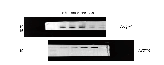
Western blot analysis of Aquaporin 4/AQP4 using anti-Aquaporin 4/AQP4 antibody (A01049).
Electrophoresis was performed on a 5-20% SDS-PAGE gel at 70V (Stacking gel) / 90V (Resolving gel) for 2-3 hours. The sample well of each lane was loaded with 30 ug of sample under reducing conditions.
Lane 1: Normal group-rat colon tissue lysates,
Lane 2: Model group-rat colon tissue lysates,
Lane 3: Triditional Chinese medicine group-rat colon tissue lysates,
Lane 4: Western medicine group-rat colon tissue lysates.
After electrophoresis, proteins were transferred to a nitrocellulose membrane at 150 mA for 50-90 minutes. Blocked the membrane with 5% non-fat milk/TBS for 1.5 hour at RT. The membrane was incubated with rabbit anti-Aquaporin 4/AQP4 antigen affinity purified polyclonal antibody (Catalog # A01049) at 1:1000 overnight at 4°C, then washed with TBS-0.1%Tween 3 times with 5 minutes each and probed with a goat anti-rabbit IgG-HRP secondary antibody for 1 hour at RT. The signal is developed using an Enhanced Chemiluminescent detection (ECL) kit (Catalog # EK1002) with ChemiDoc MP system. A specific band was detected for Aquaporin 4/AQP4 at approximately 37 kDa. The expected band size for Aquaporin 4/AQP4 is at 32 kDa.

Click image to see more details
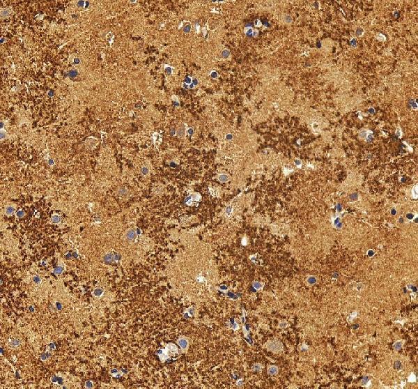
IHC analysis of Aquaporin 4/AQP4 using anti-Aquaporin 4/AQP4 antibody (A01049).
Aquaporin 4/AQP4 was detected in a paraffin-embedded section of rat brain tissue. Heat mediated antigen retrieval was performed in EDTA buffer (pH 8.0, epitope retrieval solution). The tissue section was blocked with 10% goat serum. The tissue section was then incubated with 2 μg/ml rabbit anti-Aquaporin 4/AQP4 Antibody (A01049) overnight at 4°C. Peroxidase Conjugated Goat Anti-rabbit IgG was used as secondary antibody and incubated for 30 minutes at 37°C. The tissue section was developed using HRP Conjugated Rabbit IgG Super Vision Assay Kit (Catalog # SV0002) with DAB as the chromogen.

Click image to see more details
Flow Cytometry analysis of A431 cells using anti-Aquaporin 4/AQP4 antibody (A01049).
Overlay histogram showing A431 cells stained with A01049 (Blue line). To facilitate intracellular staining, cells were fixed with 4% paraformaldehyde and permeabilized with permeabilization buffer. The cells were blocked with 10% normal goat serum. And then incubated with rabbit anti-Aquaporin 4/AQP4 Antibody (A01049, 1 μg/1x106 cells) for 30 min at 20°C. DyLight®488 conjugated goat anti-rabbit IgG (BA1127, 5-10 μg/1x106 cells) was used as secondary antibody for 30 minutes at 20°C. Isotype control antibody (Green line) was rabbit IgG (1 μg/1x106) used under the same conditions. Unlabelled sample without incubation with primary antibody and secondary antibody (Red line) was used as a blank control.
Specific Publications For Anti-Aquaporin 4/AQP4 Antibody Picoband® (A01049)
Loading publications
Recommended Resources
Here are featured tools and databases that you might find useful.
- Boster's Pathways Library
- Protein Databases
- Bioscience Research Protocol Resources
- Data Processing & Analysis Software
- Photo Editing Software
- Scientific Literature Resources
- Research Paper Management Tools
- Molecular Biology Software
- Primer Design Tools
- Bioinformatics Tools
- Phylogenetic Tree Analysis
Customer Reviews
Have you used Anti-Aquaporin 4/AQP4 Antibody Picoband®?
Share your experimental results or join a short interview to earn up to $1,000 in product credits or other rewards.
1 Reviews For Anti-Aquaporin 4/AQP4 Antibody Picoband®
This antibody is highly efficient and specific, suitable for detecting AQP4 protein in rat colon by Western blot, with only minor nonspecific bands.
Excellent
| SKU | A01049 |
|---|---|
| Application | Western Blot |
| Sample | rat colon tissue |
| Sample Processing Description | Samples were lysed in RIPA buffer containing protease inhibitor PMSF (100:1) for 10 minutes, followed by centrifugation at 12,000 rpm for 15 minutes. The supernatant was mixed with 5× loading buffer, denatured at 100°C for 10 minutes, and then loaded onto SDS-PAGE. |
| Other Reagents | Blocking buffer |
| Primary Antibody | Aquaporin 4/AQP4 Antibody Picoband® |
| Primary Incubation | 1:1000, overnight at 4 ℃ |
| Secondary Antibody | HRP Conjugated AffiniPure Goat Anti-Rabbit IgG (H+L) |
| Secondary Incubation | 1 hour in room temperature |
| Detection | Substrate: ECL, Imaging system:ChemiDoc MP |
| Results Summary | The figure shows a schematic representation of Western blot results for the target protein AQP4 and the loading control Actin in rat colon across the normal, model, traditional Chinese medicine, and western medicine groups. The target bands are clear and distinct, and the experimental results are satisfactory. |
Shiyu Zhang, LUTCM
Verified customer
Submitted 2025-12-30
Customer Q&As
Have a question?
Find answers in Q&As, reviews.
Can't find your answer?
Submit your question
