Click image to see more details
-

-
-
-
-
+2
Product Info Summary
| SKU: | PB9477 |
|---|---|
| Size: | 100 μg/vial |
| Reactive Species: | Human, Mouse, Rat |
| Host: | Rabbit |
| Application: | Flow Cytometry, IF, IHC, ICC, WB |
Customers Who Bought This Also Bought

Product info
Product Name
Anti-ARSA Antibody Picoband®
SKU/Catalog Number
PB9477
PB0501 is an alternative SKU for this antibody, used in previous lots.
Size
100 μg/vial
Form
Lyophilized
Description
Boster Bio Anti-ARSA Antibody Picoband® catalog # PB9477. Tested in Flow Cytometry, IF, IHC, ICC, WB applications. This antibody reacts with Human, Mouse, Rat. The brand Picoband indicates this is a premium antibody that guarantees superior quality, high affinity, and strong signals with minimal background in Western blot applications. Only our best-performing antibodies are designated as Picoband, ensuring unmatched performance.
Storage & Handling
Store at -20˚C for one year from date of receipt. After reconstitution, at 4˚C for one month. It can also be aliquotted and stored frozen at -20˚C for six months. Avoid repeated freeze-thaw cycles.
Cite This Product
Anti-ARSA Antibody Picoband® (Boster Biological Technology, Pleasanton CA, USA, Catalog # PB9477)
Host
Rabbit
Contents
Each vial contains antibody formulated with stabilizing components, 0.9 mg NaCl, 0.2 mg Na2HPO4, and 0.05 mg NaN3.
*This antibody is supplied in a stabilized formulation.
Compatibility with conjugation reactions depends on the chemistry of the conjugation method used.
For conjugation methods that are not compatible with the stabilizing components present in this formulation, a carrier-free antibody format is required.
Clonality
Polyclonal
Isotype
Rabbit IgG
Immunogen
A synthetic peptide corresponding to a sequence at the C-terminus of human ARSA, different from the related mouse sequence by six amino acids.
Cross-reactivity
No cross-reactivity with other proteins
Reactive Species
PB9477 is reactive to ARSA in Human, Mouse, Rat
Observed Molecular Weight
54 kDa
Calculated molecular weight
53.6 kDa
Background of ARSA
Arylsulfatase A (ARSA) is an enzyme that breaks down sulfatides, namely cerebroside 3-sulfate intocerebroside and sulfate. In humans, arylsulfatase A is encoded by the ARSA gene. ARSA is mapped to 22q13.33. The protein encoded by this gene hydrolyzes cerebroside sulfate to cerebroside and sulfate. Defects in this gene lead to metachromatic leucodystrophy (MLD), a progressive demyelination disease which results in a variety of neurological symptoms and ultimately death. Alternatively spliced transcript variants have been described for this gene.
Antibody Validation
Boster validates all antibodies on WB, IHC, ICC, Immunofluorescence, and ELISA with known positive control and negative samples to ensure specificity and high affinity, including thorough antibody incubations.
Application & Images
Applications
PB9477 is guaranteed for Flow Cytometry, IF, IHC, ICC, WB Boster Guarantee
Recommend Dilution
| Application | Dilution | Species |
|---|---|---|
| Western blot | 0.1-0.5μg/ml | Human, Mouse, Rat |
| Immunohistochemistry (Paraffin-embedded Section) | 0.5-1μg/ml | Human, Rat |
| Immunocytochemistry/Immunofluorescence | 2μg/ml | Human |
| Flow Cytometry (Fixed) | 1-3μg/1x106 cells | Human |
Tested application
Suggested blocking solution with 5% non-fat milk or BSA; (*)Recommended protein loading: 20-40 µg per lane
Use TE buffer pH 9.0 for antigen retrieval; (*) citrate buffer pH 6.0 is an alternative.
Validation Images & Assay Conditions
Click image to see more details
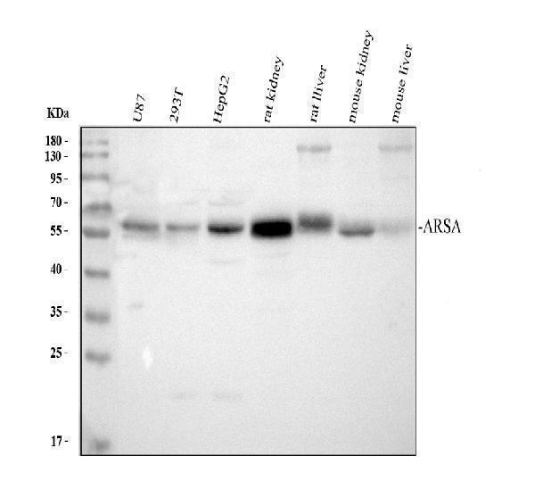
Western blot analysis of ARSA using anti-ARSA antibody (PB9477).
Electrophoresis was performed on a 10% SDS-PAGE gel at 80V (Stacking gel) / 120V (Resolving gel) for 2 hours. The sample well of each lane was loaded with 30 ug of sample under reducing conditions.
Lane 1: human U87 whole cell lysates,
Lane 2: human 293T whole cell lysates,
Lane 3: human HepG2 whole cell lysates,
Lane 4: rat kidney tissue lysates,
Lane 5: rat liver tissue lysates,
Lane 6: mouse kidney tissue lysates,
Lane 7: mouse liver tissue lysates.
After electrophoresis, proteins were transferred to a nitrocellulose membrane at 150 mA for 50-90 minutes. Blocked the membrane with 5% non-fat milk/TBS for 1.5 hour at RT. The membrane was incubated with rabbit anti-ARSA antigen affinity purified polyclonal antibody (PB9477) at 0.5 μg/mL overnight at 4°C, then washed with TBS-0.1%Tween 3 times with 5 minutes each and probed with a goat anti-rabbit IgG-HRP secondary antibody (Catalog # BA1054) at a dilution of 1:5000 for 1.5 hour at RT. The signal is developed using an ECL Plus Western Blotting Substrate (Catalog # AR1196-200) with Tanon 5200 system. A specific band was detected for ARSA at approximately 54 kDa. The expected band size for ARSA is at 54 kDa.

Click image to see more details
IHC analysis of ARSA using anti-ARSA antibody (PB9477).
ARSA was detected in paraffin-embedded section of Rat Lung Tissue. Heat mediated antigen retrieval was performed in citrate buffer (pH6, epitope retrieval solution) for 20 mins. The tissue section was blocked with 10% goat serum. The tissue section was then incubated with 1μg/ml rabbit anti-ARSA Antibody (PB9477) overnight at 4°C. Biotinylated goat anti-rabbit IgG was used as secondary antibody and incubated for 30 minutes at 37°C. The tissue section was developed using Strepavidin-Biotin-Complex (SABC)(Catalog # SA1022) with DAB as the chromogen.

Click image to see more details
IHC analysis of ARSA using anti-ARSA antibody (PB9477).
ARSA was detected in paraffin-embedded section of Human Mammary Cancer Tissue. Heat mediated antigen retrieval was performed in citrate buffer (pH6, epitope retrieval solution) for 20 mins. The tissue section was blocked with 10% goat serum. The tissue section was then incubated with 1μg/ml rabbit anti-ARSA Antibody (PB9477) overnight at 4°C. Biotinylated goat anti-rabbit IgG was used as secondary antibody and incubated for 30 minutes at 37°C. The tissue section was developed using Strepavidin-Biotin-Complex (SABC)(Catalog # SA1022) with DAB as the chromogen.

Click image to see more details
Flow Cytometry analysis of Hela cells using anti-ARSA antibody (PB9477).
Overlay histogram showing Hela cells stained with PB9477 (Blue line). To facilitate intracellular staining, cells were fixed with 4% paraformaldehyde and permeabilized with permeabilization buffer. The cells were blocked with 10% normal goat serum. And then incubated with rabbit anti-ARSA Antibody (PB9477,1μg/1x106 cells) for 30 min at 20°C. DyLight®488 conjugated goat anti-rabbit IgG (BA1127, 5-10μg/1x106 cells) was used as secondary antibody for 30 minutes at 20°C. Isotype control antibody (Green line) was rabbit IgG (1μg/1x106) used under the same conditions. Unlabelled sample without incubation with primary antibody and secondary antibody (Red line) was used as a blank control.

Click image to see more details
Flow Cytometry analysis of PC-3 cells using anti-ARSA antibody (PB9477).
Overlay histogram showing PC-3 cells stained with PB9477 (Blue line). To facilitate intracellular staining, cells were fixed with 4% paraformaldehyde and permeabilized with permeabilization buffer. The cells were blocked with 10% normal goat serum. And then incubated with rabbit anti-ARSA Antibody (PB9477,1μg/1x106 cells) for 30 min at 20°C. DyLight®488 conjugated goat anti-rabbit IgG (BA1127, 5-10μg/1x106 cells) was used as secondary antibody for 30 minutes at 20°C. Isotype control antibody (Green line) was rabbit IgG (1μg/1x106) used under the same conditions. Unlabelled sample without incubation with primary antibody and secondary antibody (Red line) was used as a blank control.

Click image to see more details
IF analysis of ARSA using anti-ARSA antibody (PB9477).
ARSA was detected in immunocytochemical section of U20S cell. Enzyme antigen retrieval was performed using IHC enzyme antigen retrieval reagent (AR0022) for 15 mins. The cells were blocked with 10% goat serum. And then incubated with 2μg/mL rabbit anti-ARSA Antibody (PB9477) overnight at 4°C. DyLight®488 Conjugated Goat Anti-Rabbit IgG (BA1127) was used as secondary antibody at 1:100 dilution and incubated for 30 minutes at 37°C. The section was counterstained with DAPI. Visualize using a fluorescence microscope and filter sets appropriate for the label used.
Specific Publications For Anti-ARSA Antibody Picoband® (PB9477)
Loading publications
Recommended Resources
Here are featured tools and databases that you might find useful.
- Boster's Pathways Library
- Protein Databases
- Bioscience Research Protocol Resources
- Data Processing & Analysis Software
- Photo Editing Software
- Scientific Literature Resources
- Research Paper Management Tools
- Molecular Biology Software
- Primer Design Tools
- Bioinformatics Tools
- Phylogenetic Tree Analysis
Customer Reviews
Have you used Anti-ARSA Antibody Picoband®?
Share your experimental results or join a short interview to earn up to $1,000 in product credits or other rewards.
0 Reviews For Anti-ARSA Antibody Picoband®
Customer Q&As
Have a question?
Find answers in Q&As, reviews.
Can't find your answer?
Submit your question
5 Customer Q&As for Anti-ARSA Antibody Picoband®
Question
Would PB9477 anti-ARSA antibody work on parafin embedded sections? If so, which fixation method do you recommend we use (PFA, paraformaldehyde, other)?
Verified Customer
Verified customer
Asked: 2019-11-13
Answer
As indicated on the product datasheet, PB9477 anti-ARSA antibody as been validated on Flow Cytometry. It is best to use PFA for fixation because it has better tissue penetration ability. PFA needs to be prepared fresh before use. Long term stored PFA turns into formalin, as the PFA molecules congregate and become formalin.
Boster Scientific Support
Answered: 2019-11-13
Question
Is a blocking peptide available for product anti-ARSA antibody (PB9477)?
Verified Customer
Verified customer
Asked: 2018-11-12
Answer
We do provide the blocking peptide for product anti-ARSA antibody (PB9477). If you would like to place an order for it please contact support@bosterbio.com and make a special request.
Boster Scientific Support
Answered: 2018-11-12
Question
We are currently using anti-ARSA antibody PB9477 for mouse tissue, and we are happy with the ICC results. The species of reactivity given in the datasheet says human, mouse, rat. Is it possible that the antibody can work on dog tissues as well?
Verified Customer
Verified customer
Asked: 2017-08-29
Answer
The anti-ARSA antibody (PB9477) has not been validated for cross reactivity specifically with dog tissues, though there is a good chance of cross reactivity. We have an innovator award program that if you test this antibody and show it works in dog you can get your next antibody for free. Please contact me if I can help you with anything.
Boster Scientific Support
Answered: 2017-08-29
Question
I was wanting to use your anti-ARSA antibody for Flow Cytometry for mouse liver on frozen tissues, but I want to know if it has been validated for this particular application. Has this antibody been validated and is this antibody a good choice for mouse liver identification?
E. Evans
Verified customer
Asked: 2013-05-13
Answer
It shows on the product datasheet, PB9477 anti-ARSA antibody has been tested for Flow Cytometry, IHC, ICC, WB on human, mouse, rat tissues. We have an innovator award program that if you test this antibody and show it works in mouse liver in IHC-frozen, you can get your next antibody for free.
Boster Scientific Support
Answered: 2013-05-13
Question
Our lab want to know about to test anti-ARSA antibody PB9477 on mouse liver for research purposes, then I may be interested in using anti-ARSA antibody PB9477 for diagnostic purposes as well. Is the antibody suitable for diagnostic purposes?
T. Walker
Verified customer
Asked: 2013-05-01
Answer
The products we sell, including anti-ARSA antibody PB9477, are only intended for research use. They would not be suitable for use in diagnostic work. If you have the means to develop a product into diagnostic use, and are interested in collaborating with us and develop our product into an IVD product, please contact us for more discussions.
Boster Scientific Support
Answered: 2013-05-01
