Click image to see more details
-

-
-
-
-
+7
Product Info Summary
| SKU: | A01552 |
|---|---|
| Size: | 0.1 mg |
| Reactive Species: | Human, Mouse |
| Host: | Rabbit |
| Application: | ELISA, IF, ICC, WB |
Customers Who Bought This Also Bought

Product info
Product Name
Anti-Bim BCL2L11 Antibody
SKU/Catalog Number
A01552
Size
0.1 mg
Form
Liquid
Description
Boster Bio Anti-Bim BCL2L11 Antibody (Catalog # A01552). Tested in ELISA, WB, ICC, IF applications. This antibody reacts with Human, Mouse.
Storage & Handling
Bim antibody can be stored at 4°C for three months and -20°C, stable for up to one year. Avoid repeated freeze-thaw cycles. Antibodies should not be exposed to prolonged high temperatures.
Cite This Product
Anti-Bim BCL2L11 Antibody (Boster Biological Technology, Pleasanton CA, USA, Catalog # A01552)
Host
Rabbit
Contents
Bim Antibody is supplied in PBS containing 0.02% sodium azide.
Clonality
Polyclonal
Isotype
IgG
Immunogen
Anti-BIM antibody was raised against a peptide corresponding to amino acids near the center of human BIM. The immunogen is located within amino acids 80-130 of BIM.
Reactive Species
A01552 is reactive to BCL2L11 in Human, Mouse
Observed Molecular Weight
68 kDa
Calculated molecular weight
22.2 kDa
Background of BCL2L11
Members in the Bcl-2 family are critical regulators of apoptosis by either inhibiting or promoting cell death. Bcl-2 homology 3 (BH3) domain is a potent death domain. BH3 domain containing pro-apoptotic proteins, including Bad, Bax, Bid, Bik, and Hrk, form a growing subclass of the Bcl-2 family. A novel BH3 domain containing protein was recently identified and designated Bim or BOD in human, mouse and rat. Bim/BOD interacts with diverse members in the pro-survival Bcl-2 sub-family including Bcl-2, Bcl-xL and Bcl-w. Bim/BOD induces apoptosis. The messenger RNA of Bim is ubiquitously expressed in multiple tissues and cell lines.
Antibody Validation
Boster validates all antibodies on WB, IHC, ICC, Immunofluorescence, and ELISA with known positive control and negative samples to ensure specificity and high affinity, including thorough antibody incubations.
Application & Images
Applications
A01552 is guaranteed for ELISA, IF, ICC, WB Boster Guarantee
Recommend Dilution
WB: 5 μg/mL; ICC: 10 μg/mL; IF: 20 μg/mL.
Antibody validated: Western Blot in human and mouse samples; Immunocytochemistry in human samples; Immunofluorescence in human samples. All other applications and species not yet tested. Optimal dilutions for each application should be determined by the researcher.
Validation Images & Assay Conditions
Click image to see more details
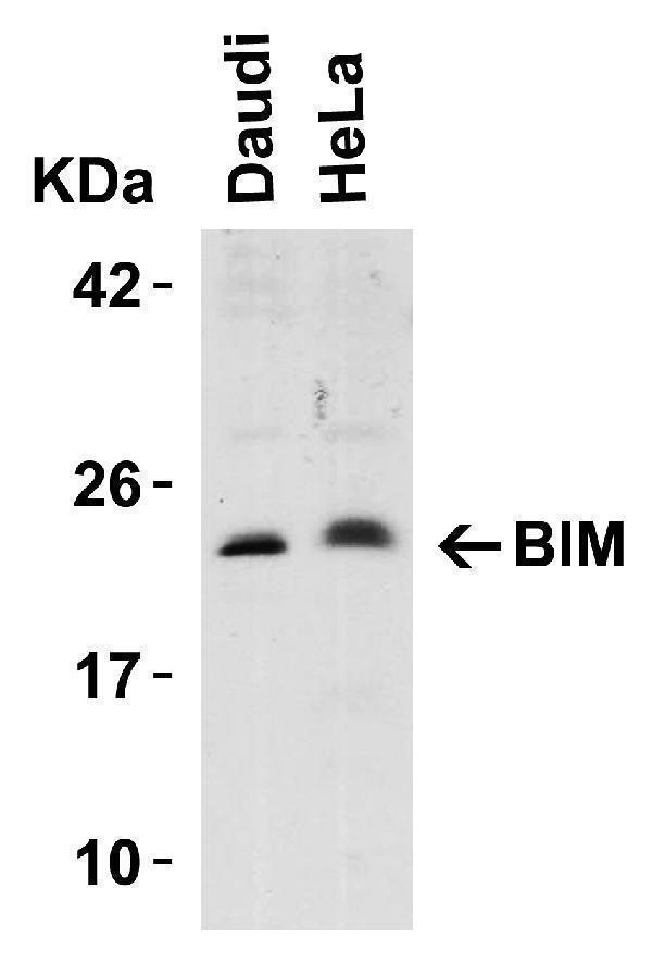
Western Blot Validation in Human Cell Lines
Loading: 15 μg of lysates per lane.
Antibodies: BIM A01552, (5 μg/mL), 1h incubation at RT in 5% NFDM/TBST.
Secondary: Goat anti-rabbit IgG HRP conjugate at 1:10000 dilution.

Click image to see more details
Independent Antibody Validation (IAV) via Protein Expression Profile in Cell Lines
Loading: 15 μg of lysates per lane.
Antibodies: BIM 2065, (0.5 μg/mL), BIM A01552, (5 μg/mL), beta-actin (1 μg/mL) and GAPDH (0.02 μg/mL), 1h incubation at RT in 5% NFDM/TBST.
Secondary: Goat anti-rabbit IgG HRP conjugate at 1:10000 dilution.

Click image to see more details
Western Blot Validation in Human Tissue
Loading: 15 μg of lysates per lane.
Antibodies: BIM A01552, (5 μg/mL), 1h incubation at RT in 5% NFDM/TBST.
Secondary: Goat anti-rabbit IgG HRP conjugate at 1:10000 dilution.
Lane 1: Human urinary bladder
Lane 2: Human pancreas

Click image to see more details
Immunofluorescence Validation of BIM in K562 Cells
Immunofluorescent analysis of 4% paraformaldehyde-fixed K562 cells labeling BIM with A01552 at 20 μg/mL, followed by goat anti-rabbit IgG secondary antibody at 1/500 dilution (red).

Click image to see more details
Immunocytochemistry Validation of BIM K562 Cells
Immunohistochemical analysis of K562 cells using anti-BIM antibody (A01552) at 10 μg/ml. Cells was fixed with formaldehyde and blocked with 10% serum for 1 h at RT; antigen retrieval was by heat mediation with a citrate buffer (pH6). Samples were incubated with primary antibody overnight at 4˚C. A goat anti-rabbit IgG H&L (HRP) at 1/250 was used as secondary. Counter stained with Hematoxylin.

Click image to see more details
Induced Expression Validation of BIM in Mouse Hippocampus (Tsuchiya et al., 2011)
The induction of Bim protein was detected by immunohistochemical analysis of mice after i.h. injection of epoxomicin with anti-BIM antibodies. Sections from epoxomicin-treated animals exhibited cells staining positive for Bim expression within the NeuN-positive population of neurons in the CA1 of the ipsilateral side. In contrast, Bim-positive cells were absent within the NeuNpositive
CA1 neurons on the contralateral side.

Click image to see more details
KD Validation of BIM in 293 Cells (Han et al., 2010)
Immunofluorescence analysis with anti-BIM antibodies was performed for BIM in 293 cells transfected with control siRNA or BIM siRNA. BIM expression was disrupted after BIM siRNA knockdown.

Click image to see more details
Regulated Expression Validation of BIM in U266 Cells (Chen et al., 2012)
Immunoblot analysis was carried out to monitor protein expression of 3 isoforms (EL, L, and S) of Bim with anti-BIM antibodies. BIM expression was up-regulated by flavopiridol treatment, which was blocked by Cycloheximide in U266 cells.

Click image to see more details
WB KD Validation of BIM in 293 Cells (Han et al., 2010)
Western blot analysis with anti-BIM antibodies was performed for BIM in 293 cells transfected with control siRNA or BIM siRNA. BIM expression was disrupted after BIM siRNA knockdown.

Click image to see more details
Localization Validation of BIM in Mouse Macrophages (Ulett et al., 2005)
Immunoblots of subcellular fractions enriched for mitochondria and cytosol were used to determine BIM protein levels with anti-BIM antibodies in J774A cells. BIM is exclusively expressed in mitochondria.

Click image to see more details
KD Validation of BIM in Human Cell Lines (Chen et al., 2009)
Human leukemia (U937 and Jurkat) and myeloma (U266) cells were stably transfected with constructs encoding shBim or a scrambled sequence (shNC). Immunoblotting was preformed to monitor expression of Bim in these cells with anti-BIM antibodies. BIM expression was disrupted after shBIM
Specific Publications For Anti-Bim BCL2L11 Antibody (A01552)
Loading publications
Recommended Resources
Here are featured tools and databases that you might find useful.
- Boster's Pathways Library
- Protein Databases
- Bioscience Research Protocol Resources
- Data Processing & Analysis Software
- Photo Editing Software
- Scientific Literature Resources
- Research Paper Management Tools
- Molecular Biology Software
- Primer Design Tools
- Bioinformatics Tools
- Phylogenetic Tree Analysis
Customer Reviews
Have you used Anti-Bim BCL2L11 Antibody?
Share your experimental results or join a short interview to earn up to $1,000 in product credits or other rewards.
0 Reviews For Anti-Bim BCL2L11 Antibody
Customer Q&As
Have a question?
Find answers in Q&As, reviews.
Can't find your answer?
Submit your question
