Click image to see more details
Product Info Summary
| SKU: | M01469-2 |
|---|---|
| Size: | 100 μl/vial |
| Reactive Species: | Human, Mouse, Rat |
| Host: | Rabbit |
| Application: | IF, IHC, ICC, WB |
Customers Who Bought This Also Bought

Product info
Product Name
Anti-BNIP3 Antibody (Monoclonal, 35B09)
SKU/Catalog Number
M01469-2
Size
100 μl/vial
Form
Liquid
Description
Boster Bio Anti-BNIP3 Antibody (Monoclonal, 35B09) catalog # M01469-2. Tested in WB, IHC, IF, ICC/IF applications. This antibody reacts with Human, Mouse, Rat.
Storage & Handling
Store at -20°C for one year. For short term storage and frequent use, store at 4°C for up to one month. Avoid repeated freeze-thaw cycles.
Cite This Product
Anti-BNIP3 Antibody (Monoclonal, 35B09) (Boster Biological Technology, Pleasanton CA, USA, Catalog # M01469-2)
Host
Rabbit
Contents
Rabbit IgG in stabilizing components, phosphate buffered saline, pH 7.4, 150mM NaCl, 0.02% sodium azide and 50% glycerol.
This antibody is supplied in a stabilized formulation.
Compatibility with conjugation reactions depends on the chemistry of the conjugation method used.
For conjugation methods that are not compatible with the stabilizing components present in this formulation, a carrier-free antibody format is required.
Clonality
Monoclonal
Clone Number
35B09
Immunogen
Synthetic peptide within human BNIP3 aa 92-128.
Reactive Species
M01469-2 is reactive to BNIP3 in Human, Mouse, Rat
Calculated molecular weight
27.8 kDa
Background of BNIP3
The Bcl-2 nineteen kilodalton interacting protein 3 (BNIP3 or NIP3), is a hypoxia-inducible proapoptotic member of the Bcl-2 family that induces cell death by associating with the mitochondria. BNIP3, expressed in skeletal muscle and in the brain at low levels, is primarily localized to the nucleus of glial cells of the normal human brain, as well as in the malignant glioma cell line U251. BNIP3 expression in the cytoplasm increases and localizes with the mitochondria, contributing to induction of cell death. Cellular protein BNIP3 interacts with E1B-19K, BCL-2, BCL-xL, and EBV-BHRF1. BNIP3 contains Bcl-2 homology 3 (BH3) domain and COOH-terminal transmembrane (TM) domain. The BH3 domain of BNIP3 mediates Bcl-2/Bcl-X(L) heterodimerization and confers pro-apoptotic activity; whereas the TM domain is critical for homodimerization, pro-apoptotic function, and mitochondrial targeting.
Antibody Validation
Boster validates all antibodies on WB, IHC, ICC, Immunofluorescence, and ELISA with known positive control and negative samples to ensure specificity and high affinity, including thorough antibody incubations.
Application & Images
Applications
M01469-2 is guaranteed for IF, IHC, ICC, WB Boster Guarantee
Assay Dilutions Recommendation
The recommendations below provide a starting point for assay optimization. The actual working concentration varies and should be decided by the user.
Western blot, 1:500-2000
Immunohistochemistry, 1:50-200
Immunofluorescence, 1:50-200
Immunocytochemistry/Immunofluorescence, 1:50-200
Positive Control
IHC: human lung cancer tissue, mouse skeletal muscle tissue, rat skeletal muscle tissue
Validation Images & Assay Conditions
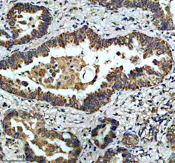
Click image to see more details
IHC analysis of BNIP3 using anti-BNIP3 antibody (M01469-2).
BNIP3 was detected in a paraffin-embedded section of human lung cancer tissue. Heat mediated antigen retrieval was performed in EDTA buffer (pH 8.0, epitope retrieval solution). The tissue section was blocked with 10% goat serum. The tissue section was then incubated with 1:50 rabbit anti-BNIP3 Antibody (M01469-2) overnight at 4°C. Peroxidase Conjugated Goat Anti-rabbit IgG was used as secondary antibody and incubated for 30 minutes at 37°C. The tissue section was developed using HRP Conjugated Rabbit IgG Super Vision Assay Kit (Catalog # SV0002) with DAB as the chromogen.

Click image to see more details
IHC analysis of BNIP3 using anti-BNIP3 antibody (M01469-2).
BNIP3 was detected in a paraffin-embedded section of mouse skeletal muscle tissue. Heat mediated antigen retrieval was performed in EDTA buffer (pH 8.0, epitope retrieval solution). The tissue section was blocked with 10% goat serum. The tissue section was then incubated with 1:50 rabbit anti-BNIP3 Antibody (M01469-2) overnight at 4°C. Peroxidase Conjugated Goat Anti-rabbit IgG was used as secondary antibody and incubated for 30 minutes at 37°C. The tissue section was developed using HRP Conjugated Rabbit IgG Super Vision Assay Kit (Catalog # SV0002) with DAB as the chromogen.

Click image to see more details
IHC analysis of BNIP3 using anti-BNIP3 antibody (M01469-2).
BNIP3 was detected in a paraffin-embedded section of rat skeletal muscle tissue. Heat mediated antigen retrieval was performed in EDTA buffer (pH 8.0, epitope retrieval solution). The tissue section was blocked with 10% goat serum. The tissue section was then incubated with 1:50 rabbit anti-BNIP3 Antibody (M01469-2) overnight at 4°C. Peroxidase Conjugated Goat Anti-rabbit IgG was used as secondary antibody and incubated for 30 minutes at 37°C. The tissue section was developed using HRP Conjugated Rabbit IgG Super Vision Assay Kit (Catalog # SV0002) with DAB as the chromogen.
Specific Publications For Anti-BNIP3 Antibody (Monoclonal, 35B09) (M01469-2)
Loading publications
Recommended Resources
Here are featured tools and databases that you might find useful.
- Boster's Pathways Library
- Protein Databases
- Bioscience Research Protocol Resources
- Data Processing & Analysis Software
- Photo Editing Software
- Scientific Literature Resources
- Research Paper Management Tools
- Molecular Biology Software
- Primer Design Tools
- Bioinformatics Tools
- Phylogenetic Tree Analysis
Customer Reviews
Have you used Anti-BNIP3 Antibody (Monoclonal, 35B09)?
Share your experimental results or join a short interview to earn up to $1,000 in product credits or other rewards.
0 Reviews For Anti-BNIP3 Antibody (Monoclonal, 35B09)
Customer Q&As
Have a question?
Find answers in Q&As, reviews.
Can't find your answer?
Submit your question
