Click image to see more details
Product Info Summary
| SKU: | A02412-1 |
|---|---|
| Size: | 100 μg/vial |
| Reactive Species: | Human |
| Host: | Rabbit |
| Application: | ELISA, Flow Cytometry, IF, ICC, WB |
Customers Who Bought This Also Bought

Product info
Product Name
Anti-BRD2 Antibody Picoband®
SKU/Catalog Number
A02412-1
Size
100 μg/vial
Form
Lyophilized
Description
Boster Bio Anti-BRD2 Antibody Picoband® catalog # A02412-1. Tested in ELISA, Flow Cytometry, IF, ICC, WB applications. This antibody reacts with Human. The brand Picoband indicates this is a premium antibody that guarantees superior quality, high affinity, and strong signals with minimal background in Western blot applications. Only our best-performing antibodies are designated as Picoband, ensuring unmatched performance.
Storage & Handling
Store at -20˚C for one year from date of receipt. After reconstitution, at 4˚C for one month. It can also be aliquotted and stored frozen at -20˚C for six months. Avoid repeated freeze-thaw cycles.
Cite This Product
Anti-BRD2 Antibody Picoband® (Boster Biological Technology, Pleasanton CA, USA, Catalog # A02412-1)
Host
Rabbit
Contents
Each vial contains 4 mg Trehalose, 0.9 mg NaCl and 0.2 mg Na2HPO4.
Clonality
Polyclonal
Isotype
Rabbit IgG
Immunogen
E.coli-derived human BRD2 recombinant protein (Position: M1-Q214).
Cross-reactivity
No cross-reactivity with other proteins.
Reactive Species
A02412-1 is reactive to BRD2 in Human
Observed Molecular Weight
110 kDa
Calculated molecular weight
88.1 kDa
Background of BRD2
Bromodomain-containing protein 2 is a protein that in humans is encoded by the BRD2 gene. It is mapped to 6p21.32. This gene encodes a transcriptional regulator that belongs to the BET (bromodomains and extra terminal domain) family of proteins. This protein associates with transcription complexes and with acetylated chromatin during mitosis, and it selectively binds to the acetylated lysine-12 residue of histone H4 via its two bromodomains. The gene maps to the major histocompatability complex (MHC) class II region on chromosome 6p21.3, but sequence comparison suggests that the protein is not involved in the immune response. This gene has been implicated in juvenile myoclonic epilepsy, a common form of epilepsy that becomes apparent in adolescence. Multiple alternatively spliced variants have been described for this gene.
Antibody Validation
Boster validates all antibodies on WB, IHC, ICC, Immunofluorescence, and ELISA with known positive control and negative samples to ensure specificity and high affinity, including thorough antibody incubations.
Application & Images
Applications
A02412-1 is guaranteed for ELISA, Flow Cytometry, IF, ICC, WB Boster Guarantee
Recommend Dilution
| Application | Dilution | Species |
|---|---|---|
| Western blot | 0.25-0.5μg/ml | Human |
| Immunocytochemistry/Immunofluorescence | 5μg/ml | Human |
| Flow Cytometry (Fixed) | 1-3μg/1x106 cells | Human |
| ELISA | 0.1-0.5μg/ml | - |
Tested application
Suggested blocking solution with 5% non-fat milk or BSA; (*)Recommended protein loading: 20-40 µg per lane
Validation Images & Assay Conditions
Click image to see more details
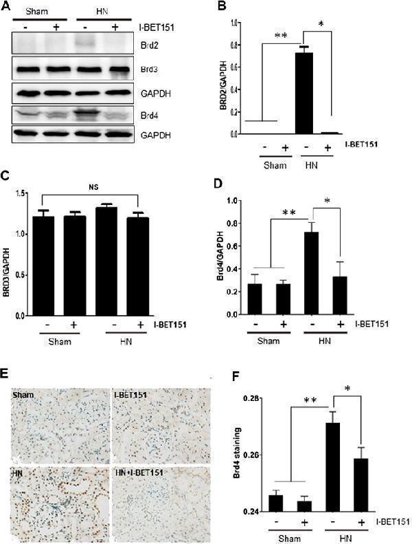
I-BET151 reduces Brd4 levels in the kidney of HN rats. (A) A rat model of HN was established and treated with I-BET151 as indicated in “Material and Methods.” After 3 weeks, kidneys were taken for immunoblot analysis for Brd2, Brd3, Brd4 or GAPDH. Expression levels of Brd2 (B) , Brd3 (C) , Brd4 (D) were quantified by densitometry analysis and then normalized with GAPDH. (E) Photomicrographs (original magnification, ×400) illustrate immunohistochemical Brd4 staining of kidney tissues. (F) Brd4 staining graphic presentation of quantitative data. Data are represented as the mean ± SEM. * p < 0.05; ** p < 0.01.
Index in PubMed under a CC BY license. PMID: 33664670

Click image to see more details
Flow Cytometry analysis of 293T cells using anti-BRD2 antibody (A02412-1).
Overlay histogram showing 293T cells stained with A02412-1 (Blue line). To facilitate intracellular staining, cells were fixed with 4% paraformaldehyde and permeabilized with permeabilization buffer. The cells were blocked with 10% normal goat serum. And then incubated with rabbit anti-BRD2 Antibody (A02412-1, 1 μg/1x106 cells) for 30 min at 20°C. DyLight®488 conjugated goat anti-rabbit IgG (BA1127, 5-10 μg/1x106 cells) was used as secondary antibody for 30 minutes at 20°C. Isotype control antibody (Green line) was rabbit IgG (1 μg/1x106) used under the same conditions. Unlabelled sample without incubation with primary antibody and secondary antibody (Red line) was used as a blank control.

Click image to see more details
Western blot analysis of BRD2 using anti-BRD2 antibody (A02412-1).
Electrophoresis was performed on a 5-20% SDS-PAGE gel at 70V (Stacking gel) / 90V (Resolving gel) for 2-3 hours. The sample well of each lane was loaded with 30 ug of sample under reducing conditions.
Lane 1: human Hela whole cell lysates,
Lane 2: human A431 whole cell lysates,
Lane 3: human Jurkat whole cell lysates,
Lane 4: human 293T whole cell lysates.
After electrophoresis, proteins were transferred to a nitrocellulose membrane at 150 mA for 50-90 minutes. Blocked the membrane with 5% non-fat milk/TBS for 1.5 hour at RT. The membrane was incubated with rabbit anti-BRD2 antigen affinity purified polyclonal antibody (Catalog # A02412-1) at 0.5 μg/mL overnight at 4°C, then washed with TBS-0.1%Tween 3 times with 5 minutes each and probed with a goat anti-rabbit IgG-HRP secondary antibody at a dilution of 1:5000 for 1.5 hour at RT. The signal is developed using an Enhanced Chemiluminescent detection (ECL) kit (Catalog # EK1002) with Tanon 5200 system. A specific band was detected for BRD2 at approximately 110 kDa. The expected band size for BRD2 is at 88 kDa.

Click image to see more details
IF analysis of BRD2 and Tubulin beta using anti-BRD2 antibody (A02412-1) and anti-Tubulin beta antibody (M05613-4).
BRD2 and Tubulin beta was detected in an immunocytochemical section of A431 cells. Enzyme antigen retrieval was performed using IHC enzyme antigen retrieval reagent (AR0022) for 15 mins. The cells were blocked with 10% goat serum. And then incubated with 5 μg/mL rabbit anti-BRD2 Antibody (A02412-1) and mouse anti-Tubulin beta Antibody (M05613-4) overnight at 4°C. DyLight®488 Conjugated Goat Anti-Rabbit IgG (BA1127) and DyLight®594 Conjugated Goat Anti-Mouse IgG (BA1141) were used as secondary antibody at 1:100 dilution and incubated for 30 minutes at 37°C. Visualize using a fluorescence microscope and filter sets appropriate for the label used.
Specific Publications For Anti-BRD2 Antibody Picoband® (A02412-1)
Loading publications
Recommended Resources
Here are featured tools and databases that you might find useful.
- Boster's Pathways Library
- Protein Databases
- Bioscience Research Protocol Resources
- Data Processing & Analysis Software
- Photo Editing Software
- Scientific Literature Resources
- Research Paper Management Tools
- Molecular Biology Software
- Primer Design Tools
- Bioinformatics Tools
- Phylogenetic Tree Analysis
Customer Reviews
Have you used Anti-BRD2 Antibody Picoband®?
Share your experimental results or join a short interview to earn up to $1,000 in product credits or other rewards.
0 Reviews For Anti-BRD2 Antibody Picoband®
Customer Q&As
Have a question?
Find answers in Q&As, reviews.
Can't find your answer?
Submit your question
