Click image to see more details
Product Info Summary
| SKU: | A00297-1 |
|---|---|
| Size: | 100 μg/vial |
| Reactive Species: | Human, Mouse, Rat |
| Host: | Rabbit |
| Application: | ELISA, WB |
Customers Who Bought This Also Bought

Product info
Product Name
Anti-c-Fos/FOS Antibody Picoband®
SKU/Catalog Number
A00297-1
Size
100 μg/vial
Form
Lyophilized
Description
Boster Bio Anti-c-Fos/FOS Antibody Picoband® catalog # A00297-1. Tested in ELISA, WB applications. This antibody reacts with Human, Mouse, Rat. The brand Picoband indicates this is a premium antibody that guarantees superior quality, high affinity, and strong signals with minimal background in Western blot applications. Only our best-performing antibodies are designated as Picoband, ensuring unmatched performance.
Storage & Handling
Store at -20˚C for one year from date of receipt. After reconstitution, at 4˚C for one month. It can also be aliquotted and stored frozen at -20˚C for six months. Avoid repeated freeze-thaw cycles.
Cite This Product
Anti-c-Fos/FOS Antibody Picoband® (Boster Biological Technology, Pleasanton CA, USA, Catalog # A00297-1)
Host
Rabbit
Contents
Each vial contains 4 mg Trehalose, 0.9 mg NaCl and 0.2 mg Na2HPO4.
Clonality
Polyclonal
Isotype
Rabbit IgG
Immunogen
E.coli-derived human c-Fos/FOS recombinant protein (Position: N45-D293).
Cross-reactivity
No cross-reactivity with other proteins.
Reactive Species
A00297-1 is reactive to FOS in Human, Mouse, Rat
Observed Molecular Weight
50-55 kDa
Calculated molecular weight
40.7 kDa
Background of FOS
The human oncogene c-fos is cellular homolog of the transforming gene of Finkel-Biskis-Jinkins (FBJ) murine osteosarcoma virus which was mapped to a single human chromosome. c-Fos is encoded by the FOS gene. FOS was the first transcription factor identified that has a critical function in regulating the development of cells destined to form and maintain the skeleton. FOS is also a major component of the activator protein-1 (AP-1) transcription factor complex, which includes members of the JUN family. c-fos is a major nuclear target for signal transduction pathways involved in the regulation of cell growth, differentiation, and transformation. Using transgenic and knockout mice, Grigoriadis et al. (1995) established a unique role for the proto-oncogene and nuclear transcription factor, Fos, in regulating the differentiation and activity of specific bone cell populations, both during normal development and in bone disease.
Antibody Validation
Boster validates all antibodies on WB, IHC, ICC, Immunofluorescence, and ELISA with known positive control and negative samples to ensure specificity and high affinity, including thorough antibody incubations.
Application & Images
Applications
A00297-1 is guaranteed for ELISA, WB Boster Guarantee
Assay Dilutions Recommendation
The recommendations below provide a starting point for assay optimization. The actual working concentration varies and should be decided by the user.
Western blot, 0.25-0.5μg/ml, Human, Mouse, Rat
ELISA, 0.1-0.5μg/ml, -
Positive Control
Suggested blocking solution with 5% non-fat milk or BSA; (*)Recommended protein loading: 20-40 µg per lane
Validation Images & Assay Conditions
Click image to see more details
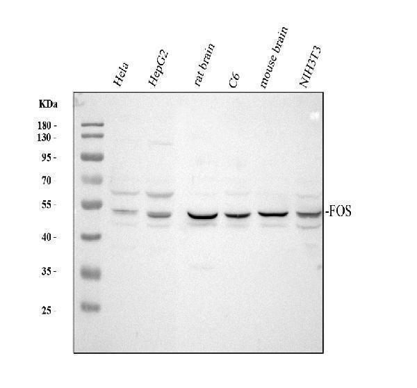
Western blot analysis of c-Fos/FOS using anti-c-Fos/FOS antibody (A00297-1).
Electrophoresis was performed on a 10% SDS-PAGE gel at 80V (Stacking gel) / 120V (Resolving gel) for 2 hours. The sample well of each lane was loaded with 30 ug of sample under reducing conditions.
Lane 1: human Hela whole cell lysates,
Lane 2: human HepG2 whole cell lysates,
Lane 3: rat brain tissue lysates,
Lane 4: rat C6 whole cell lysates,
Lane 5: mouse brain tissue lysates,
Lane 6: mouse NIH/3T3 whole cell lysates.
After Electrophoresis, proteins were transferred to a Nitrocellulose membrane at 150mA for 50-90 minutes. Blocked the membrane with 5% Non-fat Milk/ TBS for 1.5 hour at RT. The membrane was incubated with rabbit anti-c-Fos/FOS antigen affinity purified polyclonal antibody (Catalog # A00297-1) at 0.5 μg/mL overnight at 4°C, then washed with TBS-0.1%Tween 3 times with 5 minutes each and probed with a goat anti-rabbit IgG-HRP secondary antibody at a dilution of 1:5000 for 1.5 hour at RT. The signal is developed using an ECL Plus Western Blotting Substrate (Catalog # AR1196-200) with Tanon 5200 system. A specific band was detected for c-Fos/FOS at approximately 50-55 kDa. The expected band size for c-Fos/FOS is at 41 kDa.
Specific Publications For Anti-c-Fos/FOS Antibody Picoband® (A00297-1)
Loading publications
Recommended Resources
Here are featured tools and databases that you might find useful.
- Boster's Pathways Library
- Protein Databases
- Bioscience Research Protocol Resources
- Data Processing & Analysis Software
- Photo Editing Software
- Scientific Literature Resources
- Research Paper Management Tools
- Molecular Biology Software
- Primer Design Tools
- Bioinformatics Tools
- Phylogenetic Tree Analysis
Customer Reviews
Have you used Anti-c-Fos/FOS Antibody Picoband®?
Share your experimental results or join a short interview to earn up to $1,000 in product credits or other rewards.
0 Reviews For Anti-c-Fos/FOS Antibody Picoband®
Customer Q&As
Have a question?
Find answers in Q&As, reviews.
Can't find your answer?
Submit your question
