Click image to see more details
Product Info Summary
| SKU: | M00297 |
|---|---|
| Size: | 100 μl |
| Reactive Species: | Human, Mouse, Rat |
| Host: | Rabbit |
| Application: | IF, IHC, WB |
Customers Who Bought This Also Bought

Product info
Product Name
Anti-c-Fos Rabbit Monoclonal Antibody
SKU/Catalog Number
M00297
BM4864 is an alternative SKU for this antibody, used in previous lots.
Size
100 μl
Form
Liquid
Description
Boster Bio Anti-c-Fos Rabbit Monoclonal Antibody catalog # M00297. Tested in WB, IHC, IF applications. This antibody reacts with Human, Mouse, Rat.
Storage & Handling
Store at -20°C for one year. For short term storage and frequent use, store at 4°C for up to one month. Avoid repeated freeze-thaw cycles.
Cite This Product
Anti-c-Fos Rabbit Monoclonal Antibody (Boster Biological Technology, Pleasanton CA, USA, Catalog # M00297)
Host
Rabbit
Contents
Rabbit IgG in stabilizing components, phosphate buffered saline, pH 7.4, 150mM NaCl, 0.02% sodium azide and 50% glycerol.
*This antibody is supplied in a stabilized formulation.
Compatibility with conjugation reactions depends on the chemistry of the conjugation method used.
For conjugation methods that are not compatible with the stabilizing components present in this formulation, a carrier-free antibody format is required.
Clonality
Monoclonal
Clone Number
IIO-6
Isotype
Rabbit IgG
Immunogen
A synthesized peptide derived from human c-Fos
Reactive Species
M00297 is reactive to FOS in Human, Mouse, Rat
Observed Molecular Weight
41 kDa
Calculated molecular weight
40.7 kDa
Antibody Validation
Boster validates all antibodies on WB, IHC, ICC, Immunofluorescence, and ELISA with known positive control and negative samples to ensure specificity and high affinity, including thorough antibody incubations.
Application & Images
Applications
M00297 is guaranteed for IF, IHC, WB Boster Guarantee
Recommend Dilution
WB 1:500-2000
IHC 1:50-200
IF 1:50-200
Tested application
Suggested blocking solution with 5% non-fat milk or BSA; (*)Recommended protein loading: 20-40 µg per lane
Validation Images & Assay Conditions
Click image to see more details
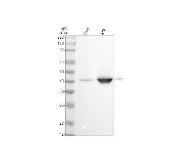
Western blot analysis of FOS using anti-FOS antibody (M00297).
Electrophoresis was performed on a 10% SDS-PAGE gel at 80V (Stacking gel) / 120V (Resolving gel) for 2 hours. The sample well of each lane was loaded with 30 ug of sample under reducing conditions.
Lane 1: human Hela whole cell lysates,
Lane 2: human RT4 whole cell lysates.
After electrophoresis, proteins were transferred to a nitrocellulose membrane at 150 mA for 50-90 minutes. Blocked the membrane with 5% non-fat milk/TBS for 1.5 hour at RT. The membrane was incubated with rabbit anti-FOS antigen affinity purified monoclonal antibody (M00297) at 1:500 overnight at 4°C, then washed with TBS-0.1%Tween 3 times with 5 minutes each and probed with a goat anti-rabbit IgG-HRP secondary antibody at a dilution of 1:5000 for 1.5 hour at RT. The signal is developed using an ECL Plus Western Blotting Substrate (Catalog # AR1196-200) with Tanon 5200 system. A specific band was detected for FOS at approximately 41 kDa. The expected band size for FOS is at 41 kDa.

Click image to see more details
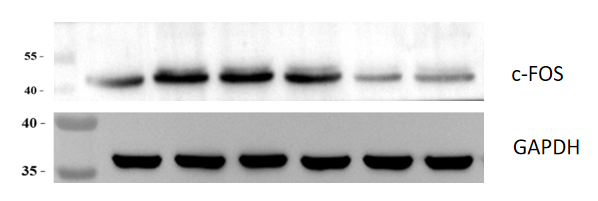
Western blot analysis of FOS using anti-FOS antibody (M00297).
Electrophoresis was performed on a 10% SDS-PAGE gel at 80V (Stacking gel) / 120V (Resolving gel) for 2 hours. The sample well of each lane was loaded with 30 ug of sample under reducing conditions.
Lane 1: mouse hippocampus tissue lysates,
Lane 2: mouse Alzheimer’s disease tissue lysates,
Lane 3-5: mouse Alzheimer’s disease hippocampi treated with increasing doses of Qianlenta herbal formula tissue lysates,
Lane 6:mouse Alzheimer’s disease hippocampi treated with positive control drug.
After electrophoresis, proteins were transferred to a nitrocellulose membrane at 150 mA for 50-90 minutes. Blocked the membrane with 5% non-fat milk/TBS for 1.5 hour at RT. The membrane was incubated with rabbit anti-FOS antigen affinity purified monoclonal antibody (M00297) at 1:2000 overnight at 4°C, then washed with TBS-0.1%Tween 3 times with 5 minutes each and probed with a goat anti-rabbit IgG-HRP secondary antibody at a dilution of 1:10000 for 1 hour at RT. The signal is developed using an ECL Plus Western Blotting Substrate (Catalog # AR1196-200) with ChemiDoc MP system. A specific band was detected for FOS at approximately 41 kDa. The expected band size for FOS is at 41 kDa.

Click image to see more details
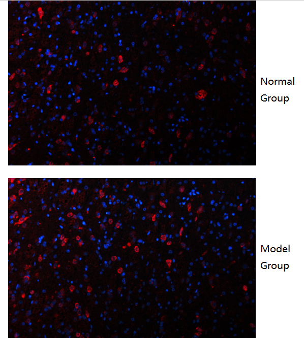
IF analysis of c-Fos using anti-c-Fos antibody (M00297).
c-Fos was detected in an immunocytochemical section of mouse brain tissue. Enzyme antigen retrieval was performed using IHC enzyme antigen retrieval reagent (AR0022) for 15 mins. The cells were blocked with 10% goat serum. And then incubated with 1:200 rabbit anti-c-Fos Antibody (M00297) overnight at 4°C. DyLight®594 Conjugated Goat Anti-Rabbit IgG (BA1142) was used as secondary antibody at 1:500 dilution and incubated for 45 minutes at 37°C. The section was counterstained with DAPI. Visualize using a fluorescence microscope and filter sets appropriate for the label used.
Specific Publications For Anti-c-Fos Rabbit Monoclonal Antibody (M00297)
Loading publications
Recommended Resources
Here are featured tools and databases that you might find useful.
- Boster's Pathways Library
- Protein Databases
- Bioscience Research Protocol Resources
- Data Processing & Analysis Software
- Photo Editing Software
- Scientific Literature Resources
- Research Paper Management Tools
- Molecular Biology Software
- Primer Design Tools
- Bioinformatics Tools
- Phylogenetic Tree Analysis
Customer Reviews
Have you used Anti-c-Fos Rabbit Monoclonal Antibody?
Share your experimental results or join a short interview to earn up to $1,000 in product credits or other rewards.
2 Reviews For Anti-c-Fos Rabbit Monoclonal Antibody
The Anti-c-FOS antibody (M00297) showed clear and specific immunofluorescence staining in mouse brain, with markedly increased c-Fos expression in neurons of the acute ischemic region compared to normal brain.
Excellent
| SKU | M00297 |
|---|---|
| Application | Immunocytochemistry |
| Sample | mouse brain |
| Sample Processing Description | Normal brain tissue and brain tissue from a cerebral infarction model |
| Other Reagents | Goat serum, DAB Chromogenic Solution, Anti-fade mounting medium |
| Primary Antibody | c-Fos Rabbit Monoclonal Antibody |
| Primary Incubation | 1:200, overnight at 4 ℃ |
| Secondary Antibody | DyLight 594 Goat Anti-Rabbit IgG (H+L) |
| Secondary Incubation | 45 min at 37℃ |
| Detection | Imaging system:Leica DM2500 |
| Results Summary | c-Fos is a marker of activated neural cells, especially neurons. During the acute phase of cerebral infarction, as a strong stress and injury stimulus, it drives c-Fos expression in the brain—particularly in the peri-infarct region—far higher than in normal resting brain. The experimental results show clear staining of positive cells, and the expression levels are consistent with expectations. |
Qihang Yang, Huazhong University of Science and Technology
Verified customer
Submitted 2026-02-12
This antibody demonstrates good specificity, with minimal nonspecific bands. The target band is clear and at the correct position, and expression differences between different groups are observable.
Excellent
| SKU | M00297 |
|---|---|
| Application | Western Blot |
| Sample | Mouse hippocampal tissue |
| Sample Processing Description | An Alzheimer’s disease (AD) model was established by injecting streptozotocin (STZ) into the lateral ventricles of mice. Hippocampal tissues were collected after treatment with four different doses of QianCengTa compound, and total protein was extracted. |
| Other Reagents | RIPA lysis buffer,Protease inhibitor,Electrophoresis buffer,Transfer buffer,Blocking buffer |
| Primary Antibody | c-Fos Rabbit Monoclonal Antibody |
| Primary Incubation | 1:2000, overnight at 4 ℃ |
| Secondary Antibody | HRP Goat Anti-Rabbit IgG |
| Secondary Incubation | 1:10000, 1 hour in room temperature |
| Detection | Substrate: ECL, Imaging system:ChemiDoc MP |
| Results Summary | c-FOS is a marker of neuronal activation under pathological conditions. Its expression is low in normal brain tissue but elevated in the AD model, and decreases following drug treatment. Lane 1 represents normal hippocampus, lane 2 the AD model hippocampus, lanes 3, 4, and 5 correspond to increasing doses of QianCengTa compound, and lane 6 is the positive control drug. The Western blot results indicate that QianCengTa compound exhibits a therapeutic effect on AD. |
Xiangnan Qin, SHUTCM
Verified customer
Submitted 2025-12-25
Customer Q&As
Have a question?
Find answers in Q&As, reviews.
Can't find your answer?
Submit your question
4 Customer Q&As for Anti-c-Fos Rabbit Monoclonal Antibody
Question
We have seen staining in rat pancreas. What should we do? Is anti-c-Fos Rabbit Monoclonal antibody supposed to stain pancreas positively?
Verified Customer
Verified customer
Asked: 2020-03-16
Answer
According to literature pancreas does express FOS. According to Uniprot.org, FOS is expressed in mucosa of stomach, colon, pancreas, leukemic t-cell, among other tissues. Regarding which tissues have FOS expression, here are a few articles citing expression in various tissues:
Colon, Pubmed ID: 14702039
Leukemic T-cell, Pubmed ID: 19690332
Pancreas, Pubmed ID: 15489334
Boster Scientific Support
Answered: 2020-03-16
Question
We want using your anti-c-Fos Rabbit Monoclonal antibody for response to gravity studies. Has this antibody been tested with western blotting on hela cell lysate? We would like to see some validation images before ordering.
Verified Customer
Verified customer
Asked: 2018-02-21
Answer
Thank you for your inquiry. This M00297 anti-c-Fos Rabbit Monoclonal antibody is tested on hela cell lysate. It is guaranteed to work for WB in human, mouse, rat. Our Boster guarantee will cover your intended experiment even if the sample type has not been be directly tested.
Boster Scientific Support
Answered: 2018-02-21
Question
My colleagues were content with the WB result of your anti-c-Fos Rabbit Monoclonal antibody. However we have observed positive staining in mucosa of stomach nucleus. endoplasmic reticulum. cytoplasm, using this antibody. Is that expected? Could you tell me where is FOS supposed to be expressed?
S. Anderson
Verified customer
Asked: 2016-03-14
Answer
From literature, mucosa of stomach does express FOS. Generally FOS expresses in nucleus. endoplasmic reticulum. cytoplasm,. Regarding which tissues have FOS expression, here are a few articles citing expression in various tissues:
Colon, Pubmed ID: 14702039
Leukemic T-cell, Pubmed ID: 19690332
Pancreas, Pubmed ID: 15489334
Boster Scientific Support
Answered: 2016-03-14
Question
We are currently using anti-c-Fos Rabbit Monoclonal antibody M00297 for rat tissue, and we are content with the WB results. The species of reactivity given in the datasheet says human, mouse, rat. Is it likely that the antibody can work on pig tissues as well?
K. Taylor
Verified customer
Asked: 2015-12-28
Answer
The anti-c-Fos Rabbit Monoclonal antibody (M00297) has not been validated for cross reactivity specifically with pig tissues, but there is a good chance of cross reactivity. We have an innovator award program that if you test this antibody and show it works in pig you can get your next antibody for free. Please contact me if I can help you with anything.
Boster Scientific Support
Answered: 2015-12-28
