Click image to see more details
-

-
-
-
-
+3
Product Info Summary
| SKU: | M00341 |
|---|---|
| Size: | 100 μl |
| Reactive Species: | Human, Mouse, Rat |
| Host: | Rabbit |
| Application: | IF, IHC, ICC, WB |
Customers Who Bought This Also Bought

Product info
Product Name
Anti-NGF/Beta Ngf Rabbit Monoclonal Antibody
SKU/Catalog Number
M00341
BM4100 is an alternative SKU for this antibody, used in previous lots.
Size
100 μl
Form
Liquid
Description
Boster Bio Anti-NGF/Beta Ngf Rabbit Monoclonal Antibody catalog # M00341. Tested in WB, IHC, ICC/IF applications. This antibody reacts with Human, Mouse, Rat.
Storage & Handling
Store at -20°C for one year. For short term storage and frequent use, store at 4°C for up to one month. Avoid repeated freeze-thaw cycles.
Cite This Product
Anti-NGF/Beta Ngf Rabbit Monoclonal Antibody (Boster Biological Technology, Pleasanton CA, USA, Catalog # M00341)
Host
Rabbit
Contents
Rabbit IgG in stabilizing components, phosphate buffered saline, pH 7.4, 150mM NaCl, 0.02% sodium azide and 50% glycerol.
*This antibody is supplied in a stabilized formulation.
Compatibility with conjugation reactions depends on the chemistry of the conjugation method used.
For conjugation methods that are not compatible with the stabilizing components present in this formulation, a carrier-free antibody format is required.
Clonality
Monoclonal
Clone Number
BDE-14
Isotype
Rabbit IgG
Immunogen
A synthesized peptide derived from human NGF
Reactive Species
M00341 is reactive to NGF in Human, Mouse, Rat
Observed Molecular Weight
35 kDa
Calculated molecular weight
27.0 kDa
Antibody Validation
Boster validates all antibodies on WB, IHC, ICC, Immunofluorescence, and ELISA with known positive control and negative samples to ensure specificity and high affinity, including thorough antibody incubations.
Application & Images
Applications
M00341 is guaranteed for IF, IHC, ICC, WB Boster Guarantee
Recommend Dilution
WB 1:500-2000
IHC 1:50-200
ICC/IF 1:50-200
Tested application
Use TE buffer pH 9.0 for antigen retrieval; (*) citrate buffer pH 6.0 is an alternative.
Validation Images & Assay Conditions
Click image to see more details
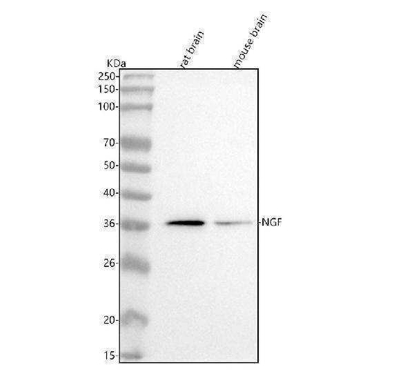
Western blot analysis of NGF using anti-NGF antibody (M00341).
Electrophoresis was performed on a 5-20% SDS-PAGE gel at 70V (Stacking gel) / 90V (Resolving gel) for 2-3 hours. The sample well of each lane was loaded with 30 ug of sample under reducing conditions.
Lane 1: rat brain tissue lysates,
Lane 2: mouse brain tissue lysates.
After electrophoresis, proteins were transferred to a nitrocellulose membrane at 150 mA for 50-90 minutes. Blocked the membrane with 5% non-fat milk/TBS for 1.5 hour at RT. The membrane was incubated with rabbit anti-NGF antigen affinity purified monoclonal antibody (Catalog # M00341) at 1:500 overnight at 4°C, then washed with TBS-0.1%Tween 3 times with 5 minutes each and probed with a goat anti-rabbit IgG-HRP secondary antibody at a dilution of 1:500 for 1.5 hour at RT. The signal is developed using an Enhanced Chemiluminescent detection (ECL) kit (Catalog # EK1002) with Tanon 5200 system. A specific band was detected for NGF at approximately 35 kDa. The expected band size for NGF is at 27 kDa.

Click image to see more details
Western blot analysis of NGF expression in (1)Mouse thyroid lysate;(2) HeLa cell lysate.

Click image to see more details
All lanes use the Antibody at 1:500 dilution for 1 hour at room temperature.

Click image to see more details
Immunohistochemical analysis of paraffin-embedded human kidney, using NGF Antibody.

Click image to see more details
Immunofluorescent analysis of SH-SY5Y cells, using NGF Antibody.

Click image to see more details
Immunofluorescent analysis using the Antibody at 1:50 dilution.

Click image to see more details
Immunofluorescent analysis using the Antibody at 1:150 dilution.
Specific Publications For Anti-NGF/Beta Ngf Rabbit Monoclonal Antibody (M00341)
Loading publications
Recommended Resources
Here are featured tools and databases that you might find useful.
- Boster's Pathways Library
- Protein Databases
- Bioscience Research Protocol Resources
- Data Processing & Analysis Software
- Photo Editing Software
- Scientific Literature Resources
- Research Paper Management Tools
- Molecular Biology Software
- Primer Design Tools
- Bioinformatics Tools
- Phylogenetic Tree Analysis
Customer Reviews
Have you used Anti-NGF/Beta Ngf Rabbit Monoclonal Antibody?
Share your experimental results or join a short interview to earn up to $1,000 in product credits or other rewards.
0 Reviews For Anti-NGF/Beta Ngf Rabbit Monoclonal Antibody
Customer Q&As
Have a question?
Find answers in Q&As, reviews.
Can't find your answer?
Submit your question
5 Customer Q&As for Anti-NGF/Beta Ngf Rabbit Monoclonal Antibody
Question
We purchased anti-NGF/Beta Ngf Rabbit Monoclonal antibody for IHC on cartilage tissue a few months ago. I am using mouse, and We are going to use the antibody for IF next. I am interested in examining cartilage tissue as well as leukocyte in our next experiment. Do you have any suggestion on which antibody would work the best for IF?
Verified Customer
Verified customer
Asked: 2019-12-09
Answer
I have checked the website and datasheets of our anti-NGF/Beta Ngf Rabbit Monoclonal antibody and I see that M00341 has been validated on mouse in both IHC and IF. Thus M00341 should work for your application. Our Boster satisfaction guarantee will cover this product for IF in mouse even if the specific tissue type has not been validated. We do have a comprehensive range of products for IF detection and you can check out our website bosterbio.com to find out more information about them.
Boster Scientific Support
Answered: 2019-12-09
Question
We are currently using anti-NGF/Beta Ngf Rabbit Monoclonal antibody M00341 for mouse tissue, and we are satisfied with the ICC results. The species of reactivity given in the datasheet says human, mouse, rat. Is it true that the antibody can work on goat tissues as well?
Verified Customer
Verified customer
Asked: 2019-09-27
Answer
The anti-NGF/Beta Ngf Rabbit Monoclonal antibody (M00341) has not been tested for cross reactivity specifically with goat tissues, though there is a good chance of cross reactivity. We have an innovator award program that if you test this antibody and show it works in goat you can get your next antibody for free. Please contact me if I can help you with anything.
Boster Scientific Support
Answered: 2019-09-27
Question
Our team were well pleased with the WB result of your anti-NGF/Beta Ngf Rabbit Monoclonal antibody. However we have seen positive staining in eye secreted. using this antibody. Is that expected? Could you tell me where is NGF supposed to be expressed?
S. Mangal
Verified customer
Asked: 2019-09-09
Answer
According to literature, eye does express NGF. Generally NGF expresses in secreted. Regarding which tissues have NGF expression, here are a few articles citing expression in various tissues:
Brain, Pubmed ID: 2374737
Eye, Pubmed ID: 15489334
Leukocyte, Pubmed ID: 2025430
Boster Scientific Support
Answered: 2019-09-09
Question
We need using your anti-NGF/Beta Ngf Rabbit Monoclonal antibody for positive regulation of neuron differentiation studies. Has this antibody been tested with western blotting on hela cell lysate? We would like to see some validation images before ordering.
E. Taylor
Verified customer
Asked: 2018-11-19
Answer
We appreciate your inquiry. This M00341 anti-NGF/Beta Ngf Rabbit Monoclonal antibody is validated on hela cell lysate. It is guaranteed to work for IF, IHC, ICC, WB in human, mouse, rat. Our Boster guarantee will cover your intended experiment even if the sample type has not been be directly tested.
Boster Scientific Support
Answered: 2018-11-19
Question
We have seen staining in rat cartilage tissue. Do you have any suggestions? Is anti-NGF/Beta Ngf Rabbit Monoclonal antibody supposed to stain cartilage tissue positively?
Verified Customer
Verified customer
Asked: 2018-08-01
Answer
According to literature cartilage tissue does express NGF. According to Uniprot.org, NGF is expressed in cartilage tissue, brain, eye, leukocyte, among other tissues. Regarding which tissues have NGF expression, here are a few articles citing expression in various tissues:
Brain, Pubmed ID: 2374737
Eye, Pubmed ID: 15489334
Leukocyte, Pubmed ID: 2025430
Boster Scientific Support
Answered: 2018-08-01
