Click image to see more details
-

-
-
-
-
+4
Product Info Summary
| SKU: | MA1028 |
|---|---|
| Size: | 100 μg/vial |
| Reactive Species: | Human |
| Host: | Mouse |
| Application: | IHC, ICC, WB |
Customers Who Bought This Also Bought

Product info
Product Name
Anti-c-Myc Antibody (Monoclonal, 9E10)
SKU/Catalog Number
MA1028
BM0238 is an alternative SKU for this antibody, used in previous lots.
Size
100 μg/vial
Form
Lyophilized
Description
Boster Bio Anti-c-Myc Antibody (Monoclonal, 9E10) catalog # MA1028. Tested in IHC, ICC, WB applications. This antibody reacts with Human.
Storage & Handling
Store at -20˚C for one year from date of receipt. After reconstitution, at 4˚C for one month. It can also be aliquotted and stored frozen at -20˚C for six months. Avoid repeated freeze-thaw cycles.
Cite This Product
Anti-c-Myc Antibody (Monoclonal, 9E10) (Boster Biological Technology, Pleasanton CA, USA, Catalog # MA1028)
Host
Mouse
Contents
Mouse IgG in stabilizing components, 1.2% sodium acetate and 0.01mg NaN3.
Clonality
Monoclonal
Clone Number
Clone: 9E10
Isotype
Mouse IgG1
Immunogen
Synthetic peptide corresponding to residues 408-439 of the human p62c-Myc protein.
Cross-reactivity
No cross-reactivity with other proteins
Reactive Species
MA1028 is reactive to Myc in Human
Observed Molecular Weight
49 kDa
Calculated molecular weight
48.9 kDa
Background of Myc
C-Myc is an oncogene that functions both in the stimulation of cell proliferation and in apoptosis. c-Myc elicits its oncogenic activity by causing immortalization, and to a lesser extent the transformation of cells, in addition to several other mechanisms. The c-MYC proto-oncogene encodes a transcription factor that is critical for cell growth and proliferation. It is one of the genes frequently altered in cancer cells in which it exhibits constitutive activity. Downregulation of c-Myc is critical for 2-Methoxyestradiol (2ME2)-induced oxidative stress and apoptosis in AML cells. And its up-regulation is important for promoting lymphocyte cell division, and demonstrating that GFP-c-Myc expression is a marker of proliferating lymphocytes in vivo.
Antibody Validation
Boster validates all antibodies on WB, IHC, ICC, Immunofluorescence, and ELISA with known positive control and negative samples to ensure specificity and high affinity, including thorough antibody incubations.
Application & Images
Applications
MA1028 is guaranteed for IHC, ICC, WB Boster Guarantee
Recommend Dilution
| Application | Dilution | Species |
|---|---|---|
| Immunohistochemistry (Paraffin-embedded Section) | 5μg/ml | Human |
| Immunocytochemistry | 1μg/ml | Human, - |
| Western blot | 1μg/ml | Human |
Tested application
Suggested blocking solution with 5% non-fat milk or BSA; (*)Recommended protein loading: 20-40 µg per lane
Use TE buffer pH 9.0 for antigen retrieval; (*) citrate buffer pH 6.0 is an alternative.
Validation Images & Assay Conditions
Click image to see more details
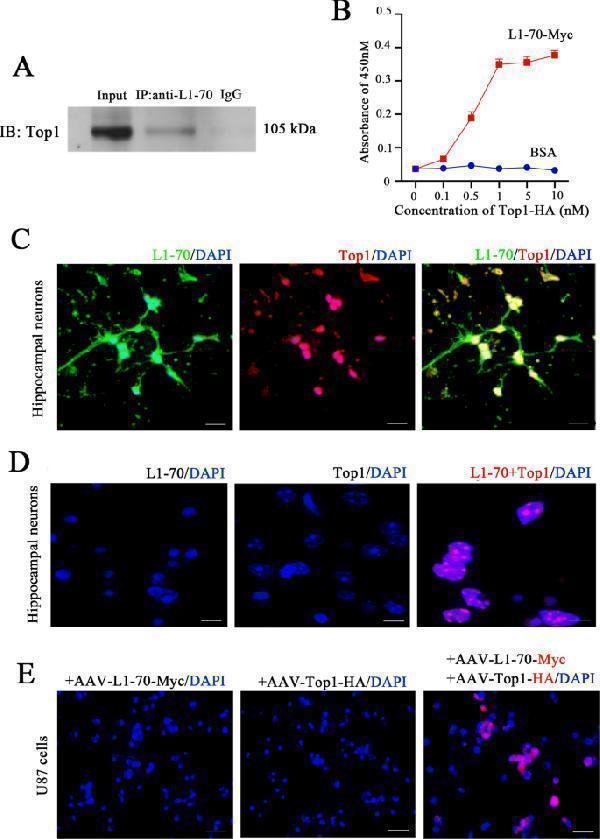
L1-70 is associated with Top1. A Co-immunoprecipitation of the hippocampus tissue homogenates from 5- to 7-day-old wild-type mice with an antibody against L1 cytoplasmic domain being used for the co-immunoprecipitation and antibody against Top1 being used for western blotting. B ELISA for measuring the interaction between L1-70 and Top1 which were prepared from genetically engineered bacteria expressing recombinant L1-70-Myc and Top1-HA. Antibodies against Myc and HA tags were used for the identification of L1-70 protein and Top1 protein. The absorbance of L1-70-myc increased with the higher concentration of Top1-HA and showed saturation at 1 nM. C Co-expression of L1-70 and Top1 in primary cultured cells from the hippocampus of wild-type newborn mice. Immunofluorescence staining for L1-70 and Top1. L1-70 (green) colocalized with Top1 (red) in the nucleus. D Proximity ligation assay (Duolink) of the interaction between endogenous L1-70 protein and Top1 protein in primary hippocampal neurons from wild-type newborn mice. L1-70/Top1 complexes are indicated by the red dots. E Proximity ligation assay (Duolink) of the interaction between recombinant L1-70-Myc protein and Top1-HA protein transduced in U87 cells (which lack expression of L1) with recombinant adeno-associated virus expressing L1-70-Myc or/and Top1-HA. Antibodies against Myc and HA tags were used in the Duolink analysis. Three groups were transduced with recombinant viruses, AAV-Top1-HA, AAV-L1-70-Myc, or both. Positive signals could be found only in U87 cells transduced with both adeno-associated viruses. Scale bar: 20 µm.
Index in PubMed under a CC BY license. PMID: 35013124

Click image to see more details
The L1-70/Top1 complex regulates MIF expression in hippocampal neurons. A Western blot analysis of the cell lysates of neuroblastoma N2a cells treated with tacrine. Antibodies against L1 cytoplasmic domain, against MIF and against β-actin were used for immunoblotting. Expression of L1 and L1-70 was induced by tacrine and accompanied by an increase in MIF expression level. B Relative full-length L1, L1-70, and MIF levels were calculated and normalized to β-actin. The data represent means ± SD, * p < 0.05, ** p < 0.01, **** p < 0.0001, one-way ANOVA with Tukey’s post hoc test, three independent experiments. C U87 cells were transduced with lentivirus, encoding a MIF promoter-driven EGFP, for 48 h. Then, AAV-L1-70-Myc , AAV-Top1-HA , and both AAV-L1-70-Myc and AAV-Top1-HA were added. After 36 h, a strong EGFP signal could be found in cells transduced with both AAV-L1-70-Myc and AAV-Top1-HA .
Index in PubMed under a CC BY license. PMID: 35013124

Click image to see more details
Comparison of the expression of lncRNA PVT1 and c-myc in GC and normal tissues by ISH and IHC. ( A ) Comparison of the expression of lncRNA PVT1 in GC and normal tissues by TMA and ISH. PVT1 staining was stronger in GC tissues. (a) PVT1 staining in Han GC tissues (40×); (b) PVT1 staining in Han GC tissues (200×, 400× in the lower right corner); (c) PVT1 staining in Han normal gastric tissues (40×); (d) PVT1 staining in Han normal gastric tissues (200×, 400× in the lower right corner); (e) PVT1 staining in Uygur GC tissues (40×); (f) PVT1 staining in Uygur GC tissues (200×, 400× in the lower right corner); (g) PVT1 staining in Uygur normal gastric tissues (40×); (h) PVT1 staining in Uygur normal gastric tissues (200×, 400× in the lower right corner). ( B ) Comparison of c-myc expression in GC and normal tissues by TMA and IHC. Staining of c-myc was stronger in GC tissues. (a) c-myc staining in Han GC tissues (40×); (b) c-myc staining in Han GC tissues (200×, 400× in the lower right corner); (c) c-myc staining in Han normal gastric tissues (40×; (d) c-myc staining in Han normal gastric tissues (200×, 400× in the lower right corner); (e) c-myc staining in Uygur GC tissues (40×); (f) c-myc staining in Uygur GC tissues (200×, 400× in the lower right corner); (g) c-myc staining in Uygur normal gastric tissues (40×); (h) c-myc staining in Uygur normal gastric tissues (200×, 400× in the lower right corner).
Index in PubMed under a CC BY license. PMID: 30679629

Click image to see more details
Decreased c-myc expression in BGC823 and AGS cells after interference with PVT1 expression. ( A ) Real time-PCR results show the endogenous PVT1 expression levels in the six GC cell lines BGC823, MGC803, MKN45, SGC7901, AGS and N87. ( B ) RNAi was used to interfere with the expression of PVT1 in BGC823 and AGS cells, and then the efficiencies of PVT1 knockdown were investigated using real time-PCR. ( C , D ) Detection of c-myc protein expression levels by western blotting in BGC823 and AGS cells after silencing of PVT1. * P < 0.05.
Index in PubMed under a CC BY license. PMID: 30679629

Click image to see more details
Western blot analysis of c-Myc using anti-c-Myc antibody (MA1028).
Electrophoresis was performed on a 5-20% SDS-PAGE gel at 70V (Stacking gel) / 90V (Resolving gel) for 2-3 hours. The sample well of each lane was loaded with 30 ug of sample under reducing conditions.
Lane 1: human Hela whole cell lysates.
After electrophoresis, proteins were transferred to a nitrocellulose membrane at 150 mA for 50-90 minutes. Blocked the membrane with 5% non-fat milk/TBS for 1.5 hour at RT. The membrane was incubated with mouse anti-c-Myc antigen affinity purified monoclonal antibody (Catalog # MA1028) at 1 μg/mL overnight at 4°C, then washed with TBS-0.1%Tween 3 times with 5 minutes each and probed with a goat anti-mouse IgG-HRP secondary antibody at a dilution of 1:10000 for 1.5 hour at RT. The signal is developed using an Enhanced Chemiluminescent detection (ECL) kit (Catalog # EK1001) with Tanon 5200 system. A specific band was detected for c-Myc at approximately 49 kDa. The expected band size for c-Myc is at 49 kDa.

Click image to see more details
IHC analysis of c-Myc using anti-c-Myc antibody (MA1028).
c-Myc was detected in paraffin-embedded section of human spleen tissues. Heat mediated antigen retrieval was performed in EDTA buffer (pH 8.0, epitope retrieval solution). The tissue section was blocked with 10% goat serum. The tissue section was then incubated with 5μg/ml mouse anti-c-Myc Antibody (MA1028) overnight at 4°C. Peroxidase Conjugated goat anti-mouse IgG was used as secondary antibody and incubated for 30 minutes at 37°C. The tissue section was developed using Strepavidin-Biotin-Complex (SABC)(Catalog # SA1021) with DAB as the chromogen.

Click image to see more details
IHC analysis of c-Myc using anti-c-Myc antibody (MA1028).
c-Myc was detected in paraffin-embedded section of human colorectal adenocarcinoma tissues. Heat mediated antigen retrieval was performed in EDTA buffer (pH 8.0, epitope retrieval solution). The tissue section was blocked with 10% goat serum. The tissue section was then incubated with 5μg/ml mouse anti-c-Myc Antibody (MA1028) overnight at 4°C. Peroxidase Conjugated goat anti-mouse IgG was used as secondary antibody and incubated for 30 minutes at 37°C. The tissue section was developed using Strepavidin-Biotin-Complex (SABC)(Catalog # SA1021) with DAB as the chromogen.

Click image to see more details
IHC analysis of c-Myc using anti-c-Myc antibody (MA1028).
c-Myc was detected in paraffin-embedded section of human thyroid cancer tissues. Heat mediated antigen retrieval was performed in EDTA buffer (pH 8.0, epitope retrieval solution). The tissue section was blocked with 10% goat serum. The tissue section was then incubated with 5μg/ml mouse anti-c-Myc Antibody (MA1028) overnight at 4°C. Peroxidase Conjugated goat anti-mouse IgG was used as secondary antibody and incubated for 30 minutes at 37°C. The tissue section was developed using Strepavidin-Biotin-Complex (SABC)(Catalog # SA1021) with DAB as the chromogen.
Specific Publications For Anti-c-Myc Antibody (Monoclonal, 9E10) (MA1028)
Loading publications
Recommended Resources
Here are featured tools and databases that you might find useful.
- Boster's Pathways Library
- Protein Databases
- Bioscience Research Protocol Resources
- Data Processing & Analysis Software
- Photo Editing Software
- Scientific Literature Resources
- Research Paper Management Tools
- Molecular Biology Software
- Primer Design Tools
- Bioinformatics Tools
- Phylogenetic Tree Analysis
Customer Reviews
Have you used Anti-c-Myc Antibody (Monoclonal, 9E10)?
Share your experimental results or join a short interview to earn up to $1,000 in product credits or other rewards.
0 Reviews For Anti-c-Myc Antibody (Monoclonal, 9E10)
Customer Q&As
Have a question?
Find answers in Q&As, reviews.
Can't find your answer?
Submit your question
17 Customer Q&As for Anti-c-Myc Antibody (Monoclonal, 9E10)
Question
I have attached the WB image, lot number and protocol we used for promyelocytic leukemia using anti-c-Myc antibody (Monoclonal, 9E10) MA1028. Please let me know if you require anything else.
Verified Customer
Verified customer
Asked: 2020-04-23
Answer
Thank you very much for the data. Our lab team are working to resolve this as quickly as possible, and we appreciate your patience and understanding! You have provided everything we needed. Please let me know if there is anything you need in the meantime.
Boster Scientific Support
Answered: 2020-04-23
Question
I see that the anti-c-Myc antibody (Monoclonal, 9E10) MA1028 works with IHC, what is the protocol used to produce the result images on the product page?
Verified Customer
Verified customer
Asked: 2020-02-07
Answer
You can find protocols for IHC on the "support/technical resources" section of our navigation menu. If you have any further questions, please send an email to support@bosterbio.com
Boster Scientific Support
Answered: 2020-02-07
Question
Will MA1028 anti-c-Myc antibody (Monoclonal, 9E10) work on parafin embedded sections? If so, which fixation method do you recommend we use (PFA, paraformaldehyde, other)?
Verified Customer
Verified customer
Asked: 2019-11-19
Answer
As indicated on the product datasheet, MA1028 anti-c-Myc antibody (Monoclonal, 9E10) as been validated on IHC. It is best to use PFA for fixation because it has better tissue penetration ability. PFA needs to be prepared fresh before use. Long term stored PFA turns into formalin, as the PFA molecules congregate and become formalin.
Boster Scientific Support
Answered: 2019-11-19
Question
you antibody to test anti-c-Myc antibody (Monoclonal, 9E10) MA1028 on human promyelocytic leukemia for research purposes, then I may be interested in using anti-c-Myc antibody (Monoclonal, 9E10) MA1028 for diagnostic purposes as well. Is the antibody suitable for diagnostic purposes?
R. Li
Verified customer
Asked: 2019-09-27
Answer
The products we sell, including anti-c-Myc antibody (Monoclonal, 9E10) MA1028, are only intended for research use. They would not be suitable for use in diagnostic work. If you have the means to develop a product into diagnostic use, and are interested in collaborating with us and develop our product into an IVD product, please contact us for more discussions.
Boster Scientific Support
Answered: 2019-09-27
Question
We bought anti-c-Myc antibody (Monoclonal, 9E10) for WB on cervix a few years ago. I am using human, and We intend to use the antibody for IHC next. I am interested in examining cervix as well as cervix carcinoma erythroleukemia in our next experiment. Could you please give me some suggestion on which antibody would work the best for IHC?
Verified Customer
Verified customer
Asked: 2019-09-24
Answer
I have checked the website and datasheets of our anti-c-Myc antibody (Monoclonal, 9E10) and it seems that MA1028 has been tested on human in both WB and IHC. Thus MA1028 should work for your application. Our Boster satisfaction guarantee will cover this product for IHC in human even if the specific tissue type has not been validated. We do have a comprehensive range of products for IHC detection and you can check out our website bosterbio.com to find out more information about them.
Boster Scientific Support
Answered: 2019-09-24
Question
I appreciate helping with my inquiry over the phone. Here are the WB image, lot number and protocol we used for promyelocytic leukemia using anti-c-Myc antibody (Monoclonal, 9E10) MA1028. Let me know if you need anything else.
Verified Customer
Verified customer
Asked: 2019-06-26
Answer
I appreciate the data. You have provided everything we needed. Our lab team are working to resolve your inquiry as quickly as possible, and we appreciate your patience and understanding! Please let me know if there is anything you need in the meantime.
Boster Scientific Support
Answered: 2019-06-26
Question
Will anti-c-Myc antibody (Monoclonal, 9E10) MA1028 work for IHC with promyelocytic leukemia?
Verified Customer
Verified customer
Asked: 2019-05-24
Answer
According to the expression profile of promyelocytic leukemia, MYC is highly expressed in promyelocytic leukemia. So, it is likely that anti-c-Myc antibody (Monoclonal, 9E10) MA1028 will work for IHC with promyelocytic leukemia.
Boster Scientific Support
Answered: 2019-05-24
Question
Is a blocking peptide available for product anti-c-Myc antibody (Monoclonal, 9E10) (MA1028)?
Verified Customer
Verified customer
Asked: 2018-04-09
Answer
We do provide the blocking peptide for product anti-c-Myc antibody (Monoclonal, 9E10) (MA1028). If you would like to place an order for it please contact support@bosterbio.com and make a special request.
Boster Scientific Support
Answered: 2018-04-09
Question
Is this MA1028 anti-c-Myc antibody (Monoclonal, 9E10) reactive to the isotypes of MYC?
Verified Customer
Verified customer
Asked: 2018-02-09
Answer
The immunogen of MA1028 anti-c-Myc antibody (Monoclonal, 9E10) is Synthetic peptide corresponding to residues 408-439 of the human p62c-Myc protein. Could you tell me which isotype you are interested in so I can help see if the immunogen is part of this isotype?
Boster Scientific Support
Answered: 2018-02-09
Question
Is there a BSA free version of anti-c-Myc antibody (Monoclonal, 9E10) MA1028 available?
A. Zhao
Verified customer
Asked: 2017-12-21
Answer
We appreciate your recent telephone inquiry. I can confirm that some lots of this anti-c-Myc antibody (Monoclonal, 9E10) MA1028 are BSA free. For now, these lots are available and we can make a BSA free formula for you free of charge. It will take 3 extra days to prepare. If you require this antibody BSA free again in future, please do not hesitate to contact me and I will be pleased to check which lots we have in stock that are BSA free.
Boster Scientific Support
Answered: 2017-12-21
Question
My question regarding product MA1028, anti-c-Myc antibody (Monoclonal, 9E10). I was wondering if it would be possible to conjugate this antibody with biotin. I would need it to be without BSA or sodium azide. I am planning on using a buffer exchange of sodium azide with PBS only. Would there be problems for me to conjugate the antibody and store it in -20 degrees in small aliquots?
Verified Customer
Verified customer
Asked: 2017-12-15
Answer
It is not recommended storing this antibody with PBS buffer only in -20 degrees. If you want to store it in -20 degrees it is best to add some cryoprotectant like glycerol. If you want carrier free MA1028 anti-c-Myc antibody (Monoclonal, 9E10), we can provide it to you in a special formula with trehalose and/or glycerol. These molecules will not interfere with conjugation chemistry and provide a good level of protection for the antibody from degradation. Please be sure to specify this in your purchase order.
Boster Scientific Support
Answered: 2017-12-15
Question
Will anti-c-Myc antibody (Monoclonal, 9E10) MA1028 work on zebrafish IHC with cervix?
C. Wu
Verified customer
Asked: 2017-03-29
Answer
Our lab technicians have not validated anti-c-Myc antibody (Monoclonal, 9E10) MA1028 on zebrafish. You can run a BLAST between zebrafish and the immunogen sequence of anti-c-Myc antibody (Monoclonal, 9E10) MA1028 to see if they may cross-react. If the sequence homology is close, then you can perform a pilot test. Keep in mind that since we have not validated zebrafish samples, this use of the antibody is not covered by our guarantee. However we have an innovator award program that if you test this antibody and show it works in zebrafish cervix in IHC, you can get your next antibody for free.
Boster Scientific Support
Answered: 2017-03-29
Question
We need using your anti-c-Myc antibody (Monoclonal, 9E10) for response to gamma radiation studies. Has this antibody been tested with western blotting on cardiac muscle tissue? We would like to see some validation images before ordering.
N. Dhar
Verified customer
Asked: 2016-12-30
Answer
Thank you for your inquiry. This MA1028 anti-c-Myc antibody (Monoclonal, 9E10) is validated on cardiac muscle tissue. It is guaranteed to work for IHC, ICC, WB in human. Our Boster guarantee will cover your intended experiment even if the sample type has not been be directly tested.
Boster Scientific Support
Answered: 2016-12-30
Question
I was wanting to use your anti-c-Myc antibody (Monoclonal, 9E10) for IHC for human promyelocytic leukemia on frozen tissues, but I want to know if it has been tested for this particular application. Has this antibody been tested and is this antibody a good choice for human promyelocytic leukemia identification?
J. Roberts
Verified customer
Asked: 2015-05-14
Answer
As indicated on the product datasheet, MA1028 anti-c-Myc antibody (Monoclonal, 9E10) has been tested for IHC, ICC, WB on human tissues. We have an innovator award program that if you test this antibody and show it works in human promyelocytic leukemia in IHC-frozen, you can get your next antibody for free.
Boster Scientific Support
Answered: 2015-05-14
Question
We are currently using anti-c-Myc antibody (Monoclonal, 9E10) MA1028 for human tissue, and we are satisfied with the WB results. The species of reactivity given in the datasheet says human. Is it true that the antibody can work on zebrafish tissues as well?
K. Banerjee
Verified customer
Asked: 2014-11-24
Answer
The anti-c-Myc antibody (Monoclonal, 9E10) (MA1028) has not been validated for cross reactivity specifically with zebrafish tissues, but there is a good chance of cross reactivity. We have an innovator award program that if you test this antibody and show it works in zebrafish you can get your next antibody for free. Please contact me if I can help you with anything.
Boster Scientific Support
Answered: 2014-11-24
Question
We have observed staining in human cervix. What should we do? Is anti-c-Myc antibody (Monoclonal, 9E10) supposed to stain cervix positively?
G. Yang
Verified customer
Asked: 2014-09-05
Answer
From what I have seen in literature cervix does express MYC. From what I have seen in Uniprot.org, MYC is expressed in thoracic mammary gland, uterus, cervix, placenta testis, promyelocytic leukemia, cervix carcinoma, cervix carcinoma erythroleukemia, among other tissues. Regarding which tissues have MYC expression, here are a few articles citing expression in various tissues:
Cervix carcinoma, Pubmed ID: 17081983, 18669648, 20068231
Cervix carcinoma, and Erythroleukemia, Pubmed ID: 23186163
Cervix, Placenta, and Testis, Pubmed ID: 15489334
Promyelocytic leukemia, Pubmed ID: 3540591
Uterus, Pubmed ID: 14702039
Boster Scientific Support
Answered: 2014-09-05
Question
We were satisfied with the WB result of your anti-c-Myc antibody (Monoclonal, 9E10). However we have been able to see positive staining in cervix carcinoma erythroleukemia nucleoplasm using this antibody. Is that expected? Could you tell me where is MYC supposed to be expressed?
C. Thomas
Verified customer
Asked: 2013-01-22
Answer
According to literature, cervix carcinoma erythroleukemia does express MYC. Generally MYC expresses in nucleus, nucleoplasm. Regarding which tissues have MYC expression, here are a few articles citing expression in various tissues:
Cervix carcinoma, Pubmed ID: 17081983, 18669648, 20068231
Cervix carcinoma, and Erythroleukemia, Pubmed ID: 23186163
Cervix, Placenta, and Testis, Pubmed ID: 15489334
Promyelocytic leukemia, Pubmed ID: 3540591
Uterus, Pubmed ID: 14702039
Boster Scientific Support
Answered: 2013-01-22
