This website uses cookies to ensure you get the best experience on our website.
- Table of Contents
16 Citations 17 Q&As
19 Citations 5 Q&As
5 Citations 3 Q&As
8 Citations 5 Q&As
5 Citations 5 Q&As
5 Citations 4 Q&As
8 Citations 14 Q&As
4 Citations
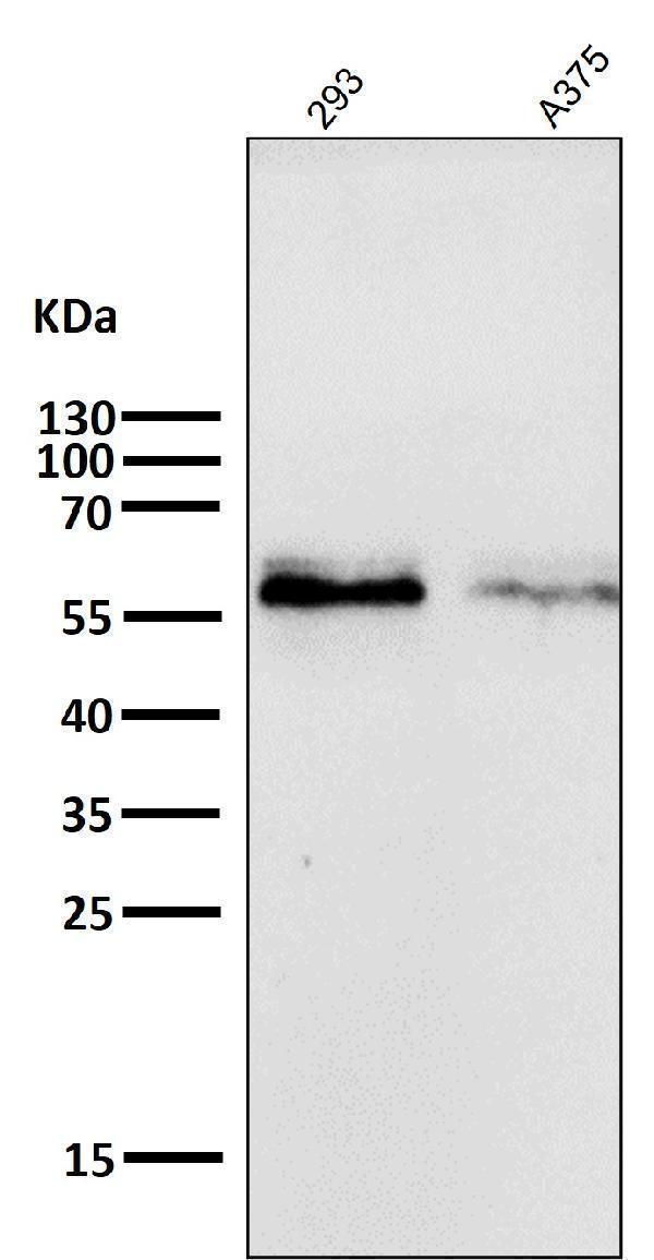
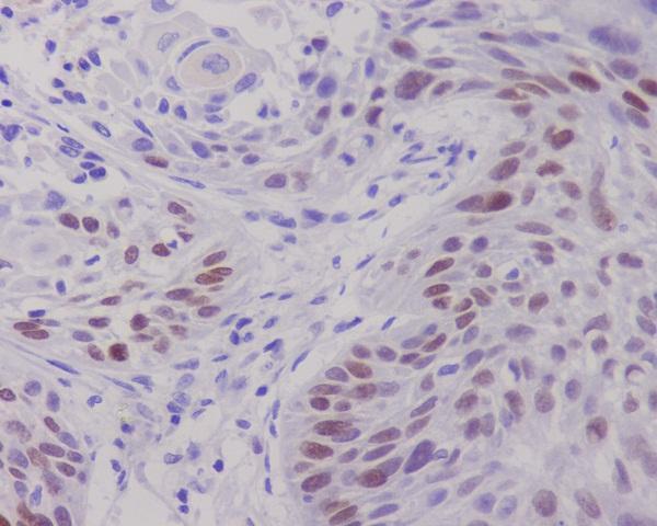
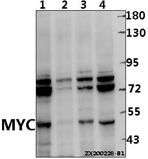
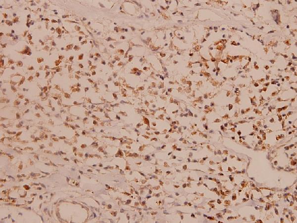
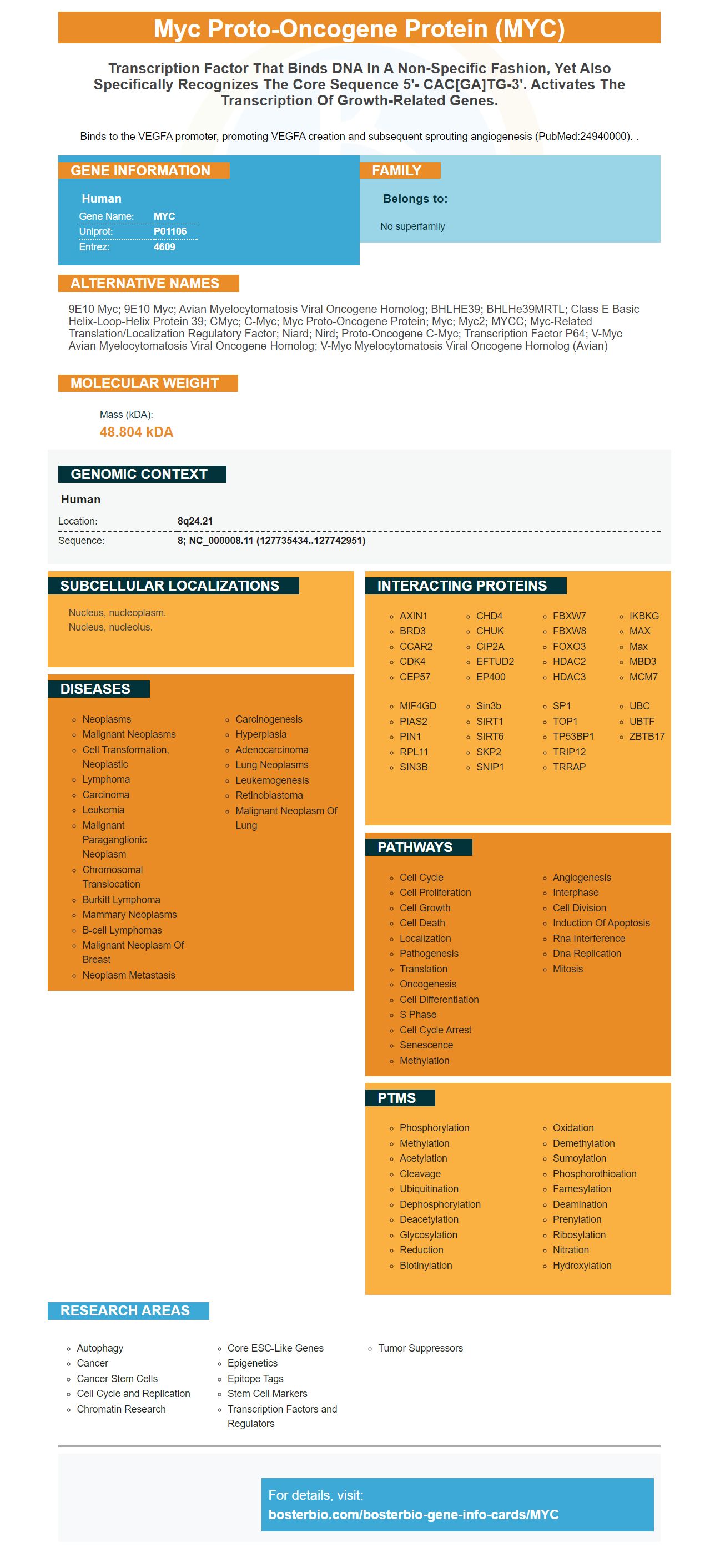
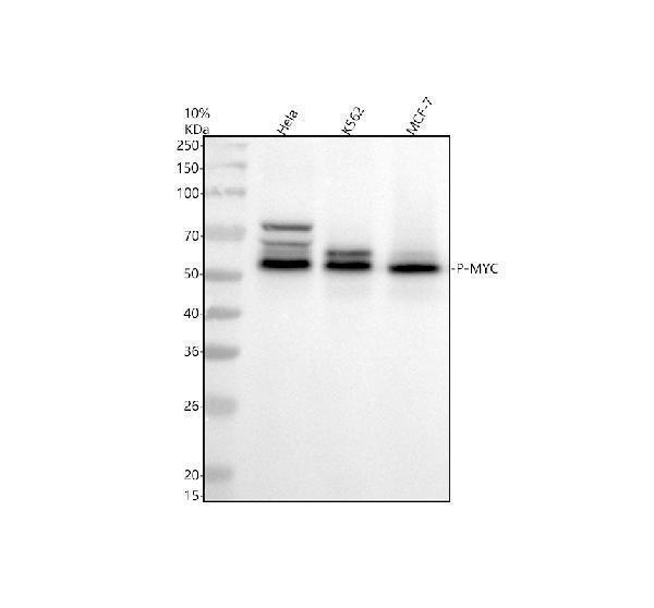
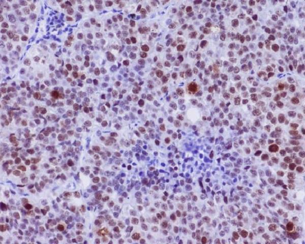
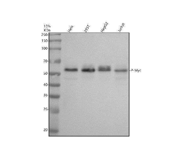
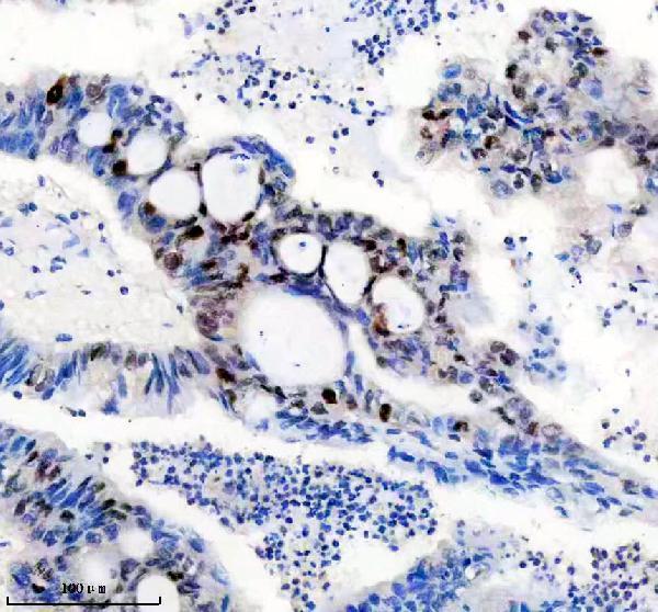
Facts about Myc proto-oncogene protein.
Binds to the VEGFA promoter, promoting VEGFA creation and subsequent sprouting angiogenesis (PubMed:24940000). .
Human | |
---|---|
Gene Name: | MYC |
Uniprot: | P01106 |
Entrez: | 4609 |
Belongs to: |
---|
No superfamily |
9E10 Myc; 9E10 Myc; avian myelocytomatosis viral oncogene homolog; BHLHE39; bHLHe39MRTL; Class E basic helix-loop-helix protein 39; cMyc; c-Myc; myc proto-oncogene protein; Myc; Myc2; MYCC; myc-related translation/localization regulatory factor; Niard; Nird; Proto-oncogene c-Myc; Transcription factor p64; v-myc avian myelocytomatosis viral oncogene homolog; v-myc myelocytomatosis viral oncogene homolog (avian)
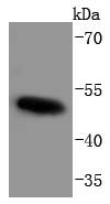
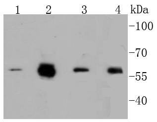
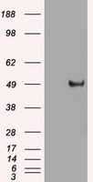
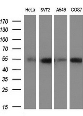
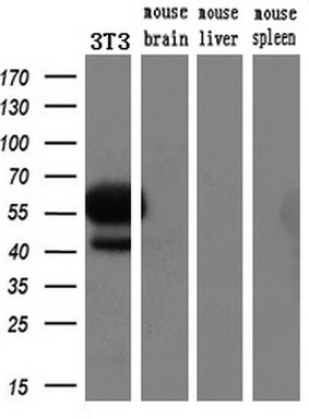
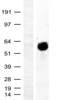
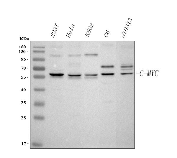
Mass (kDA):
48.804 kDA
Human | |
---|---|
Location: | 8q24.21 |
Sequence: | 8; NC_000008.11 (127735434..127742951) |
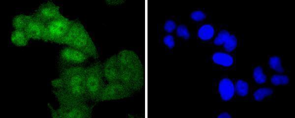
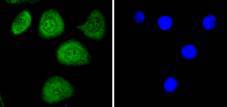
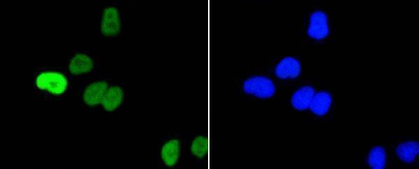
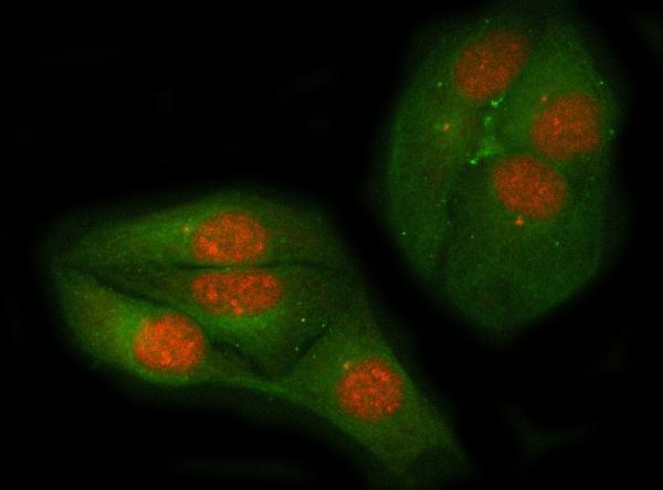
Nucleus, nucleoplasm. Nucleus, nucleolus.
The best way to know about the most effective applications of the MYC marker is to learn about the different kinds of applications and samples that utilize the MYC protein. The marker can be utilized in a variety of samples, including those from rare species. The MYC protein is frequently used to identify mutations or other alterations in the body’s cells.
This study was conducted to study the markers cMyc in a set of pancreatic cancers. The tumors were treated with formalin before being sent for histology analysis to the Hopital Nord. The slides stained with H&E were evaluated for the penetrability of the histological hallmarks. The pathological score for the entire tumor was determined by looking at the predominant character of the tumor.
The RRx-001 MYC inhibitor is an erythrophagoimmunotherapeutic, a novel hypoxia-selective epigenetic agent. It causes oxidative stress under hypoxic environments and blocks global hypermethylation. Additionally, it restores the function of tumor suppressor genes. Its anti-tumor properties in a myeloma-xenograft model has been demonstrated, and it is currently being tested using conventional myeloma antimyeloma treatments.
The MYC-low subgroup of pancreatic tumors has a high degree of expression of biological processes that reflect the differentiated state of the tumor. MYC high tumors produce PDX with high proliferative indice. For the 55 PDX analyzed, IHC-based Ki67 staining revealed that they were significantly enriched in the epithelial compartment. These tumors are characterized by a high level of proliferation.
The expression of RANKL is an important regulator of this phenotypic marker. It also functions as an important transcriptional regulator for c-myc. Thus, the c-Myc expression levels in Boster bio is controlled by RANKL in osteoclastic differentiation. This study supports the function of TRAF6 in osteoclastic differentiation. However, further work is required to fully understand its precise role in osteoclastic differentiation.
Utilizing an array of MYC-high and -low patients, a standardized method can detect MYC high and -low tumors. To confirm the results, Affymetrix data was used on four MYC high and four MYC low patients. The gene sets were normalized to 28S-ribosomalRNA (rRNA) and their proportions were calculated using the Ct method. The median expression ratio was used to compare the MYC-high and MYC-low profiles.
The proto-oncogene, c-Myc, codes for a transcription factor involved in numerous aspects of cellular function. It also forms a heterodimer along with the protein Max. Myc and Max work together to regulate the expression of genes and the process of apoptosis. The c-Myc gene is located in nearly every replicating cell. It is decreased during the final differentiation.
The HA produced by Boster Bio is a high-quality recombinant antigen or protein produced by an efficient process of gene synthesis. This is a cost-effective alternative for custom polyclonal antibody production. You can use this system to produce antibodies for rare species and discontinued antigens. The production process is made simpler and economies of scale are among the major advantages. The package includes the target sequence, as well as expression of the recombinant protein or antigen.
Expression vectors are the basic tools in biotechnology. These viruses or plasmids have the genes needed for expression of genes. When used in biotechnology applications, these vectors commandeer the process of synthesising proteins in cells. A good expression vector consists of all the elements needed for gene expression in the host organism. Some of these elements include promoters, the correct translation initiation sequence, and an ribosomal binding location. Expression vectors also have the appropriate sequences that are based on the host chosen. For instance the prokaryotic vector would include the Shine Dalgarno sequence whereas Eukaryotes would contain the Kozak consensus sequence.
Furthermore, expression vectors contain regulatory sequences acting as promoters or enhancers, that allow the gene cloned by the expression vector to be created. This allows for efficient protein production by producing messenger RNA which is then translated into the protein. Protein expression can be either strictly controlled or permanently controlled. The most popular host for this process is Escherichia coli. However, other types of cells can also be used.
Expression vectors are used to introduce genes into animals and plants to boost their growth. Beta-carotene can be introduced into rice plants in order to produce golden rice. Certain expression vectors can introduce genes that produce an insecticide called Bttoxin. This eliminates the need for chemical pesticides. Similarly, expression vectors can extend the ripening time of tomatoes by altering the plants to reduce the amount of rot.
Escherichiacoli and Saccharomyces cerevisiae are both able to perform a wide variety of biotechnology applications. These bacteria make a wide range of proteins, making them an ideal option for research on gene expression. The yeast two-hybrid can trigger transcription of a reporter genes when a protein interacts to its environment. This way there are many different proteins can be synthesized.
The MYC marker is used in IHC workflows in the majority of labs. It allows the visualization of distributions of cellular components within cells or tissues. Different IHC methods are used to identify antigens. It is important that you select the right method for the type of specimen you are testing and test sensitivity, as these factors can impact antigen presentation. The IHC workflow is also influenced by the preparation of the sample. The quality of the sample will determine the efficiency of antigen exhibition.
The detection system must determine the method by which the primary antibody will be diluted. Polymer detection systems require a lower concentration. Some sites for antibody manufacturers suggest high concentrations. In all cases, the starting concentration should not exceed 5ug/ml for mouse antibodies and 2ug/ml rabbit antibodies. Background staining can occur when this occurs. It is recommended that you conduct negative control tests using several antibodies when performing an IHC workflow.
The presence of MYC in a sample can be determined by immunohistochemistry. FISH uses an antibody against the MYC gene in order to examine the MYC copy numbers at a cellular level. FISH requires FFPE samples. This can be challenging because of the high level of experimental artifacts. As such, immunohistochemistry should be used in these instances, since FFPE samples are more likely to contain this protein.
With the aid of a reliable guideline, a thorough IHC procedure shouldn't be difficult to follow. BosterBio's MYC primer provides detailed instructions and a diagram of the workflow that includes the recommended the reagents. It also includes troubleshooting suggestions including how to resolve weak staining. It also contains a step-by–step troubleshooting guide. You can refer to the Boster bio guide If you have any questions.
PMID: 6414718 by Battey J., et al. The human c-myc oncogene: structural consequences of translocation into the IgH locus in Burkitt lymphoma.
PMID: 6321164 by Bernard O., et al. Sequence of the murine and human cellular myc oncogenes and two modes of myc transcription resulting from chromosome translocation in B lymphoid tumours.
*Showing only the more recent 20. More publications can be found for each product on its corresponding product page