Click image to see more details
-

-
-
-
-
+3
Product Info Summary
| SKU: | PB9045 |
|---|---|
| Size: | 100 μg/vial |
| Reactive Species: | Human, Mouse, Rat |
| Host: | Rabbit |
| Application: | IP, IHC, WB |
Customers Who Bought This Also Bought

Product info
Product Name
Anti-Calbindin/CALB1 Antibody Picoband®
SKU/Catalog Number
PB9045
Size
100 μg/vial
Form
Lyophilized
Description
Boster Bio Anti-Calbindin/CALB1 Antibody Picoband® catalog # PB9045. Tested in IP, IHC, WB applications. This antibody reacts with Human, Mouse, Rat. The brand Picoband indicates this is a premium antibody that guarantees superior quality, high affinity, and strong signals with minimal background in Western blot applications. Only our best-performing antibodies are designated as Picoband, ensuring unmatched performance.
Storage & Handling
Store at -20˚C for one year from date of receipt. After reconstitution, at 4˚C for one month. It can also be aliquotted and stored frozen at -20˚C for six months. Avoid repeated freeze-thaw cycles.
Cite This Product
Anti-Calbindin/CALB1 Antibody Picoband® (Boster Biological Technology, Pleasanton CA, USA, Catalog # PB9045)
Host
Rabbit
Contents
Each vial contains 4 mg Trehalose, 0.9 mg NaCl and 0.2 mg Na2HPO4.
Clonality
Polyclonal
Isotype
Rabbit IgG
Immunogen
E.coli-derived human Calbindin recombinant protein (Position: A2-E175). Human Calbindin shares 99% amino acid (aa) sequence identity with both mouse and rat Calbindin.
Cross-reactivity
No cross-reactivity with other proteins
Reactive Species
PB9045 is reactive to CALB1 in Human, Mouse, Rat
Observed Molecular Weight
26 kDa
Calculated molecular weight
30.0 kDa
Background of CALB1
Calbindin is a calcium-binding protein belonging to the troponin C superfamily. Calretinin is expressed in central and peripheral nervous system and in many normal and pathological tissues. The rat and human calretinin exhibit 98% sequence homology and 91% homology to many other species. Two calcium binding proteins, calbindin and calretinin, have been reported to be expressed in abundance in Purkinje cells and other cell types in the cerebellum.
Antibody Validation
Boster validates all antibodies on WB, IHC, ICC, Immunofluorescence, and ELISA with known positive control and negative samples to ensure specificity and high affinity, including thorough antibody incubations.
Application & Images
Applications
PB9045 is guaranteed for IP, IHC, WB Boster Guarantee
Recommend Dilution
| Application | Dilution | Species |
|---|---|---|
| Western blot | 0.1-0.5μg/ml | Human, Mouse, Rat |
| Immunohistochemistry (Paraffin-embedded Section) | 0.5-1μg/ml | Mouse, Rat |
| Immunoprecipitation | 0.5-2 μg/ml | Human |
Tested application
Suggested blocking solution with 5% non-fat milk or BSA; (*)Recommended protein loading: 20-40 µg per lane
Use TE buffer pH 9.0 for antigen retrieval; (*) citrate buffer pH 6.0 is an alternative.
Validation Images & Assay Conditions
Click image to see more details
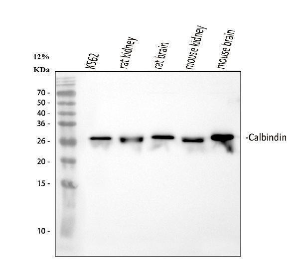
Western blot analysis of Calbindin using anti-Calbindin antibody (PB9045).
Electrophoresis was performed on a 5-20% SDS-PAGE gel at 70V (Stacking gel) / 90V (Resolving gel) for 2-3 hours. The sample well of each lane was loaded with 30 ug of sample under reducing conditions.
Lane 1: human K562 whole cell lysates,
Lane 2: rat kidney tissue lysates,
Lane 3: rat brain tissue lysates,
Lane 4: mouse kidney tissue lysates,
Lane 5: mouse brain tissue lysates.
After electrophoresis, proteins were transferred to a nitrocellulose membrane at 150 mA for 50-90 minutes. Blocked the membrane with 5% non-fat milk/TBS for 1.5 hour at RT. The membrane was incubated with rabbit anti-Calbindin antigen affinity purified polyclonal antibody (Catalog # PB9045) at 0.5 μg/mL overnight at 4°C, then washed with TBS-0.1%Tween 3 times with 5 minutes each and probed with a goat anti-rabbit IgG-HRP secondary antibody at a dilution of 1:5000 for 1.5 hour at RT. The signal is developed using an Enhanced Chemiluminescent detection (ECL) kit (Catalog # EK1002) with Tanon 5200 system. A specific band was detected for Calbindin at approximately 26 kDa. The expected band size for Calbindin is at 30 kDa.

Click image to see more details
IHC analysis of Calbindin using anti-Calbindin antibody (PB9045).
Calbindin was detected in a paraffin-embedded section of mouse kidney tissue. Heat mediated antigen retrieval was performed in EDTA buffer (pH 8.0, epitope retrieval solution). The tissue section was blocked with 10% goat serum. The tissue section was then incubated with 2 μg/ml rabbit anti-Calbindin Antibody (PB9045) overnight at 4°C. Peroxidase Conjugated Goat Anti-rabbit IgG was used as secondary antibody and incubated for 30 minutes at 37°C. The tissue section was developed using HRP Conjugated Rabbit IgG Super Vision Assay Kit (Catalog # SV0002) with DAB as the chromogen.

Click image to see more details
IHC analysis of Calbindin/CALB1 using anti-Calbindin/CALB1 antibody (PB9045).
Calbindin/CALB1 was detected in a paraffin-embedded section of human kidney tissue. Heat mediated antigen retrieval was performed in EDTA buffer (pH 8.0, epitope retrieval solution). The tissue section was blocked with 10% goat serum. The tissue section was then incubated with 2 μg/ml rabbit anti-Calbindin/CALB1 Antibody (PB9045) overnight at 4°C. Peroxidase Conjugated Goat Anti-rabbit IgG was used as secondary antibody and incubated for 30 minutes at 37°C. The tissue section was developed using HRP Conjugated Rabbit IgG Super Vision Assay Kit (Catalog # SV0002) with DAB as the chromogen.

Click image to see more details
IHC analysis of Calbindin/CALB1 using anti-Calbindin/CALB1 antibody (PB9045).
Calbindin/CALB1 was detected in a paraffin-embedded section of human cerebellum tissue. Heat mediated antigen retrieval was performed in EDTA buffer (pH 8.0, epitope retrieval solution). The tissue section was blocked with 10% goat serum. The tissue section was then incubated with 2 μg/ml rabbit anti-Calbindin/CALB1 Antibody (PB9045) overnight at 4°C. Peroxidase Conjugated Goat Anti-rabbit IgG was used as secondary antibody and incubated for 30 minutes at 37°C. The tissue section was developed using HRP Conjugated Rabbit IgG Super Vision Assay Kit (Catalog # SV0002) with DAB as the chromogen.

Click image to see more details
IHC analysis of Calbindin using anti-Calbindin antibody (PB9045).
Calbindin was detected in a paraffin-embedded section of rat kidney tissue. Heat mediated antigen retrieval was performed in EDTA buffer (pH 8.0, epitope retrieval solution). The tissue section was blocked with 10% goat serum. The tissue section was then incubated with 2 μg/ml rabbit anti-Calbindin Antibody (PB9045) overnight at 4°C. Peroxidase Conjugated Goat Anti-rabbit IgG was used as secondary antibody and incubated for 30 minutes at 37°C. The tissue section was developed using HRP Conjugated Rabbit IgG Super Vision Assay Kit (Catalog # SV0002) with DAB as the chromogen.

Click image to see more details
Immunoprecipitating (IP) Calbindin in K562 whole cell lysate.
Western blot analysis of Calbindin using anti-Calbindin antibody (PB9045);
Lane 1: K562 whole cell lysates (30ug);
Lane 2: Rabbit control IgG instead of anti-Calbindin antibody in K562 whole cell lysate;
Lane 3: anti-Calbindin antibody (2μg) + K562 whole cell lysate (500μg).
After electrophoresis, proteins were transferred to a membrane. Then the membrane was incubated with rabbit anti-Calbindin antigen affinity purified polyclonal antibody (PB9045) at a dilution of 0.5 μg/mL and probed with a goat anti-rabbit IgG-HRP secondary antibody (Catalog # BA1054). The signal is developed using ECL Plus Western Blotting Substrate (Catalog # AR1196-200). A specific band was detected for Calbindin at approximately 26 kDa. The expected band size for Calbindin is at 30 kDa.
Specific Publications For Anti-Calbindin/CALB1 Antibody Picoband® (PB9045)
Loading publications
Recommended Resources
Here are featured tools and databases that you might find useful.
- Boster's Pathways Library
- Protein Databases
- Bioscience Research Protocol Resources
- Data Processing & Analysis Software
- Photo Editing Software
- Scientific Literature Resources
- Research Paper Management Tools
- Molecular Biology Software
- Primer Design Tools
- Bioinformatics Tools
- Phylogenetic Tree Analysis
Customer Reviews
Have you used Anti-Calbindin/CALB1 Antibody Picoband®?
Share your experimental results or join a short interview to earn up to $1,000 in product credits or other rewards.
0 Reviews For Anti-Calbindin/CALB1 Antibody Picoband®
Customer Q&As
Have a question?
Find answers in Q&As, reviews.
Can't find your answer?
Submit your question
4 Customer Q&As for Anti-Calbindin/CALB1 Antibody Picoband®
Question
We need using your anti-Calbindin/CALB1 antibody for metanephric part of ureteric bud development studies. Has this antibody been tested with western blotting on siha cells? We would like to see some validation images before ordering.
Verified Customer
Verified customer
Asked: 2020-01-30
Answer
Thank you for your inquiry. This PB9045 anti-Calbindin/CALB1 antibody is tested on intestinal cancer tissue, tissue lysate, mammary cancer tissue, mouse kidney tissue, rat kidney tissue, brain tissue, siha cells, hela cells. It is guaranteed to work for Flow Cytometry, IHC, ICC, WB in human, mouse, rat. Our Boster guarantee will cover your intended experiment even if the sample type has not been be directly tested.
Boster Scientific Support
Answered: 2020-01-30
Question
Our lab used your anti-Calbindin/CALB1 antibody for Flow Cytometry on caudate nucleus a few months ago. I am using mouse, and I plan to use the antibody for IHC next. I am interested in examining caudate nucleus as well as kidney lung in our next experiment. Could you please give me some suggestion on which antibody would work the best for IHC?
D. Singh
Verified customer
Asked: 2019-05-20
Answer
I have checked the website and datasheets of our anti-Calbindin/CALB1 antibody and it seems that PB9045 has been validated on mouse in both Flow Cytometry and IHC. Thus PB9045 should work for your application. Our Boster satisfaction guarantee will cover this product for IHC in mouse even if the specific tissue type has not been validated. We do have a comprehensive range of products for IHC detection and you can check out our website bosterbio.com to find out more information about them.
Boster Scientific Support
Answered: 2019-05-20
Question
We have observed staining in human amygdala. Are there any suggestions? Is anti-Calbindin/CALB1 antibody supposed to stain amygdala positively?
Verified Customer
Verified customer
Asked: 2017-08-14
Answer
From literature amygdala does express CALB1. From Uniprot.org, CALB1 is expressed in caudate nucleus, brain, amygdala, cerebellum substantia nigra, kidney lung, among other tissues. Regarding which tissues have CALB1 expression, here are a few articles citing expression in various tissues:
Amygdala, Cerebellum, and Substantia nigra, Pubmed ID: 14702039
Brain, Pubmed ID: 2565876, 3691519
Kidney, and Lung, Pubmed ID: 15489334
Boster Scientific Support
Answered: 2017-08-14
Question
We are currently using anti-Calbindin/CALB1 antibody PB9045 for human tissue, and we are happy with the WB results. The species of reactivity given in the datasheet says human, mouse, rat. Is it true that the antibody can work on bovine tissues as well?
A. Thomas
Verified customer
Asked: 2015-11-25
Answer
The anti-Calbindin/CALB1 antibody (PB9045) has not been tested for cross reactivity specifically with bovine tissues, but there is a good chance of cross reactivity. We have an innovator award program that if you test this antibody and show it works in bovine you can get your next antibody for free. Please contact me if I can help you with anything.
Boster Scientific Support
Answered: 2015-11-25
