Click image to see more details
-

-
-
-
-
+1
Product Info Summary
| SKU: | PB9925 |
|---|---|
| Size: | 100 μg/vial |
| Reactive Species: | Human, Mouse, Rat |
| Host: | Rabbit |
| Application: | Flow Cytometry, IF, IHC, ICC, WB |
Customers Who Bought This Also Bought

Product info
Product Name
Anti-Catalase Antibody Picoband®
SKU/Catalog Number
PB9925
PB0971 is an alternative SKU for this antibody, used in previous lots.
Size
100 μg/vial
Form
Lyophilized
Description
Boster Bio Anti-Catalase Antibody Picoband® catalog # PB9925. Tested in Flow Cytometry, IF, IHC, ICC, WB applications. This antibody reacts with Human, Mouse, Rat. The brand Picoband indicates this is a premium antibody that guarantees superior quality, high affinity, and strong signals with minimal background in Western blot applications. Only our best-performing antibodies are designated as Picoband, ensuring unmatched performance.
Storage & Handling
Store at -20˚C for one year from date of receipt. After reconstitution, at 4˚C for one month. It can also be aliquotted and stored frozen at -20˚C for six months. Avoid repeated freeze-thaw cycles.
Cite This Product
Anti-Catalase Antibody Picoband® (Boster Biological Technology, Pleasanton CA, USA, Catalog # PB9925)
Host
Rabbit
Contents
Each vial contains 4 mg Trehalose, 0.9 mg NaCl and 0.2 mg Na2HPO4.
Clonality
Polyclonal
Isotype
Rabbit IgG
Immunogen
E. coli-derived human Catalase recombinant protein (Position: E344-L527). Human Catalase shares 85.3% and 82.6% amino acid (aa) sequence identity with mouse and rat Catalase, respectively.
Cross-reactivity
No cross-reactivity with other proteins
Reactive Species
PB9925 is reactive to CAT in Human, Mouse, Rat
Observed Molecular Weight
60 kDa
Calculated molecular weight
59.8 kDa
Background of CAT
Catalase is a key antioxidant enzyme in the bodies defensing against oxidative stress. It is also a heme enzyme that is present in the peroxisome of nearly all aerobic cells. Catalase converts the reactive oxygen species hydrogen peroxide to water and oxygen and thereby mitigates the toxic effects of hydrogen peroxide. Oxidative stress is hypothesized to play a role in the development of many chronic or late-onset diseases such as diabetes, asthma, Alzheimer's disease, systemic lupus erythematosus, rheumatoid arthritis, and cancers. Polymorphisms in this gene have been associated with decreases in catalase activity but, to date, acatalasemia is the only disease known to be caused by this gene.
Antibody Validation
Boster validates all antibodies on WB, IHC, ICC, Immunofluorescence, and ELISA with known positive control and negative samples to ensure specificity and high affinity, including thorough antibody incubations.
Application & Images
Applications
PB9925 is guaranteed for Flow Cytometry, IF, IHC, ICC, WB Boster Guarantee
Recommend Dilution
| Application | Dilution | Species |
|---|---|---|
| Western blot | 0.1-0.5μg/ml | Human, Mouse, Rat |
| Immunohistochemistry (Paraffin-embedded Section) | 2-5μg/ml | Human |
| Immunocytochemistry/Immunofluorescence | 5μg/ml | Human |
| Flow Cytometry (Fixed) | 1-3μg/1x106 cells | Human |
Tested application
Suggested blocking solution with 5% non-fat milk or BSA; (*)Recommended protein loading: 20-40 µg per lane
Use TE buffer pH 9.0 for antigen retrieval; (*) citrate buffer pH 6.0 is an alternative.
Validation Images & Assay Conditions
Click image to see more details
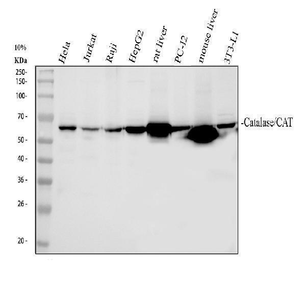
Western blot analysis of Catalase using anti-Catalase antibody (PB9925).
Electrophoresis was performed on a 10% SDS-PAGE gel at 80V (Stacking gel) / 120V (Resolving gel) for 2 hours. The sample well of each lane was loaded with 30 ug of sample under reducing conditions.
Lane 1: human Hela whole cell lysates,
Lane 2: human Jurkat whole cell lysates,
Lane 3: human Raji whole cell lysates,
Lane 4: human HepG2 whole cell lysates,
Lane 5: rat liver tissue lysates,
Lane 6: rat PC-12 whole cell lysates,
Lane 7: mouse liver tissue lysates,
Lane 6: mouse 3T3-L1 whole cell lysates.
After electrophoresis, proteins were transferred to a nitrocellulose membrane at 150 mA for 50-90 minutes. Blocked the membrane with 5% non-fat milk/TBS for 1.5 hour at RT. The membrane was incubated with rabbit anti-Catalase antigen affinity purified polyclonal antibody (PB9925) at 0.5 μg/mL overnight at 4°C, then washed with TBS-0.1%Tween 3 times with 5 minutes each and probed with a goat anti-rabbit IgG-HRP secondary antibody (Catalog # BA1054) at a dilution of 1:5000 for 1.5 hour at RT. The signal is developed using an ECL Plus Western Blotting Substrate (Catalog # AR1196-200) with Tanon 5200 system. A specific band was detected for Catalase at approximately 60 kDa. The expected band size for Catalase is at 60 kDa.

Click image to see more details
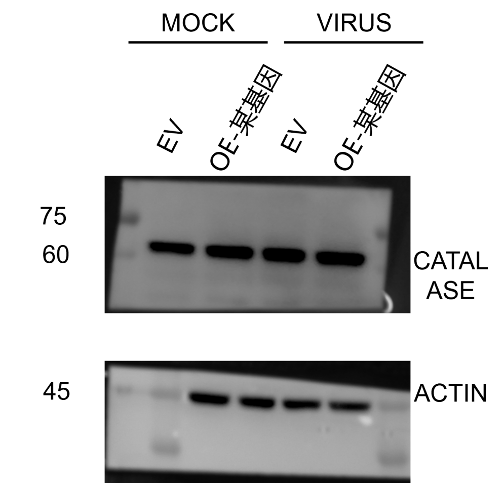
Western blot analysis of Catalase using anti-Catalase antibody (PB9925).
Electrophoresis was performed on a 10% SDS-PAGE gel at 80V (Stacking gel) / 120V (Resolving gel) for 2 hours. The sample well of each lane was loaded with 30 ug of sample under reducing conditions.
Lane 1: Mock group-EV-Monkey MARC-145 whole cell lysates,
Lane 2: Mock group-OE-Monkey MARC-145 whole cell lysates,
Lane 3: Virus group-EV-Monkey MARC-145 whole cell lysates,
Lane 4: Virus group-OE-Monkey MARC-145 whole cell lysates,.
After electrophoresis, proteins were transferred to a nitrocellulose membrane at 150 mA for 50-90 minutes. Blocked the membrane with 5% non-fat milk/TBS for 1.5 hour at RT. The membrane was incubated with rabbit anti-Catalase antigen affinity purified polyclonal antibody (PB9925) at 1:1000 overnight at 4°C, then washed with TBS-0.1%Tween 3 times with 5 minutes each and probed with a HRP-conjugated anti-rabbit IgG secondary antibody at a dilution of for 1 hour at RT. The signal is developed using an ECL Plus Western Blotting Substrate (Catalog # AR1196-200) with Unitec Cambridge system. A specific band was detected for Catalase at approximately 63 kDa. The expected band size for Catalase is at 60 kDa.

Click image to see more details
IHC analysis of Catalase using anti-Catalase antibody (PB9925).
Catalase was detected in a paraffin-embedded section of human liver cancer tissue. Heat mediated antigen retrieval was performed in EDTA buffer (pH 8.0, epitope retrieval solution). The tissue section was blocked with 10% goat serum. The tissue section was then incubated with 2 μg/ml rabbit anti-Catalase Antibody (PB9925) overnight at 4°C. Peroxidase Conjugated Goat Anti-rabbit IgG was used as secondary antibody and incubated for 30 minutes at 37°C. The tissue section was developed using HRP Conjugated Rabbit IgG Super Vision Assay Kit (Catalog # SV0002) with DAB as the chromogen.

Click image to see more details
IF analysis of Catalase using anti-Catalase antibody (PB9925).
Catalase was detected in an immunocytochemical section of U2OS cells. Enzyme antigen retrieval was performed using IHC enzyme antigen retrieval reagent (AR0022) for 15 mins. The cells were blocked with 10% goat serum. And then incubated with 5 μg/mL rabbit anti-Catalase Antibody (PB9925) overnight at 4°C. Fluoro488 Conjugated Goat Anti-Rabbit IgG (BA1127) was used as secondary antibody at 1:500 dilution and incubated for 30 minutes at 37°C. The section was counterstained with DAPI. Visualize using a fluorescence microscope and filter sets appropriate for the label used.

Click image to see more details
Flow Cytometry analysis of Jurkat cells using anti-Catalase antibody (PB9925).
Overlay histogram showing Jurkat cells stained with PB9925 (Blue line). To facilitate intracellular staining, cells were fixed with 4% paraformaldehyde and permeabilized with permeabilization buffer. The cells were blocked with 10% normal goat serum. And then incubated with rabbit anti-Catalase Antibody (PB9925, 1 μg/1x106 cells) for 30 min at 20°C. DyLight®488 conjugated goat anti-rabbit IgG (BA1127, 5-10 μg/1x106 cells) was used as secondary antibody for 30 minutes at 20°C. Isotype control antibody (Green line) was rabbit IgG (1 μg/1x106) used under the same conditions. Unlabelled sample without incubation with primary antibody and secondary antibody (Red line) was used as a blank control.
Specific Publications For Anti-Catalase Antibody Picoband® (PB9925)
Loading publications
Recommended Resources
Here are featured tools and databases that you might find useful.
- Boster's Pathways Library
- Protein Databases
- Bioscience Research Protocol Resources
- Data Processing & Analysis Software
- Photo Editing Software
- Scientific Literature Resources
- Research Paper Management Tools
- Molecular Biology Software
- Primer Design Tools
- Bioinformatics Tools
- Phylogenetic Tree Analysis
Customer Reviews
Have you used Anti-Catalase Antibody Picoband®?
Share your experimental results or join a short interview to earn up to $1,000 in product credits or other rewards.
1 Reviews For Anti-Catalase Antibody Picoband®
This antibody is highly specific and efficient, suitable for Western blot detection of Catalase protein in MARC-145 cells, with only minor nonspecific bands.
Excellent
| SKU | PB9925 |
|---|---|
| Application | Western Blot |
| Sample | MARC-145 cells |
| Sample Processing Description | Cells or tissues were lysed in RIPA buffer supplemented with protease inhibitor PMSF (100:1) for 10 minutes. The lysate was centrifuged at 12,000 rpm for 15 minutes, and the supernatant was collected. Samples were mixed with 5× loading buffer and denatured at 100 °C for 10 minutes before loading onto SDS-PAGE. |
| Other Reagents | Blocking buffer |
| Primary Antibody | Catalase Antibody Picoband® |
| Primary Incubation | 1:1000, overnight at 4 ℃ |
| Secondary Antibody | Anti-rabbit IgG secondary antibody conjugated with horseradish peroxidase (HRP) |
| Secondary Incubation | 1:10000, 1 hour in room temperature |
| Detection | Substrate: ECL, Imaging system:ChemiDoc MP |
| Results Summary | The figure shows a schematic representation of Western blot results for the target protein Catalase and the loading control Actin in MARC-145 cells under normal and post-infection conditions. The target bands are clear and well-defined, and the experimental results are satisfactory. |
Yuchao Yan, Tianjin University
Verified customer
Submitted 2025-12-25
Customer Q&As
Have a question?
Find answers in Q&As, reviews.
Can't find your answer?
Submit your question
16 Customer Q&As for Anti-Catalase Antibody Picoband®
Question
My team were happy with the WB result of your anti-Catalase antibody. However we have observed positive staining in liver peroxisome. using this antibody. Is that expected? Could you tell me where is CAT supposed to be expressed?
Verified Customer
Verified customer
Asked: 2020-01-22
Answer
According to literature, liver does express CAT. Generally CAT expresses in peroxisome. Regarding which tissues have CAT expression, here are a few articles citing expression in various tissues:
Brain, and Cajal-Retzius cell, Pubmed ID: 2308162
Cervix carcinoma, and Erythroleukemia, Pubmed ID: 23186163
Erythrocyte, Pubmed ID: 7882369
Eye, Pubmed ID: 15489334
Fibroblast, Pubmed ID: 6548744
Kidney, Pubmed ID: 3755526
Liver, Pubmed ID: 11728823, 24275569
Placenta, and Uterus, Pubmed ID: 14702039
Platelet, Pubmed ID: 12665801, 18088087
Boster Scientific Support
Answered: 2020-01-22
Question
We have been able to see staining in human placenta uterus. Any tips? Is anti-Catalase antibody supposed to stain placenta uterus positively?
Verified Customer
Verified customer
Asked: 2019-12-19
Answer
From literature placenta uterus does express CAT. From Uniprot.org, CAT is expressed in bone marrow, kidney, liver, placenta uterus, eye, platelet, erythrocyte, fibroblast, brain cajal-retzius cell, cervix carcinoma erythroleukemia, among other tissues. Regarding which tissues have CAT expression, here are a few articles citing expression in various tissues:
Brain, and Cajal-Retzius cell, Pubmed ID: 2308162
Cervix carcinoma, and Erythroleukemia, Pubmed ID: 23186163
Erythrocyte, Pubmed ID: 7882369
Eye, Pubmed ID: 15489334
Fibroblast, Pubmed ID: 6548744
Kidney, Pubmed ID: 3755526
Liver, Pubmed ID: 11728823, 24275569
Placenta, and Uterus, Pubmed ID: 14702039
Platelet, Pubmed ID: 12665801, 18088087
Boster Scientific Support
Answered: 2019-12-19
Question
My question regarding product PB9925, anti-Catalase antibody. I was wondering if it would be possible to conjugate this antibody with biotin. I would need it to be without BSA or sodium azide. I am planning on using a buffer exchange of sodium azide with PBS only. Would there be problems for me to conjugate the antibody and store it in -20 degrees in small aliquots?
Verified Customer
Verified customer
Asked: 2019-08-09
Answer
It is not recommended storing this antibody with PBS buffer only in -20 degrees. If you want to store it in -20 degrees it is best to add some cryoprotectant like glycerol. If you want carrier free PB9925 anti-Catalase antibody, we can provide it to you in a special formula with trehalose and/or glycerol. These molecules will not interfere with conjugation chemistry and provide a good level of protection for the antibody from degradation. Please be sure to specify this in your purchase order.
Boster Scientific Support
Answered: 2019-08-09
Question
My question regards using your anti-Catalase antibody for protein targeting to peroxisome studies. Has this antibody been tested with western blotting on mouse liver? We would like to see some validation images before ordering.
D. Jha
Verified customer
Asked: 2019-08-08
Answer
Thank you for your inquiry. This PB9925 anti-Catalase antibody is validated on mouse liver, a549 whole cell lysates, siha cells. It is guaranteed to work for Flow Cytometry, IF, IHC-P, IHC-F, ICC, WB in human, mouse, rat. Our Boster guarantee will cover your intended experiment even if the sample type has not been be directly tested.
Boster Scientific Support
Answered: 2019-08-08
Question
Does anti-Catalase antibody PB9925 work for Flow Cytometry with platelet?
Verified Customer
Verified customer
Asked: 2019-08-05
Answer
According to the expression profile of platelet, CAT is highly expressed in platelet. So, it is likely that anti-Catalase antibody PB9925 will work for Flow Cytometry with platelet.
Boster Scientific Support
Answered: 2019-08-05
Question
Is a blocking peptide available for product anti-Catalase antibody (PB9925)?
Verified Customer
Verified customer
Asked: 2019-07-26
Answer
We do provide the blocking peptide for product anti-Catalase antibody (PB9925). If you would like to place an order for it please contact support@bosterbio.com and make a special request.
Boster Scientific Support
Answered: 2019-07-26
Question
I was wanting to use to test anti-Catalase antibody PB9925 on rat platelet for research purposes, then I may be interested in using anti-Catalase antibody PB9925 for diagnostic purposes as well. Is the antibody suitable for diagnostic purposes?
Verified Customer
Verified customer
Asked: 2019-06-17
Answer
The products we sell, including anti-Catalase antibody PB9925, are only intended for research use. They would not be suitable for use in diagnostic work. If you have the means to develop a product into diagnostic use, and are interested in collaborating with us and develop our product into an IVD product, please contact us for more discussions.
Boster Scientific Support
Answered: 2019-06-17
Question
Would PB9925 anti-Catalase antibody work on parafin embedded sections? If so, which fixation method do you recommend we use (PFA, paraformaldehyde, other)?
Verified Customer
Verified customer
Asked: 2017-11-23
Answer
As indicated on the product datasheet, PB9925 anti-Catalase antibody as been tested on Flow Cytometry. It is best to use PFA for fixation because it has better tissue penetration ability. PFA needs to be prepared fresh before use. Long term stored PFA turns into formalin, as the PFA molecules congregate and become formalin.
Boster Scientific Support
Answered: 2017-11-23
Question
I see that the anti-Catalase antibody PB9925 works with Flow Cytometry, what is the protocol used to produce the result images on the product page?
Verified Customer
Verified customer
Asked: 2017-08-23
Answer
You can find protocols for Flow Cytometry on the "support/technical resources" section of our navigation menu. If you have any further questions, please send an email to support@bosterbio.com
Boster Scientific Support
Answered: 2017-08-23
Question
See attached the WB image, lot number and protocol we used for platelet using anti-Catalase antibody PB9925. Please let me know if you require anything else.
Verified Customer
Verified customer
Asked: 2017-07-24
Answer
Thank you very much for the data. Our lab team are working to resolve this as quickly as possible, and we appreciate your patience and understanding! You have provided everything we needed. Please let me know if there is anything you need in the meantime.
Boster Scientific Support
Answered: 2017-07-24
Question
Is this PB9925 anti-Catalase antibody reactive to the isotypes of CAT?
Verified Customer
Verified customer
Asked: 2017-05-23
Answer
The immunogen of PB9925 anti-Catalase antibody is E. coli-derived human Catalase recombinant protein (Position: E344-L527). Human Catalase shares 85.3% and 82.6% amino acid (aa) sequence identity with mouse and rat Catalase, respectively. Could you tell me which isotype you are interested in so I can help see if the immunogen is part of this isotype?
Boster Scientific Support
Answered: 2017-05-23
Question
I was wanting to use your anti-Catalase antibody for Flow Cytometry for rat platelet on frozen tissues, but I want to know if it has been tested for this particular application. Has this antibody been tested and is this antibody a good choice for rat platelet identification?
P. Wu
Verified customer
Asked: 2016-07-19
Answer
It shows on the product datasheet, PB9925 anti-Catalase antibody has been validated for Flow Cytometry, IF, IHC-P, IHC-F, ICC, WB on human, mouse, rat tissues. We have an innovator award program that if you test this antibody and show it works in rat platelet in IHC-frozen, you can get your next antibody for free.
Boster Scientific Support
Answered: 2016-07-19
Question
Do you have a BSA free version of anti-Catalase antibody PB9925 available?
K. Bhatt
Verified customer
Asked: 2015-01-21
Answer
Thanks for your recent telephone inquiry. I can confirm that some lots of this anti-Catalase antibody PB9925 are BSA free. For now, these lots are available and we can make a BSA free formula for you free of charge. It will take 3 extra days to prepare. If you require this antibody BSA free again in future, please do not hesitate to contact me and I will be pleased to check which lots we have in stock that are BSA free.
Boster Scientific Support
Answered: 2015-01-21
Question
Thanks for helping with my inquiry over the phone. Here are the WB image, lot number and protocol we used for platelet using anti-Catalase antibody PB9925. Let me know if you need anything else.
V. Li
Verified customer
Asked: 2014-12-22
Answer
I appreciate the data. You have provided everything we needed. Our lab team are working to resolve your inquiry as quickly as possible, and we appreciate your patience and understanding! Please let me know if there is anything you need in the meantime.
Boster Scientific Support
Answered: 2014-12-22
Question
We ordered your anti-Catalase antibody for IHC-F on liver in a previous project. I am using human, and I plan to use the antibody for WB next. We want examining liver as well as kidney in our next experiment. Could you please give me some suggestion on which antibody would work the best for WB?
J. Krishna
Verified customer
Asked: 2014-08-14
Answer
I took a look at the website and datasheets of our anti-Catalase antibody and it appears that PB9925 has been validated on human in both IHC-F and WB. Thus PB9925 should work for your application. Our Boster satisfaction guarantee will cover this product for WB in human even if the specific tissue type has not been validated. We do have a comprehensive range of products for WB detection and you can check out our website bosterbio.com to find out more information about them.
Boster Scientific Support
Answered: 2014-08-14
Question
We are currently using anti-Catalase antibody PB9925 for mouse tissue, and we are well pleased with the IHC-F results. The species of reactivity given in the datasheet says human, mouse, rat. Is it likely that the antibody can work on pig tissues as well?
K. Li
Verified customer
Asked: 2014-03-28
Answer
The anti-Catalase antibody (PB9925) has not been tested for cross reactivity specifically with pig tissues, though there is a good chance of cross reactivity. We have an innovator award program that if you test this antibody and show it works in pig you can get your next antibody for free. Please contact me if I can help you with anything.
Boster Scientific Support
Answered: 2014-03-28
