Click image to see more details
Product Info Summary
| SKU: | A02373-1 |
|---|---|
| Size: | 100 μl/vial |
| Reactive Species: | Human, Mouse, Rat |
| Host: | Rabbit |
| Application: | ELISA, IP, IHC, WB |
Customers Who Bought This Also Bought

Product info
Product Name
Anti-Cingulin/CGN Antibody
SKU/Catalog Number
A02373-1
Size
100 μl/vial
Form
Liquid
Description
Boster Bio Anti-Cingulin/CGN Antibody catalog # A02373-1. Tested in WB, IHC, IP, ELISA applications. This antibody reacts with Human, Mouse, Rat.
Storage & Handling
12 months from date of receipt,-20℃ as supplied. 6 months 2 to 8℃ after reconstitution. Avoid repeated freezing and thawing.
Cite This Product
Anti-Cingulin/CGN Antibody (Boster Biological Technology, Pleasanton CA, USA, Catalog # A02373-1)
Host
Rabbit
Contents
500 μg/ml antibody with PBS, 0.02% NaN3, 1 mg stabilizing protein and 50% glycerol
*This antibody is supplied in a stabilized formulation.
Compatibility with conjugation reactions depends on the chemistry of the conjugation method used.
For conjugation methods that are not compatible with the stabilizing components present in this formulation, a carrier-free antibody format is required.
Clonality
Polyclonal
Immunogen
E.coli-derived human Cingulin/CGN recombinant protein (Position: E3-G347).
Reactive Species
A02373-1 is reactive to CGN in Human, Mouse, Rat
Observed Molecular Weight
150 kDa
Calculated molecular weight
136.4 kDa
Background of CGN
Cingulin is a component of the cytoplasmic plaque of tight junction, and is localized on the cytoplasmic face of tight junctions of polarized epithelia and some endothelia. Cingulin is a homodimer, each subunit containing a globular "head" domain, a coiled-coil "rod" domain and a small globular "tail" region. Cingulin can interact interact with ZO-1, ZO-2, ZO-3, actin, and myosin. It probably plays a role in the formation and regulation of the tight junction paracellular permeability barrier.
Antibody Validation
Boster validates all antibodies on WB, IHC, ICC, Immunofluorescence, and ELISA with known positive control and negative samples to ensure specificity and high affinity, including thorough antibody incubations.
Application & Images
Applications
A02373-1 is guaranteed for ELISA, IP, IHC, WB Boster Guarantee
Assay Dilutions Recommendation
The recommendations below provide a starting point for assay optimization. The actual working concentration varies and should be decided by the user.
Western blot, 1:500-2000
Immunohistochemistry, 1:50-400
ImmunoPrecipitation, 1:250-300
ELISA, 1:100-1000
Positive Control
WB: human 293T whole cell, human MCF-7 whole cell, human Jurkat whole cell
Validation Images & Assay Conditions
Click image to see more details
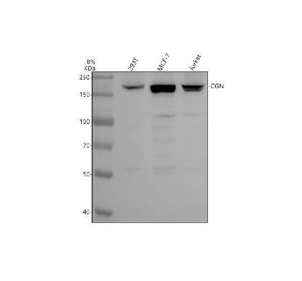
Western blot analysis of Cingulin/CGN using anti-Cingulin/CGN antibody (A02373-1).
Electrophoresis was performed on a 8% SDS-PAGE gel at 80V (Stacking gel) / 120V (Resolving gel) for 2 hours. The sample well of each lane was loaded with 30 ug of sample under reducing conditions.
Lane 1: human 293T whole cell lysates,
Lane 2: human MCF-7 whole cell lysates,
Lane 3: human Jurkat whole cell lysates.
After electrophoresis, proteins were transferred to a nitrocellulose membrane at 150 mA for 50-90 minutes. Blocked the membrane with 5% non-fat milk/TBS for 1.5 hour at RT. The membrane was incubated with rabbit anti-Cingulin/CGN antigen affinity purified polyclonal antibody (A02373-1) at 1:1000 overnight at 4°C, then washed with TBS-0.1%Tween 3 times with 5 minutes each and probed with a goat anti-rabbit IgG-HRP secondary antibody at a dilution of 1:5000 for 1.5 hour at RT. The signal is developed using an ECL Plus Western Blotting Substrate (Catalog # AR1196-200) with Tanon 5200 system. A specific band was detected for Cingulin/CGN at approximately 150 kDa. The expected band size for Cingulin/CGN is at 136 kDa.

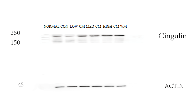
Click image to see more details
Western blot analysis of Cingulin/CGN using anti-Cingulin/CGN antibody (A02373-1).
Electrophoresis was performed on a 8% SDS-PAGE gel at 80V (Stacking gel) / 120V (Resolving gel) for 2 hours. The sample well of each lane was loaded with 30 ug of sample under reducing conditions.
Lane 1: normal group-rat colon tissue lysates,
Lane 2: control group-model rat colon tissue lysates,
Lane 3: low dose Chinese medicine group-model rat colon tissue lysates,
Lane 4: medium dose Chinese medicine group-model rat colon tissue lysates,
Lane 5: high dose Chinese medicine group-model rat colon tissue lysates,
Lane 6: western medicine group-model rat colon tissue lysates.
After electrophoresis, proteins were transferred to a nitrocellulose membrane at 150 mA for 50-90 minutes. Blocked the membrane with 5% non-fat milk/TBS for 1.5 hour at RT. The membrane was incubated with rabbit anti-Cingulin/CGN antigen affinity purified polyclonal antibody (A02373-1) at 1:1000 overnight at 4°C, then washed with TBS-0.1%Tween 3 times with 5 minutes each and probed with a goat anti-rabbit IgG-HRP secondary antibody at a dilution of 1:5000 for 1 hour at RT. The signal is developed using an ECL Plus Western Blotting Substrate (Catalog # AR1196-200) with ChemiDoc MP system. The expected band size for Cingulin/CGN is at 136 kDa.
Specific Publications For Anti-Cingulin/CGN Antibody (A02373-1)
Loading publications
Recommended Resources
Here are featured tools and databases that you might find useful.
- Boster's Pathways Library
- Protein Databases
- Bioscience Research Protocol Resources
- Data Processing & Analysis Software
- Photo Editing Software
- Scientific Literature Resources
- Research Paper Management Tools
- Molecular Biology Software
- Primer Design Tools
- Bioinformatics Tools
- Phylogenetic Tree Analysis
Customer Reviews
Have you used Anti-Cingulin/CGN Antibody?
Share your experimental results or join a short interview to earn up to $1,000 in product credits or other rewards.
1 Reviews For Anti-Cingulin/CGN Antibody
WB results showed that Cingulin/CGN (A02373-1) was downregulated in the rat colon model group and restored after Chinese medicine treatment, with the high-dose group showing the best effect, and the target bands were clear and specific.
Excellent
| SKU | A02373-1 |
|---|---|
| Application | Western Blot |
| Sample | rat colon tissue |
| Sample Processing Description | Rat colon tissues were lysed in RIPA buffer containing PMSF (100:1) for 10 min, centrifuged at 12,000 rpm for 15 min, and the supernatant was mixed with 5× loading buffer, denatured at 100°C for 10 min, and then loaded onto SDS-PAGE. |
| Other Reagents | 5% non-fat milk |
| Primary Antibody | Cingulin/CGN Antibody |
| Primary Incubation | 1:1000, overnight at 4 ℃ |
| Secondary Antibody | HRP Conjugated AffiniPure Goat Anti-Rabbit IgG (H+L) (BA1054) |
| Secondary Incubation | 1:5000, 1 h in RT |
| Detection | Substrate: ECL substrate, Image system:ChemiDoc MP |
| Results Summary | The figure shows WB results of Cingulin/CGN and the internal control Actin in rat colon across different groups; expression was decreased in the model group and restored after Chinese medicine treatment, with the high-dose group showing the best effect, and the target bands were clear and specific, indicating satisfactory results. |
Shiyu Zhang, Liaoning University of Traditional Chinese Medicine
Verified customer
Submitted 2026-03-04
Customer Q&As
Have a question?
Find answers in Q&As, reviews.
Can't find your answer?
Submit your question
