Click image to see more details
-

-
-
-
-
+14
Product Info Summary
| SKU: | M02226 |
|---|---|
| Size: | 100 μl |
| Reactive Species: | Human, Mouse, Rat |
| Host: | Rabbit |
| Application: | IF, IHC, ICC, WB |
Customers Who Bought This Also Bought

Product info
Product Name
Anti-Collagen VI COL6A1 Rabbit Monoclonal Antibody
SKU/Catalog Number
M02226
BM4748 is an alternative SKU for this antibody, used in previous lots.
Size
100 μl
Form
Liquid
Description
Boster Bio Anti-Collagen VI COL6A1 Rabbit Monoclonal Antibody catalog # M02226. Tested in WB, IHC, ICC/IF applications. This antibody reacts with Human, Mouse, Rat.
Storage & Handling
Store at -20°C for one year. For short term storage and frequent use, store at 4°C for up to one month. Avoid repeated freeze-thaw cycles.
Cite This Product
Anti-Collagen VI COL6A1 Rabbit Monoclonal Antibody (Boster Biological Technology, Pleasanton CA, USA, Catalog # M02226)
Host
Rabbit
Contents
Rabbit IgG in stabilizing components, phosphate buffered saline, pH 7.4, 150mM NaCl, 0.02% sodium azide and 50% glycerol.
*This antibody is supplied in a stabilized formulation.
Compatibility with conjugation reactions depends on the chemistry of the conjugation method used.
For conjugation methods that are not compatible with the stabilizing components present in this formulation, a carrier-free antibody format is required.
Clonality
Monoclonal
Clone Number
HGG-3
Isotype
Rabbit IgG
Immunogen
A synthesized peptide derived from human Collagen VI
Reactive Species
M02226 is reactive to COL6A1 in Human, Mouse, Rat
Observed Molecular Weight
147 kDa
Calculated molecular weight
108.5 kDa
Antibody Validation
Boster validates all antibodies on WB, IHC, ICC, Immunofluorescence, and ELISA with known positive control and negative samples to ensure specificity and high affinity, including thorough antibody incubations.
Application & Images
Applications
M02226 is guaranteed for IF, IHC, ICC, WB Boster Guarantee
Recommend Dilution
WB 1:500-2000
IHC 1:50-200
ICC/IF 1:50-200
Tested application
Suggested blocking solution with 5% non-fat milk or BSA; (*)Recommended protein loading: 20-40 µg per lane
Use TE buffer pH 9.0 for antigen retrieval; (*) citrate buffer pH 6.0 is an alternative.
Validation Images & Assay Conditions
Click image to see more details
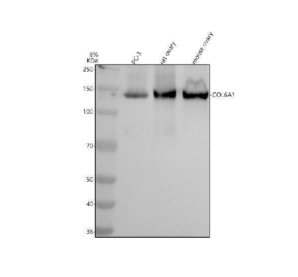
Western blot analysis of Collagen Type VI/COL6A1 using anti-Collagen Type VI/COL6A1 antibody (M02226).
Electrophoresis was performed on a 8% SDS-PAGE gel at 80V (Stacking gel) / 120V (Resolving gel) for 2 hours. The sample well of each lane was loaded with 30 ug of sample under reducing conditions.
Lane 1: human PC-3 whole cell lysates,
Lane 2: rat ovary tissue lysates,
Lane 3: mouse ovary tissue lysates.
After electrophoresis, proteins were transferred to a nitrocellulose membrane at 150 mA for 50-90 minutes. Blocked the membrane with 5% non-fat milk/TBS for 1.5 hour at RT. The membrane was incubated with rabbit anti-Collagen Type VI/COL6A1 antigen affinity purified monoclonal antibody (M02226) at 1: 1000 overnight at 4°C, then washed with TBS-0.1%Tween 3 times with 5 minutes each and probed with a goat anti-rabbit IgG-HRP secondary antibody at a dilution of 1:5000 for 1.5 hour at RT. The signal is developed using an ECL Plus Western Blotting Substrate (Catalog # AR1196-200) with Tanon 5200 system. A specific band was detected for Collagen Type VI/COL6A1 at approximately 147 kDa. The expected band size for Collagen Type VI/COL6A1 is at 109 kDa.

Click image to see more details
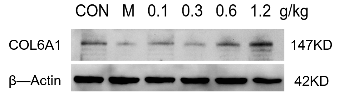
Western blot analysis of COL6A1 using anti-COL6A1 antibody (M02226).
Electrophoresis was performed on a 5-20% SDS-PAGE gel at 80V (Stacking gel) / 120V (Resolving gel) for 2 hours. The sample well of each lane was loaded with 30 ug of sample under reducing conditions.
Lane 1: control group-Mouse hippocampus tissue lysates,
Lane 2: model group-Mouse hippocampus tissue lysates,
Lane 3: Drug treatment (0.1g/kg) – Mouse hippocampus tissue lysates,
Lane 4: Drug treatment (0.3g/kg) – Mouse hippocampus tissue lysates,
Lane 5: Drug treatment (0.6g/kg) – Mouse hippocampus tissue lysates,
Lane 6: Drug treatment (1.2g/kg) – Mouse hippocampus tissue lysates.
After electrophoresis, proteins were transferred to a nitrocellulose membrane at 150 mA for 50-90 minutes. Blocked the membrane with 5% non-fat milk/TBS for 1.5 hour at RT. The membrane was incubated with rabbit anti-COL6A1 antigen affinity purified monoclonal antibody (M02226) at 1:1000 overnight at 4°C, then washed with TBS-0.1%Tween 3 times with 5 minutes each and probed with a goat anti-rabbit IgG-HRP secondary antibody (Catalog # BA1054) at a dilution of 1:5000 for 1 hour at RT. The signal is developed using an ECL Plus Western Blotting Substrate (Catalog # AR1196-200) with ChemiDoc MP system. A specific band was detected for COL6A1 at approximately 147 kDa. The expected band size for COL6A1 is at 108 kDa.

Click image to see more details
IHC analysis of Collagen Type VI/COL6A1 using anti-Collagen Type VI/COL6A1 antibody (M02226).
Collagen Type VI/COL6A1 was detected in a paraffin-embedded section of mouse kidney tissue. Heat mediated antigen retrieval was performed in EDTA buffer (pH 8.0, epitope retrieval solution). The tissue section was blocked with 10% goat serum. The tissue section was then incubated with 1: 50 rabbit anti-Collagen Type VI/COL6A1 Antibody (M02226) overnight at 4°C. Peroxidase Conjugated Goat Anti-rabbit IgG was used as secondary antibody and incubated for 30 minutes at 37°C. The tissue section was developed using HRP Conjugated Rabbit IgG Super Vision Assay Kit (Catalog # SV0002) with DAB as the chromogen.

Click image to see more details
IHC analysis of Collagen Type VI/COL6A1 using anti-Collagen Type VI/COL6A1 antibody (M02226).
Collagen Type VI/COL6A1 was detected in a paraffin-embedded section of mouse lung tissue. Heat mediated antigen retrieval was performed in EDTA buffer (pH 8.0, epitope retrieval solution). The tissue section was blocked with 10% goat serum. The tissue section was then incubated with 1: 50 rabbit anti-Collagen Type VI/COL6A1 Antibody (M02226) overnight at 4°C. Peroxidase Conjugated Goat Anti-rabbit IgG was used as secondary antibody and incubated for 30 minutes at 37°C. The tissue section was developed using HRP Conjugated Rabbit IgG Super Vision Assay Kit (Catalog # SV0002) with DAB as the chromogen.

Click image to see more details
IHC analysis of Collagen Type VI/COL6A1 using anti-Collagen Type VI/COL6A1 antibody (M02226).
Collagen Type VI/COL6A1 was detected in a paraffin-embedded section of mouse spleen tissue. Heat mediated antigen retrieval was performed in EDTA buffer (pH 8.0, epitope retrieval solution). The tissue section was blocked with 10% goat serum. The tissue section was then incubated with 1: 50 rabbit anti-Collagen Type VI/COL6A1 Antibody (M02226) overnight at 4°C. Peroxidase Conjugated Goat Anti-rabbit IgG was used as secondary antibody and incubated for 30 minutes at 37°C. The tissue section was developed using HRP Conjugated Rabbit IgG Super Vision Assay Kit (Catalog # SV0002) with DAB as the chromogen.

Click image to see more details
IHC analysis of Collagen Type VI/COL6A1 using anti-Collagen Type VI/COL6A1 antibody (M02226).
Collagen Type VI/COL6A1 was detected in a paraffin-embedded section of mouse liver tissue. Heat mediated antigen retrieval was performed in EDTA buffer (pH 8.0, epitope retrieval solution). The tissue section was blocked with 10% goat serum. The tissue section was then incubated with 1: 50 rabbit anti-Collagen Type VI/COL6A1 Antibody (M02226) overnight at 4°C. Peroxidase Conjugated Goat Anti-rabbit IgG was used as secondary antibody and incubated for 30 minutes at 37°C. The tissue section was developed using HRP Conjugated Rabbit IgG Super Vision Assay Kit (Catalog # SV0002) with DAB as the chromogen.

Click image to see more details
IHC analysis of Collagen Type VI/COL6A1 using anti-Collagen Type VI/COL6A1 antibody (M02226).
Collagen Type VI/COL6A1 was detected in a paraffin-embedded section of rat kidney tissue. Heat mediated antigen retrieval was performed in EDTA buffer (pH 8.0, epitope retrieval solution). The tissue section was blocked with 10% goat serum. The tissue section was then incubated with 1: 50 rabbit anti-Collagen Type VI/COL6A1 Antibody (M02226) overnight at 4°C. Peroxidase Conjugated Goat Anti-rabbit IgG was used as secondary antibody and incubated for 30 minutes at 37°C. The tissue section was developed using HRP Conjugated Rabbit IgG Super Vision Assay Kit (Catalog # SV0002) with DAB as the chromogen.

Click image to see more details
IHC analysis of Collagen Type VI/COL6A1 using anti-Collagen Type VI/COL6A1 antibody (M02226).
Collagen Type VI/COL6A1 was detected in a paraffin-embedded section of rat lung tissue. Heat mediated antigen retrieval was performed in EDTA buffer (pH 8.0, epitope retrieval solution). The tissue section was blocked with 10% goat serum. The tissue section was then incubated with 1: 50 rabbit anti-Collagen Type VI/COL6A1 Antibody (M02226) overnight at 4°C. Peroxidase Conjugated Goat Anti-rabbit IgG was used as secondary antibody and incubated for 30 minutes at 37°C. The tissue section was developed using HRP Conjugated Rabbit IgG Super Vision Assay Kit (Catalog # SV0002) with DAB as the chromogen.

Click image to see more details
IHC analysis of Collagen Type VI/COL6A1 using anti-Collagen Type VI/COL6A1 antibody (M02226).
Collagen Type VI/COL6A1 was detected in a paraffin-embedded section of rat spleen tissue. Heat mediated antigen retrieval was performed in EDTA buffer (pH 8.0, epitope retrieval solution). The tissue section was blocked with 10% goat serum. The tissue section was then incubated with 1: 50 rabbit anti-Collagen Type VI/COL6A1 Antibody (M02226) overnight at 4°C. Peroxidase Conjugated Goat Anti-rabbit IgG was used as secondary antibody and incubated for 30 minutes at 37°C. The tissue section was developed using HRP Conjugated Rabbit IgG Super Vision Assay Kit (Catalog # SV0002) with DAB as the chromogen.

Click image to see more details
IHC analysis of Collagen Type VI/COL6A1 using anti-Collagen Type VI/COL6A1 antibody (M02226).
Collagen Type VI/COL6A1 was detected in a paraffin-embedded section of rat liver tissue. Heat mediated antigen retrieval was performed in EDTA buffer (pH 8.0, epitope retrieval solution). The tissue section was blocked with 10% goat serum. The tissue section was then incubated with 1: 50 rabbit anti-Collagen Type VI/COL6A1 Antibody (M02226) overnight at 4°C. Peroxidase Conjugated Goat Anti-rabbit IgG was used as secondary antibody and incubated for 30 minutes at 37°C. The tissue section was developed using HRP Conjugated Rabbit IgG Super Vision Assay Kit (Catalog # SV0002) with DAB as the chromogen.

Click image to see more details
IHC analysis of Collagen Type VI/COL6A1 using anti-Collagen Type VI/COL6A1 antibody (M02226).
Collagen Type VI/COL6A1 was detected in a paraffin-embedded section of human placenta tissue. Heat mediated antigen retrieval was performed in EDTA buffer (pH 8.0, epitope retrieval solution). The tissue section was blocked with 10% goat serum. The tissue section was then incubated with 1: 50 rabbit anti-Collagen Type VI/COL6A1 Antibody (M02226) overnight at 4°C. Peroxidase Conjugated Goat Anti-rabbit IgG was used as secondary antibody and incubated for 30 minutes at 37°C. The tissue section was developed using HRP Conjugated Rabbit IgG Super Vision Assay Kit (Catalog # SV0002) with DAB as the chromogen.

Click image to see more details
IHC analysis of Collagen Type VI/COL6A1 using anti-Collagen Type VI/COL6A1 antibody (M02226).
Collagen Type VI/COL6A1 was detected in a paraffin-embedded section of human lung cancer tissue. Heat mediated antigen retrieval was performed in EDTA buffer (pH 8.0, epitope retrieval solution). The tissue section was blocked with 10% goat serum. The tissue section was then incubated with 1: 50 rabbit anti-Collagen Type VI/COL6A1 Antibody (M02226) overnight at 4°C. Peroxidase Conjugated Goat Anti-rabbit IgG was used as secondary antibody and incubated for 30 minutes at 37°C. The tissue section was developed using HRP Conjugated Rabbit IgG Super Vision Assay Kit (Catalog # SV0002) with DAB as the chromogen.

Click image to see more details
IHC analysis of Collagen Type VI/COL6A1 using anti-Collagen Type VI/COL6A1 antibody (M02226).
Collagen Type VI/COL6A1 was detected in a paraffin-embedded section of human lymphoma tissue. Heat mediated antigen retrieval was performed in EDTA buffer (pH 8.0, epitope retrieval solution). The tissue section was blocked with 10% goat serum. The tissue section was then incubated with 1: 50 rabbit anti-Collagen Type VI/COL6A1 Antibody (M02226) overnight at 4°C. Peroxidase Conjugated Goat Anti-rabbit IgG was used as secondary antibody and incubated for 30 minutes at 37°C. The tissue section was developed using HRP Conjugated Rabbit IgG Super Vision Assay Kit (Catalog # SV0002) with DAB as the chromogen.

Click image to see more details
IHC analysis of Collagen Type VI/COL6A1 using anti-Collagen Type VI/COL6A1 antibody (M02226).
Collagen Type VI/COL6A1 was detected in a paraffin-embedded section of human ovarian cancer tissue. Heat mediated antigen retrieval was performed in EDTA buffer (pH 8.0, epitope retrieval solution). The tissue section was blocked with 10% goat serum. The tissue section was then incubated with 1: 50 rabbit anti-Collagen Type VI/COL6A1 Antibody (M02226) overnight at 4°C. Peroxidase Conjugated Goat Anti-rabbit IgG was used as secondary antibody and incubated for 30 minutes at 37°C. The tissue section was developed using HRP Conjugated Rabbit IgG Super Vision Assay Kit (Catalog # SV0002) with DAB as the chromogen.

Click image to see more details
IHC analysis of Collagen Type VI/COL6A1 using anti-Collagen Type VI/COL6A1 antibody (M02226).
Collagen Type VI/COL6A1 was detected in a paraffin-embedded section of human breast cancer tissue. Heat mediated antigen retrieval was performed in EDTA buffer (pH 8.0, epitope retrieval solution). The tissue section was blocked with 10% goat serum. The tissue section was then incubated with 1: 50 rabbit anti-Collagen Type VI/COL6A1 Antibody (M02226) overnight at 4°C. Peroxidase Conjugated Goat Anti-rabbit IgG was used as secondary antibody and incubated for 30 minutes at 37°C. The tissue section was developed using HRP Conjugated Rabbit IgG Super Vision Assay Kit (Catalog # SV0002) with DAB as the chromogen.

Click image to see more details
IHC analysis of Collagen Type VI/COL6A1 using anti-Collagen Type VI/COL6A1 antibody (M02226).
Collagen Type VI/COL6A1 was detected in a paraffin-embedded section of human esophageal cancer tissue. Heat mediated antigen retrieval was performed in EDTA buffer (pH 8.0, epitope retrieval solution). The tissue section was blocked with 10% goat serum. The tissue section was then incubated with 1: 50 rabbit anti-Collagen Type VI/COL6A1 Antibody (M02226) overnight at 4°C. Peroxidase Conjugated Goat Anti-rabbit IgG was used as secondary antibody and incubated for 30 minutes at 37°C. The tissue section was developed using HRP Conjugated Rabbit IgG Super Vision Assay Kit (Catalog # SV0002) with DAB as the chromogen.

Click image to see more details
IHC analysis of Collagen Type VI/COL6A1 using anti-Collagen Type VI/COL6A1 antibody (M02226).
Collagen Type VI/COL6A1 was detected in a paraffin-embedded section of human liver cancer tissue. Heat mediated antigen retrieval was performed in EDTA buffer (pH 8.0, epitope retrieval solution). The tissue section was blocked with 10% goat serum. The tissue section was then incubated with 1: 50 rabbit anti-Collagen Type VI/COL6A1 Antibody (M02226) overnight at 4°C. Peroxidase Conjugated Goat Anti-rabbit IgG was used as secondary antibody and incubated for 30 minutes at 37°C. The tissue section was developed using HRP Conjugated Rabbit IgG Super Vision Assay Kit (Catalog # SV0002) with DAB as the chromogen.

Click image to see more details
IHC analysis of Collagen Type VI/COL6A1 using anti-Collagen Type VI/COL6A1 antibody (M02226).
Collagen Type VI/COL6A1 was detected in a paraffin-embedded section of human colon cancer tissue. Heat mediated antigen retrieval was performed in EDTA buffer (pH 8.0, epitope retrieval solution). The tissue section was blocked with 10% goat serum. The tissue section was then incubated with 1: 50 rabbit anti-Collagen Type VI/COL6A1 Antibody (M02226) overnight at 4°C. Peroxidase Conjugated Goat Anti-rabbit IgG was used as secondary antibody and incubated for 30 minutes at 37°C. The tissue section was developed using HRP Conjugated Rabbit IgG Super Vision Assay Kit (Catalog # SV0002) with DAB as the chromogen.
Specific Publications For Anti-Collagen VI COL6A1 Rabbit Monoclonal Antibody (M02226)
Loading publications
Recommended Resources
Here are featured tools and databases that you might find useful.
- Boster's Pathways Library
- Protein Databases
- Bioscience Research Protocol Resources
- Data Processing & Analysis Software
- Photo Editing Software
- Scientific Literature Resources
- Research Paper Management Tools
- Molecular Biology Software
- Primer Design Tools
- Bioinformatics Tools
- Phylogenetic Tree Analysis
Customer Reviews
Have you used Anti-Collagen VI COL6A1 Rabbit Monoclonal Antibody?
Share your experimental results or join a short interview to earn up to $1,000 in product credits or other rewards.
1 Reviews For Anti-Collagen VI COL6A1 Rabbit Monoclonal Antibody
Western blot analysis was performed using the COL6A1 antibody to detect COL6A1 protein expression in the mouse hippocampus.
Excellent
| SKU | M02226 |
|---|---|
| Application | Western Blot |
| Sample | Mouse hippocampus tissue |
| Sample Processing Description | The mouse hippocampus was lysed in RIPA buffer supplemented with a protease inhibitor cocktail. After protein quantification, samples were mixed with 5× protein loading buffer and denatured by heating at 100°C for 10 minutes. Five microliters of each protein sample were loaded per lane onto SDS-PAGE. |
| Primary Antibody | Anti-Collagen VI COL6A1 Rabbit Monoclonal Antibody |
| Primary Incubation | overnight at 4 ℃ |
| Secondary Antibody | HRP-conjugated Anti-Rabbit IgG Secondary Antibody |
| Secondary Incubation | 1 hour in room temperature |
| Detection | Substrate: Ultra-sensitive ECL luminescent reagent (Cat# AR1191), Imaging system:ChemiDoc MP |
| Results Summary | Western blot analysis was performed using the COL6A1 antibody to detect COL6A1 protein expression in the mouse hippocampus. Although minor non-specific bands were observed, they did not affect the trend analysis, indicating that the antibody is suitable for detecting the target protein in this tissue. |
Changyang Yu, LUTCM
Verified customer
Submitted 2025-11-06
Customer Q&As
Have a question?
Find answers in Q&As, reviews.
Can't find your answer?
Submit your question
4 Customer Q&As for Anti-Collagen VI COL6A1 Rabbit Monoclonal Antibody
Question
We are currently using anti-Collagen VI Rabbit Monoclonal antibody M02226 for rat tissue, and we are well pleased with the IHC-F results. The species of reactivity given in the datasheet says human, mouse, rat. Is it possible that the antibody can work on goat tissues as well?
Verified Customer
Verified customer
Asked: 2020-01-28
Answer
The anti-Collagen VI Rabbit Monoclonal antibody (M02226) has not been tested for cross reactivity specifically with goat tissues, though there is a good chance of cross reactivity. We have an innovator award program that if you test this antibody and show it works in goat you can get your next antibody for free. Please contact me if I can help you with anything.
Boster Scientific Support
Answered: 2020-01-28
Question
We appreciate helping with my inquiry over the phone. Here are the WB image, lot number and protocol we used for placenta using anti-Collagen VI Rabbit Monoclonal antibody M02226. Let me know if you need anything else.
Verified Customer
Verified customer
Asked: 2019-12-09
Answer
We appreciate the data. You have provided everything we needed. Our lab team are working to resolve your inquiry as quickly as possible, and we appreciate your patience and understanding! Please let me know if there is anything you need in the meantime.
Boster Scientific Support
Answered: 2019-12-09
Question
Our lab want to know about to test anti-Collagen VI Rabbit Monoclonal antibody M02226 on mouse placenta for research purposes, then I may be interested in using anti-Collagen VI Rabbit Monoclonal antibody M02226 for diagnostic purposes as well. Is the antibody suitable for diagnostic purposes?
Verified Customer
Verified customer
Asked: 2018-08-02
Answer
The products we sell, including anti-Collagen VI Rabbit Monoclonal antibody M02226, are only intended for research use. They would not be suitable for use in diagnostic work. If you have the means to develop a product into diagnostic use, and are interested in collaborating with us and develop our product into an IVD product, please contact us for more discussions.
Boster Scientific Support
Answered: 2018-08-02
Question
Is a blocking peptide available for product anti-Collagen VI Rabbit Monoclonal antibody (M02226)?
Verified Customer
Verified customer
Asked: 2018-06-29
Answer
We do provide the blocking peptide for product anti-Collagen VI Rabbit Monoclonal antibody (M02226). If you would like to place an order for it please contact support@bosterbio.com and make a special request.
Boster Scientific Support
Answered: 2018-06-29
