Click image to see more details
Product Info Summary
| SKU: | A03564-5 |
|---|---|
| Size: | 100 μg/vial |
| Reactive Species: | Human, Mouse, Rat |
| Host: | Rabbit |
| Application: | ELISA, IP, WB |
Customers Who Bought This Also Bought

Product info
Product Name
Anti-CREB3 Antibody Picoband®
SKU/Catalog Number
A03564-5
Size
100 μg/vial
Form
Lyophilized
Description
Boster Bio Anti-CREB3 Antibody Picoband® catalog # A03564-5. Tested in WB, IP, ELISA applications. This antibody reacts with Human, Mouse, Rat. The brand Picoband indicates this is a premium antibody that guarantees superior quality, high affinity, and strong signals with minimal background in Western blot applications. Only our best-performing antibodies are designated as Picoband, ensuring unmatched performance.
Storage & Handling
At -20°C for one year from date of receipt. After reconstitution, at 4°C for one month. It can also be aliquotted and stored frozen at -20°C for six months. Avoid repeated freezing and thawing.
Cite This Product
Anti-CREB3 Antibody Picoband® (Boster Biological Technology, Pleasanton CA, USA, Catalog # A03564-5)
Host
Rabbit
Contents
Each vial contains 4 mg Trehalose, 0.9 mg NaCl, 0.2 mg Na2HPO4.
Clonality
Polyclonal
Immunogen
E.coli-derived human CREB3 recombinant protein (Position: M1-K352). Human CREB3 shares 67.1% amino acid (aa) sequence identity with mouse CREB3.
Reactive Species
A03564-5 is reactive to CREB3 in Human, Mouse, Rat
Observed Molecular Weight
41 kDa
Calculated molecular weight
41.4 kDa
Background of CREB3
This gene encodes a transcription factor that is a member of the leucine zipper family of DNA binding proteins. This protein binds to the cAMP-response element and regulates cell proliferation. The protein interacts with host cell factor C1, which also associates with the herpes simplex virus (HSV) protein VP16 that induces transcription of HSV immediate-early genes. This protein and VP16 both bind to the same site on host cell factor C1. It is thought that the interaction between this protein and host cell factor C1 plays a role in the establishment of latency during HSV infection. This protein also plays a role in leukocyte migration, tumor suppression, and endoplasmic reticulum stress-associated protein degradation. Additional transcript variants have been identified, but their biological validity has not been determined.
Antibody Validation
Boster validates all antibodies on WB, IHC, ICC, Immunofluorescence, and ELISA with known positive control and negative samples to ensure specificity and high affinity, including thorough antibody incubations.
Application & Images
Applications
A03564-5 is guaranteed for ELISA, IP, WB Boster Guarantee
Recommend Dilution
| Application | Dilution | Species |
|---|---|---|
| Western blot | 0.25-0.5 μg/ml | Human, Mouse, Rat |
| Immunoprecipitation | 0.5-2 μg/ml | Human |
| ELISA | 0.1-0.5 μg/ml |
Validation Images & Assay Conditions
Click image to see more details
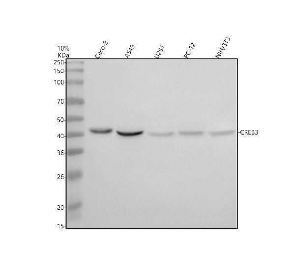
Western blot analysis of CREB3 using anti-CREB3 antibody (A03564-5).
Electrophoresis was performed on a 10% SDS-PAGE gel at 80V (Stacking gel) / 120V (Resolving gel) for 2 hours. The sample well of each lane was loaded with 30 ug of sample under reducing conditions.
Lane 1: human Caco-2 whole cell lysates,
Lane 2: human A549 whole cell lysates,
Lane 3: human U251 whole cell lysates
Lane 4: rat PC-12 whole cell lysates,
Lane 5: mouse NIH/3T3 whole cell lysates.
After electrophoresis, proteins were transferred to a nitrocellulose membrane at 150 mA for 50-90 minutes. Blocked the membrane with 5% non-fat milk/TBS for 1.5 hour at RT. The membrane was incubated with rabbit anti-CREB3 antigen affinity purified polyclonal antibody (A03564-5) at 0.5 μg/mL overnight at 4°C, then washed with TBS-0.1%Tween 3 times with 5 minutes each and probed with a goat anti-rabbit IgG-HRP secondary antibody at a dilution of 1:5000 for 1.5 hour at RT. The signal is developed using an ECL Plus Western Blotting Substrate (Catalog # AR1196-200) with Tanon 5200 system. A specific band was detected for CREB3 at approximately 41 kDa. The expected band size for CREB3 is at 41 kDa.

Click image to see more details
Immunoprecipitating CREB3 in A549 whole cell lysate.
Western blot analysis of CREB3 using anti-CREB3 antibody (A03564-5).
Lane 1: A549 whole cell lysates (30ug),
Lane 2: Rabbit control IgG instead of anti-CREB3 antibody in A549 whole cell lysate,
Lane 3: anti-CREB3 antibody (2μg) + A549 whole cell lysate (500μg).
After electrophoresis, proteins were transferred to a membrane. Then the membrane was incubated with rabbit anti-CREB3 antigen affinity purified polyclonal antibody (A03564-5) at a dilution of 0.5 μg/mL and probed with a goat anti-rabbit IgG-HRP secondary antibody (Catalog # BA1054). The signal is developed using ECL Plus Western Blotting Substrate (Catalog # AR1197). A specific band was detected for CREB3 at approximately 45 kDa. The expected band size for CREB3 is at 41 kDa.
Specific Publications For Anti-CREB3 Antibody Picoband® (A03564-5)
Loading publications
Recommended Resources
Here are featured tools and databases that you might find useful.
- Boster's Pathways Library
- Protein Databases
- Bioscience Research Protocol Resources
- Data Processing & Analysis Software
- Photo Editing Software
- Scientific Literature Resources
- Research Paper Management Tools
- Molecular Biology Software
- Primer Design Tools
- Bioinformatics Tools
- Phylogenetic Tree Analysis
Customer Reviews
Have you used Anti-CREB3 Antibody Picoband®?
Share your experimental results or join a short interview to earn up to $1,000 in product credits or other rewards.
0 Reviews For Anti-CREB3 Antibody Picoband®
Customer Q&As
Have a question?
Find answers in Q&As, reviews.
Can't find your answer?
Submit your question
