Click image to see more details
Product Info Summary
| SKU: | A00629-4 |
|---|---|
| Size: | 100 μg/vial |
| Reactive Species: | Human, Mouse, Rat |
| Host: | Rabbit |
| Application: | Flow Cytometry, WB |
Customers Who Bought This Also Bought

Product info
Product Name
Anti-CRF/CRH Antibody Picoband®
SKU/Catalog Number
A00629-4
Size
100 μg/vial
Form
Lyophilized
Description
Boster Bio Anti-CRF-CRH Antibody Picoband® catalog # A00629-4. Tested in WB, Flow Cytometry applications. This antibody reacts with Human, Mouse, Rat. The brand Picoband indicates this is a premium antibody that guarantees superior quality, high affinity, and strong signals with minimal background in Western blot applications. Only our best-performing antibodies are designated as Picoband, ensuring unmatched performance.
Storage & Handling
At -20°C for one year from date of receipt. After reconstitution, at 4°C for one month. It can also be aliquotted and stored frozen at -20°C for six months. Avoid repeated freezing and thawing.
Cite This Product
Anti-CRF/CRH Antibody Picoband® (Boster Biological Technology, Pleasanton CA, USA, Catalog # A00629-4)
Host
Rabbit
Contents
Each vial contains 4 mg Trehalose, 0.9 mg NaCl, 0.2 mg Na2HPO4.
Clonality
Polyclonal
Immunogen
A synthetic peptide corresponding to a sequence at the C-terminus of human CRF/CRH, identical to the related mouse and rat sequences.
Reactive Species
A00629-4 is reactive to CRH in Human, Mouse, Rat
Calculated molecular weight
21.4 kDa
Background of CRH
This gene encodes a member of the corticotropin-releasing factor family. The encoded preproprotein is proteolytically processed to generate the mature neuropeptide hormone. In response to stress, this hormone is secreted by the paraventricular nucleus (PVN) of the hypothalamus, binds to corticotropin releasing hormone receptors and stimulates the release of adrenocorticotropic hormone from the pituitary gland. Marked reduction in this protein has been observed in association with Alzheimer's disease. Autosomal recessive hypothalamic corticotropin deficiency has multiple and potentially fatal metabolic consequences including hypoglycemia and hepatitis. In addition to production in the hypothalamus, this protein is also synthesized in peripheral tissues, such as T lymphocytes, and is highly expressed in the placenta. In the placenta it is a marker that determines the length of gestation and the timing of parturition and delivery. A rapid increase in circulating levels of the hormone occurs at the onset of parturition, suggesting that, in addition to its metabolic functions, this protein may act as a trigger for parturition.
Antibody Validation
Boster validates all antibodies on WB, IHC, ICC, Immunofluorescence, and ELISA with known positive control and negative samples to ensure specificity and high affinity, including thorough antibody incubations.
Application & Images
Applications
A00629-4 is guaranteed for Flow Cytometry, WB Boster Guarantee
Recommend Dilution
| Application | Dilution | Species |
|---|---|---|
| Western Blot (WB) | 0.25-0.5 μg/ml | Human, Mouse, Rat |
| Flow Cytometry (FC) | 1-3 μg/1x106 cells | Human |
Validation Images & Assay Conditions
Click image to see more details
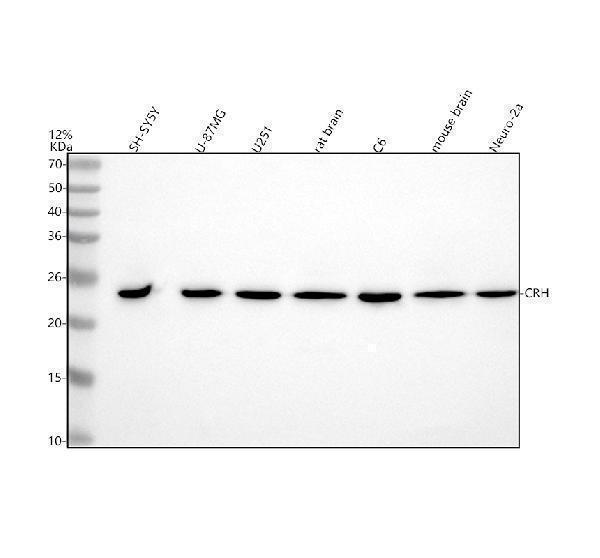
Western blot analysis of CRF/CRH using anti-CRF/CRH antibody (A00629-4).
Electrophoresis was performed on a 5-20% SDS-PAGE gel at 70V (Stacking gel) / 90V (Resolving gel) for 2-3 hours. The sample well of each lane was loaded with 30 ug of sample under reducing conditions.
Lane 1: human SH-SY5Y whole cell lysates,
Lane 2: human U-87MG whole cell lysates,
Lane 3: human U251 whole cell lysates,
Lane 4: rat brain tissue lysates,
Lane 5: rat C6 whole cell lysates,
Lane 6: mouse brain tissue lysates,
Lane 7: mouse Neuro-2a whole cell lysates,
After electrophoresis, proteins were transferred to a nitrocellulose membrane at 150 mA for 50-90 minutes. Blocked the membrane with 5% non-fat milk/TBS for 1.5 hour at RT. The membrane was incubated with rabbit anti-CRF/CRH antigen affinity purified polyclonal antibody (A00629-4) at 0.5 μg/mL overnight at 4°C, then washed with TBS-0.1%Tween 3 times with 5 minutes each and probed with a goat anti-rabbit IgG-HRP secondary antibody at a dilution of 1:5000 for 1.5 hour at RT. The signal is developed using an Enhanced Chemiluminescent detection (ECL) kit (Catalog # EK1002) with Tanon 5200 system. A specific band was detected for CRF/CRH at approximately 25 kDa. The expected band size for CRF/CRH is at 21 kDa.

Click image to see more details
Flow Cytometry analysis of U251 cells using anti-CRF/CRH antibody (A00629-4).
Overlay histogram showing U251 cells stained with A00629-4 (Blue line). The cells were fixed with 4% paraformaldehyde and blocked with 10% normal goat serum. And then incubated with rabbit anti-CRF/CRH Antibody (A00629-4, 1 μg/1x106 cells) for 30 min at 20°C. DyLight®488 conjugated goat anti-rabbit IgG (BA1127, 5-10 μg/1x106 cells) was used as secondary antibody for 30 minutes at 20°C. Isotype control antibody (Green line) was rabbit IgG (1 μg/1x106) used under the same conditions. Unlabelled sample without incubation with primary antibody and secondary antibody (Red line) was used as a blank control.
Specific Publications For Anti-CRF/CRH Antibody Picoband® (A00629-4)
Loading publications
Recommended Resources
Here are featured tools and databases that you might find useful.
- Boster's Pathways Library
- Protein Databases
- Bioscience Research Protocol Resources
- Data Processing & Analysis Software
- Photo Editing Software
- Scientific Literature Resources
- Research Paper Management Tools
- Molecular Biology Software
- Primer Design Tools
- Bioinformatics Tools
- Phylogenetic Tree Analysis
Customer Reviews
Have you used Anti-CRF/CRH Antibody Picoband®?
Share your experimental results or join a short interview to earn up to $1,000 in product credits or other rewards.
0 Reviews For Anti-CRF/CRH Antibody Picoband®
Customer Q&As
Have a question?
Find answers in Q&As, reviews.
Can't find your answer?
Submit your question
