Click image to see more details
-

-
-
-
-
+12
Product Info Summary
| SKU: | A01421 |
|---|---|
| Size: | 100 μg/vial |
| Reactive Species: | Human, Mouse, Rat |
| Host: | Rabbit |
| Application: | IF, IHC, WB |
Customers Who Bought This Also Bought

Product info
Product Name
Anti-Cytokeratin 8/KRT8 Antibody Picoband®
SKU/Catalog Number
A01421
Size
100 μg/vial
Form
Lyophilized
Description
Boster Bio Anti-Cytokeratin 8/KRT8 Antibody Picoband® catalog # A01421. Tested in IHC, WB applications. This antibody reacts with Human, Mouse, Rat. The brand Picoband indicates this is a premium antibody that guarantees superior quality, high affinity, and strong signals with minimal background in Western blot applications. Only our best-performing antibodies are designated as Picoband, ensuring unmatched performance.
Storage & Handling
Store at -20˚C for one year from date of receipt. After reconstitution, at 4˚C for one month. It can also be aliquotted and stored frozen at -20˚C for six months. Avoid repeated freeze-thaw cycles.
Cite This Product
Anti-Cytokeratin 8/KRT8 Antibody Picoband® (Boster Biological Technology, Pleasanton CA, USA, Catalog # A01421)
Host
Rabbit
Contents
Each vial contains 4mg Trehalose, 0.9mg NaCl and 0.2mg Na2HPO4.
Clonality
Polyclonal
Isotype
Rabbit IgG
Immunogen
E.coli-derived human Cytokeratin 8 recombinant protein (Position: D107-K325). Human Cytokeratin 8 shares 95.4% and 94.5% amino acid (aa) sequence identity with mouse and rat Cytokeratin 8, respectively.
Cross-reactivity
No cross-reactivity with other proteins
Reactive Species
A01421 is reactive to KRT8 in Human, Mouse, Rat
Observed Molecular Weight
54 kDa, 56 kDa
Calculated molecular weight
53.7 kDa
Background of KRT8
Keratin, type II cytoskeletal 8, also known as cytokeratin-8 (CK-8) or keratin-8 (K8) is a keratin protein that is encoded in humans by the KRT8 gene. This gene is a member of the type II keratin family clustered on the long arm of chromosome 12. Type I and type II keratins heteropolymerize to form intermediate-sized filaments in the cytoplasm of epithelial cells. The product of this gene typically dimerizes with keratin 18 to form an intermediate filament in simple single-layered epithelial cells. This protein plays a role in maintaining cellular structural integrity and also functions in signal transduction and cellular differentiation. Mutations in this gene cause cryptogenic cirrhosis. Alternatively spliced transcript variants have been found for this gene.
Antibody Validation
Boster validates all antibodies on WB, IHC, ICC, Immunofluorescence, and ELISA with known positive control and negative samples to ensure specificity and high affinity, including thorough antibody incubations.
Application & Images
Applications
A01421 is guaranteed for IF, IHC, WB Boster Guarantee
Recommend Dilution
| Application | Dilution | Species |
|---|---|---|
| Immunohistochemistry (Paraffin-embedded Section) | 0.5-1μg/ml | Human, Mouse, Rat |
| Western blot | 0.1-0.5μg/ml | Human, Mouse, Rat |
Tested application
Suggested blocking solution with 5% non-fat milk or BSA; (*)Recommended protein loading: 20-40 µg per lane
Use TE buffer pH 9.0 for antigen retrieval; (*) citrate buffer pH 6.0 is an alternative.
Validation Images & Assay Conditions
Click image to see more details
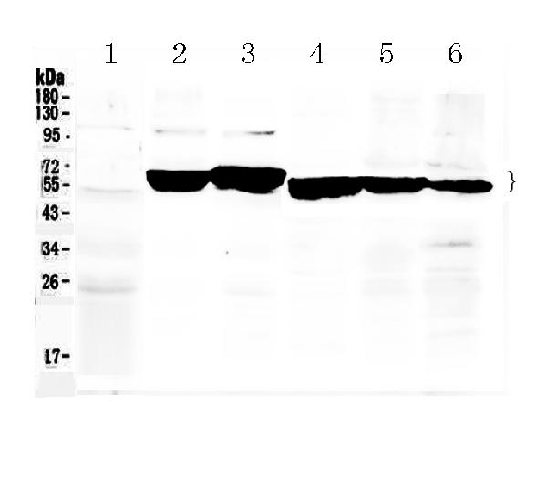
Western blot analysis of Cytokeratin 8 using anti-Cytokeratin 8 antibody (A01421).
Electrophoresis was performed on a 5-20% SDS-PAGE gel at 70V (Stacking gel) / 90V (Resolving gel) for 2-3 hours. The sample well of each lane was loaded with 50ug of sample under reducing conditions.
Lane 1: rat liver tissue lysates,
Lane 2: mouse ovary tissue lysates,
Lane 3: mouse liver tissue lysates,
Lane 4: MCF-7 whole cell lysates,
Lane 5: A549 whole cell lysates,
Lane 6: HELA whole Cell lysates.
After Electrophoresis, proteins were transferred to a Nitrocellulose membrane at 150mA for 50-90 minutes. Blocked the membrane with 5% Non-fat Milk/ TBS for 1.5 hour at RT. The membrane was incubated with rabbit anti-Cytokeratin 8 antigen affinity purified polyclonal antibody (Catalog # A01421) at 0.5 μg/mL overnight at 4°C, then washed with TBS-0.1%Tween 3 times with 5 minutes each and probed with a goat anti-rabbit IgG-HRP secondary antibody at a dilution of 1:10000 for 1.5 hour at RT. The signal is developed using an Enhanced Chemiluminescent detection (ECL) kit (Catalog # EK1002) with Tanon 5200 system. A specific band was detected for Cytokeratin 8 at approximately 54KD, 56KD. The expected band size for Cytokeratin 8 is at 54KD.

Click image to see more details
IHC analysis of Cytokeratin 8 using anti-Cytokeratin 8 antibody (A01421).
Cytokeratin 8 was detected in paraffin-embedded section of mouse liver tissues. Heat mediated antigen retrieval was performed in citrate buffer (pH6, epitope retrieval solution) for 20 mins. The tissue section was blocked with 10% goat serum. The tissue section was then incubated with 1μg/ml rabbit anti-Cytokeratin 8 Antibody (A01421) overnight at 4°C. Biotinylated goat anti-rabbit IgG was used as secondary antibody and incubated for 30 minutes at 37°C. The tissue section was developed using Strepavidin-Biotin-Complex (SABC)(Catalog # SA1022) with DAB as the chromogen.

Click image to see more details
IHC analysis of Cytokeratin 8 using anti-Cytokeratin 8 antibody (A01421).
Cytokeratin 8 was detected in paraffin-embedded section of rat intestine tissues. Heat mediated antigen retrieval was performed in citrate buffer (pH6, epitope retrieval solution) for 20 mins. The tissue section was blocked with 10% goat serum. The tissue section was then incubated with 1μg/ml rabbit anti-Cytokeratin 8 Antibody (A01421) overnight at 4°C. Biotinylated goat anti-rabbit IgG was used as secondary antibody and incubated for 30 minutes at 37°C. The tissue section was developed using Strepavidin-Biotin-Complex (SABC)(Catalog # SA1022) with DAB as the chromogen.

Click image to see more details
IHC analysis of Cytokeratin 8 using anti-Cytokeratin 8 antibody (A01421).
Cytokeratin 8 was detected in paraffin-embedded section of human intestinal cancer tissues. Heat mediated antigen retrieval was performed in citrate buffer (pH6, epitope retrieval solution) for 20 mins. The tissue section was blocked with 10% goat serum. The tissue section was then incubated with 1μg/ml rabbit anti-Cytokeratin 8 Antibody (A01421) overnight at 4°C. Biotinylated goat anti-rabbit IgG was used as secondary antibody and incubated for 30 minutes at 37°C. The tissue section was developed using Strepavidin-Biotin-Complex (SABC)(Catalog # SA1022) with DAB as the chromogen.

Click image to see more details
IHC analysis of Cytokeratin 8/KRT8 using anti-Cytokeratin 8/KRT8 antibody (A01421).
Cytokeratin 8/KRT8 was detected in a paraffin-embedded section of human stomach cancer tissue. Heat mediated antigen retrieval was performed in EDTA buffer (pH 8.0, epitope retrieval solution). The tissue section was blocked with 10% goat serum. The tissue section was then incubated with 1 μg/ml rabbit anti-Cytokeratin 8/KRT8 Antibody (A01421) for 30 minutes at 37°C. HRP-AffiniPure Goat Anti-Rabbit IgG was used as secondary antibody and incubated for 30 minutes at 37°C. The tissue section was developed using HRP Conjugated Rabbit IgG Super Vision Assay Kit (Catalog # SV0002) with DAB as the chromogen.

Click image to see more details
IHC analysis of Cytokeratin 8/KRT8 using anti-Cytokeratin 8/KRT8 antibody (A01421).
Cytokeratin 8/KRT8 was detected in a paraffin-embedded section of human endometrial cancer tissue. Heat mediated antigen retrieval was performed in EDTA buffer (pH 8.0, epitope retrieval solution). The tissue section was blocked with 10% goat serum. The tissue section was then incubated with 1 μg/ml rabbit anti-Cytokeratin 8/KRT8 Antibody (A01421) for 30 minutes at 37°C. HRP-AffiniPure Goat Anti-Rabbit IgG was used as secondary antibody and incubated for 30 minutes at 37°C. The tissue section was developed using HRP Conjugated Rabbit IgG Super Vision Assay Kit (Catalog # SV0002) with DAB as the chromogen.

Click image to see more details
IHC analysis of Cytokeratin 8/KRT8 using anti-Cytokeratin 8/KRT8 antibody (A01421).
Cytokeratin 8/KRT8 was detected in a paraffin-embedded section of human ovarian cancer tissue. Heat mediated antigen retrieval was performed in EDTA buffer (pH 8.0, epitope retrieval solution). The tissue section was blocked with 10% goat serum. The tissue section was then incubated with 1 μg/ml rabbit anti-Cytokeratin 8/KRT8 Antibody (A01421) for 30 minutes at 37°C. HRP-AffiniPure Goat Anti-Rabbit IgG was used as secondary antibody and incubated for 30 minutes at 37°C. The tissue section was developed using HRP Conjugated Rabbit IgG Super Vision Assay Kit (Catalog # SV0002) with DAB as the chromogen.

Click image to see more details
IHC analysis of Cytokeratin 8/KRT8 using anti-Cytokeratin 8/KRT8 antibody (A01421).
Cytokeratin 8/KRT8 was detected in a paraffin-embedded section of human thyroid cancer tissue. Heat mediated antigen retrieval was performed in EDTA buffer (pH 8.0, epitope retrieval solution). The tissue section was blocked with 10% goat serum. The tissue section was then incubated with 1 μg/ml rabbit anti-Cytokeratin 8/KRT8 Antibody (A01421) for 30 minutes at 37°C. HRP-AffiniPure Goat Anti-Rabbit IgG was used as secondary antibody and incubated for 30 minutes at 37°C. The tissue section was developed using HRP Conjugated Rabbit IgG Super Vision Assay Kit (Catalog # SV0002) with DAB as the chromogen.

Click image to see more details
IHC analysis of Cytokeratin 8/KRT8 using anti-Cytokeratin 8/KRT8 antibody (A01421).
Cytokeratin 8/KRT8 was detected in a paraffin-embedded section of human pancreas cancer tissue. Heat mediated antigen retrieval was performed in EDTA buffer (pH 8.0, epitope retrieval solution). The tissue section was blocked with 10% goat serum. The tissue section was then incubated with 1 μg/ml rabbit anti-Cytokeratin 8/KRT8 Antibody (A01421) for 30 minutes at 37°C. HRP-AffiniPure Goat Anti-Rabbit IgG was used as secondary antibody and incubated for 30 minutes at 37°C. The tissue section was developed using HRP Conjugated Rabbit IgG Super Vision Assay Kit (Catalog # SV0002) with DAB as the chromogen.

Click image to see more details
IHC analysis of Cytokeratin 8/KRT8 using anti-Cytokeratin 8/KRT8 antibody (A01421).
Cytokeratin 8/KRT8 was detected in a paraffin-embedded section of human appendix cancer tissue. Heat mediated antigen retrieval was performed in EDTA buffer (pH 8.0, epitope retrieval solution). The tissue section was blocked with 10% goat serum. The tissue section was then incubated with 1 μg/ml rabbit anti-Cytokeratin 8/KRT8 Antibody (A01421) for 30 minutes at 37°C. HRP-AffiniPure Goat Anti-Rabbit IgG was used as secondary antibody and incubated for 30 minutes at 37°C. The tissue section was developed using HRP Conjugated Rabbit IgG Super Vision Assay Kit (Catalog # SV0002) with DAB as the chromogen.

Click image to see more details
IF analysis of Cytokeratin 8/KRT8 using anti-Cytokeratin 8/KRT8 antibody (A01421).
Cytokeratin 8/KRT8 was detected in a paraffin-embedded section of human appendix tissue. Heat mediated antigen retrieval was performed in EDTA buffer (pH 8.0, epitope retrieval solution). The tissue section was blocked with 10% goat serum. The tissue section was then incubated with 5 μg/mL rabbit anti-Cytokeratin 8/KRT8 Antibody (A01421) overnight at 4°C. DyLight 594 Conjugated AffiniPure Goat Anti-rabbit IgG(H+L) (BA1142) was used as secondary antibody at 1:100 dilution and incubated for 30 minutes at 37°C. The section was counterstained with DAPI. Visualize using a fluorescence microscope and filter sets appropriate for the label used.

Click image to see more details
IF analysis of Cytokeratin 8/KRT8 using anti-Cytokeratin 8/KRT8 antibody (A01421).
Cytokeratin 8/KRT8 was detected in a paraffin-embedded section of human placenta tissue. Heat mediated antigen retrieval was performed in EDTA buffer (pH 8.0, epitope retrieval solution). The tissue section was blocked with 10% goat serum. The tissue section was then incubated with 5 μg/mL rabbit anti-Cytokeratin 8/KRT8 Antibody (A01421) overnight at 4°C. DyLight 594 Conjugated AffiniPure Goat Anti-rabbit IgG(H+L) (BA1142) was used as secondary antibody at 1:100 dilution and incubated for 30 minutes at 37°C. The section was counterstained with DAPI. Visualize using a fluorescence microscope and filter sets appropriate for the label used.

Click image to see more details
IF analysis of Cytokeratin 8/KRT8 using anti-Cytokeratin 8/KRT8 antibody (A01421).
Cytokeratin 8/KRT8 was detected in a paraffin-embedded section of rat bladder cancer tissue. Heat mediated antigen retrieval was performed in EDTA buffer (pH 8.0, epitope retrieval solution). The tissue section was blocked with 10% goat serum. The tissue section was then incubated with 5 μg/mL rabbit anti-Cytokeratin 8/KRT8 Antibody (A01421) overnight at 4°C. DyLight 594 Conjugated AffiniPure Goat Anti-rabbit IgG(H+L) (BA1142) was used as secondary antibody at 1:100 dilution and incubated for 30 minutes at 37°C. The section was counterstained with DAPI. Visualize using a fluorescence microscope and filter sets appropriate for the label used.

Click image to see more details
IF analysis of Cytokeratin 8/KRT8 using anti-Cytokeratin 8/KRT8 antibody (A01421).
Cytokeratin 8/KRT8 was detected in a paraffin-embedded section of human thyroid cancer tissue. Heat mediated antigen retrieval was performed in EDTA buffer (pH 8.0, epitope retrieval solution). The tissue section was blocked with 10% goat serum. The tissue section was then incubated with 5 μg/mL rabbit anti-Cytokeratin 8/KRT8 Antibody (A01421) overnight at 4°C. DyLight 594 Conjugated AffiniPure Goat Anti-rabbit IgG(H+L) (BA1142) was used as secondary antibody at 1:100 dilution and incubated for 30 minutes at 37°C. The section was counterstained with DAPI. Visualize using a fluorescence microscope and filter sets appropriate for the label used.

Click image to see more details
IF analysis of Cytokeratin 8/KRT8 using anti-Cytokeratin 8/KRT8 antibody (A01421).
Cytokeratin 8/KRT8 was detected in a paraffin-embedded section of human ovarian cancer tissue. Heat mediated antigen retrieval was performed in EDTA buffer (pH 8.0, epitope retrieval solution). The tissue section was blocked with 10% goat serum. The tissue section was then incubated with 5 μg/mL rabbit anti-Cytokeratin 8/KRT8 Antibody (A01421) overnight at 4°C. DyLight 594 Conjugated AffiniPure Goat Anti-rabbit IgG(H+L) (BA1142) was used as secondary antibody at 1:100 dilution and incubated for 30 minutes at 37°C. The section was counterstained with DAPI. Visualize using a fluorescence microscope and filter sets appropriate for the label used.
Specific Publications For Anti-Cytokeratin 8/KRT8 Antibody Picoband® (A01421)
Loading publications
Recommended Resources
Here are featured tools and databases that you might find useful.
- Boster's Pathways Library
- Protein Databases
- Bioscience Research Protocol Resources
- Data Processing & Analysis Software
- Photo Editing Software
- Scientific Literature Resources
- Research Paper Management Tools
- Molecular Biology Software
- Primer Design Tools
- Bioinformatics Tools
- Phylogenetic Tree Analysis
Customer Reviews
Have you used Anti-Cytokeratin 8/KRT8 Antibody Picoband®?
Share your experimental results or join a short interview to earn up to $1,000 in product credits or other rewards.
0 Reviews For Anti-Cytokeratin 8/KRT8 Antibody Picoband®
Customer Q&As
Have a question?
Find answers in Q&As, reviews.
Can't find your answer?
Submit your question
