Click image to see more details
-

-
-
-
-
+4
Product Info Summary
| SKU: | PB9174 |
|---|---|
| Size: | 100 μg/vial |
| Reactive Species: | Human, Mouse, Rat |
| Host: | Rabbit |
| Application: | IHC, WB |
Customers Who Bought This Also Bought

Product info
Product Name
Anti-Decorin/DCN Antibody Picoband®
SKU/Catalog Number
PB9174
Size
100 μg/vial
Form
Lyophilized
Description
Boster Bio Anti-Decorin/DCN Antibody Picoband® catalog # PB9174. Tested in IHC, WB applications. This antibody reacts with Human, Mouse, Rat. The brand Picoband indicates this is a premium antibody that guarantees superior quality, high affinity, and strong signals with minimal background in Western blot applications. Only our best-performing antibodies are designated as Picoband, ensuring unmatched performance.
Storage & Handling
Store at -20˚C for one year from date of receipt. After reconstitution, at 4˚C for one month. It can also be aliquotted and stored frozen at -20˚C for six months. Avoid repeated freeze-thaw cycles.
Cite This Product
Anti-Decorin/DCN Antibody Picoband® (Boster Biological Technology, Pleasanton CA, USA, Catalog # PB9174)
Host
Rabbit
Contents
Each vial contains antibody formulated with stabilizing components, 0.9mg NaCl, 0.2mg Na2HPO4, 0.01mg NaN3.
*This antibody is supplied in a stabilized formulation.
Compatibility with conjugation reactions depends on the chemistry of the conjugation method used.
For conjugation methods that are not compatible with the stabilizing components present in this formulation, a carrier-free antibody format is required.
Clonality
Polyclonal
Isotype
Rabbit IgG
Immunogen
E.coli-derived human Decorin recombinant protein (Position: D31-K359). Human Decorin shares 80% and 77% amino acid (aa) sequences identity with mouse and rat Decorin, respectively.
Cross-reactivity
No cross-reactivity with other proteins
Reactive Species
PB9174 is reactive to DCN in Human, Mouse, Rat
Observed Molecular Weight
70 kDa
Calculated molecular weight
39.7 kDa
Background of DCN
Decorin is a protein that in humans is encoded by the DCN gene. This gene is mapped to 12q21.3. It belongs to the small leucine-rich proteoglycan (SLRP) family and consists of a protein core containing leucine repeats with a glycosaminoglycan (GAG) chain consisting of either chondroitin sulfate (CS) or dermatan sulfate (DS). Decorin is a small cellular or pericellular matrix proteoglycan and is closely related in structure to biglycan protein. This protein is a component of connective tissue, binds to type I collagen fibrils, and plays a role in matrix assembly. And it also may play a role in epithelial/mesenchymal interactions during organ development and shaping. Decorin has been shown to have anti-tumorigenic properties in an experimental murine tumor model and is capable of suppressing the growth of various tumor cell lines.
Antibody Validation
Boster validates all antibodies on WB, IHC, ICC, Immunofluorescence, and ELISA with known positive control and negative samples to ensure specificity and high affinity, including thorough antibody incubations.
Application & Images
Applications
PB9174 is guaranteed for IHC, WB Boster Guarantee
Recommend Dilution
| Application | Dilution | Species |
|---|---|---|
| Western blot | 0.1-0.5μg/ml | Human, Mouse, Rat |
| Immunohistochemistry (Paraffin-embedded Section) | 2-5μg/ml | Human |
Tested application
Suggested blocking solution with 5% non-fat milk or BSA; (*)Recommended protein loading: 20-40 µg per lane
Use TE buffer pH 9.0 for antigen retrieval; (*) citrate buffer pH 6.0 is an alternative.
Validation Images & Assay Conditions
Click image to see more details
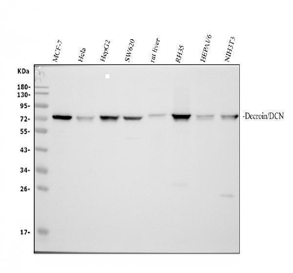
Western blot analysis of Decorin using anti-Decorin antibody (PB9174).
Electrophoresis was performed on a 10% SDS-PAGE gel at 80V (Stacking gel) / 120V (Resolving gel) for 2 hours. The sample well of each lane was loaded with 30 ug of sample under reducing conditions.
Lane 1: human MCF-7 whole cell lysates,
Lane 2: human Hela whole cell lysates,
Lane 3: human HepG2 whole cell lysates,
Lane 4: human SW620 whole cell lysates,
Lane 5: rat liver tissue lysates,
Lane 6: rat RH35 whole cell lysates,
Lane 7: mouse HEPA1-6 whole cell lysates,
Lane 8: mouse NIH/3T3 whole cell lysates.
After electrophoresis, proteins were transferred to a nitrocellulose membrane at 150 mA for 50-90 minutes. Blocked the membrane with 5% non-fat milk/TBS for 1.5 hour at RT. The membrane was incubated with rabbit anti-Decorin antigen affinity purified polyclonal antibody (PB9174) at 0.5 μg/mL overnight at 4°C, then washed with TBS-0.1%Tween 3 times with 5 minutes each and probed with a goat anti-rabbit IgG-HRP secondary antibody at a dilution of 1:5000 for 1.5 hour at RT. The signal is developed using an ECL Plus Western Blotting Substrate (Catalog # AR1196-200) with Tanon 5200 system. A specific band was detected for Decorin at approximately 70 kDa. The expected band size for Decorin is at 40 kDa.

Click image to see more details
IHC analysis of Decorin using anti-Decorin antibody (PB9174).
Decorin was detected in a paraffin-embedded section of human placenta tissue. Heat mediated antigen retrieval was performed in EDTA buffer (pH 8.0, epitope retrieval solution). The tissue section was blocked with 10% goat serum. The tissue section was then incubated with 2 μg/ml rabbit anti-Decorin Antibody (PB9174) overnight at 4°C. Peroxidase Conjugated Goat Anti-rabbit IgG was used as secondary antibody and incubated for 30 minutes at 37°C. The tissue section was developed using HRP Conjugated Rabbit IgG Super Vision Assay Kit (Catalog # SV0002) with DAB as the chromogen.

Click image to see more details
The AKT inhibitor MK2206 abolished the effects induced by overexpression of decorin. ( A ) The tube formation test; the photographs were taken at a magnification of 100×. ( B ) The cell wound healing test; the photographs were taken at a magnification of 100×. ( C ) The apoptosis assay. ( D ) CCK8 assessment. ( E , F ) The expression of VEGF, Bcl2, and Bax and the phosphorylation of AKT and AP 1. All data are presented as the mean ± SEM. *p < 0.05; **p < 0.01. # p < 0.05, ## p < 0.01, compared to Con. & p < 0.05, && p < 0.01, compared to HG + GFP (HG). $ p < 0.05, $$ p < 0.01, compared to HG + DCN.
Index in PubMed under a CC BY license. PMID: 28290552

Click image to see more details
The IGF1R antibody (αIGF1R) blocked the effects induced by overexpression of decorin. ( A , B ) The tube formation test; the photographs were taken at a magnification of 100×. ( C , D ) The cell wound healing test; the photographs were taken at a magnification of 100×. ( E , F ) The apoptosis assay. ( G ) CCK8 assessment. ( H , I ) The expression of VEGF, Bcl2, and Bax and the phosphorylation of AKT and AP 1. All data are presented as the mean ± SEM. *p < 0.05; **p < 0.01. # p < 0.05, ## p < 0.01, compared to Con. & p < 0.05, && p < 0.01, compared to HG + GFP. $ p < 0.05, $$ p < 0.01, compared to HG + DCN.
Index in PubMed under a CC BY license. PMID: 28290552

Click image to see more details
Overexpression of decorin activated the IGF1R-AKT-AP-1 pathway. ( A , B ) The phosphorylation of AKT was decreased by HG treatment in a time-dependent manner. ( C – E ) The expression of IGF1R, Bcl2, and Bax and the phosphorylation of AKT. ( F , G ) The phosphorylation of AP-1. All data are presented as the mean ± SEM. *p < 0.05; **p < 0.01. # p < 0.05, compared to Con. & p < 0.05, && p < 0.01, compared to HG + GFP.
Index in PubMed under a CC BY license. PMID: 28290552

Click image to see more details
Overexpression of decorin ameliorated the angiogenesis impaired by high glucose (HG). ( A – C ) The expression of decorin and VEGF. ( D , E ) The tube formation test; the photographs were taken at a magnification of 100×. ( F , G ) The cell wound healing test; the photographs were taken in a magnification of 100×. ( H , I ) The apoptosis rate analyzed by an Annexin V–FITC kit through flow cytometry. ( J ) The proliferative ability assessed with a CCK8 assay. All data are presented as the mean ± SEM. *p < 0.05; **p < 0.01.
Index in PubMed under a CC BY license. PMID: 28290552

Click image to see more details
Overexpression of decorin increased angiogenesis in the diabetic hearts. ( A – C ) The expression of decorin and VEGF. ( D ) The VEGF concentration in the plasma. ( E ) The capillary density using immunochemistry with a CD31 antibody. The photographs were taken at a magnification of 100× and zoomed at a magnification of 200×. The arrows show the capillary stained with the CD31 antibody. ( F ) The number of vessels counted from the immunochemistry staining. All data are presented as the mean ± SEM. *p < 0.05; **p < 0.01.
Index in PubMed under a CC BY license. PMID: 28290552

Click image to see more details
Overexpression of decorin improved cardiac function in diabetic cardiomyopathy. ( A ) The oral glucose tolerance test (OGTT). ( B ) The left ventricular ejection fraction (EF) evaluated by echocardiography. ( C ) The fraction shortening (FS) evaluated by echocardiography. ( D , E ) The left ventricular wall thickness (including the interventricular septum (IVS) and the posterior wall (PW)) and the internal dimension of the left ventricle (LVID). ( F – H ) The hemodynamic function was evaluated by the cardiac catheter system, including the left ventricular end-diastolic pressure (LVEDP), as well as the Dp/dt maximum and minimum. All data are presented as the mean ± SEM. *p < 0.05; **p < 0.01.
Index in PubMed under a CC BY license. PMID: 28290552
Specific Publications For Anti-Decorin/DCN Antibody Picoband® (PB9174)
Loading publications
Recommended Resources
Here are featured tools and databases that you might find useful.
- Boster's Pathways Library
- Protein Databases
- Bioscience Research Protocol Resources
- Data Processing & Analysis Software
- Photo Editing Software
- Scientific Literature Resources
- Research Paper Management Tools
- Molecular Biology Software
- Primer Design Tools
- Bioinformatics Tools
- Phylogenetic Tree Analysis
Customer Reviews
Have you used Anti-Decorin/DCN Antibody Picoband®?
Share your experimental results or join a short interview to earn up to $1,000 in product credits or other rewards.
0 Reviews For Anti-Decorin/DCN Antibody Picoband®
Customer Q&As
Have a question?
Find answers in Q&As, reviews.
Can't find your answer?
Submit your question
1 Customer Q&As for Anti-Decorin/DCN Antibody Picoband®
Question
We are currently using anti-Decorin/DCN antibody PB9174 for rat tissue, and we are satisfied with the WB results. The species of reactivity given in the datasheet says human, rat. Is it true that the antibody can work on feline tissues as well?
K. Anderson
Verified customer
Asked: 2019-04-18
Answer
The anti-Decorin/DCN antibody (PB9174) has not been tested for cross reactivity specifically with feline tissues, though there is a good chance of cross reactivity. We have an innovator award program that if you test this antibody and show it works in feline you can get your next antibody for free. Please contact me if I can help you with anything.
Boster Scientific Support
Answered: 2019-04-18
