Click image to see more details
Product Info Summary
| SKU: | A11735 |
|---|---|
| Size: | 0.1 mg |
| Reactive Species: | Human |
| Host: | Rabbit |
| Application: | ELISA, IHC-P, WB |
Customers Who Bought This Also Bought

Product info
Product Name
Anti-DEPDC1B Antibody
SKU/Catalog Number
A11735
Size
0.1 mg
Form
Liquid
Description
Boster Bio Anti-DEPDC1B Antibody (Catalog # A11735). Tested in ELISA, WB, IHC-P applications. This antibody reacts with Human.
Storage & Handling
DEPDC1B antibody can be stored at 4°C for three months and -20°C, stable for up to one year.
Cite This Product
Anti-DEPDC1B Antibody (Boster Biological Technology, Pleasanton CA, USA, Catalog # A11735)
Host
Rabbit
Contents
DEPDC1B antibody is supplied in PBS containing 0.02% sodium azide.
Clonality
Polyclonal
Isotype
IgG
Immunogen
DEPDC1B antibody was raised against a 19 amino acid peptide near the carboxy terminus of human DEPDC1B. The immunogen is located within the last 50 amino acids of DEPDC1B.
Cross-reactivity
DEPDC1B antibody is human specific. At least two isoforms are known to exist; this antibody will only detect the largest isoform. DEPDC1B antibody is predicted to not cross-react with DEPDC1A.
Reactive Species
A11735 is reactive to DEPDC1B in Human
Observed Molecular Weight
68 kDa
Calculated molecular weight
61.8 kDa
Background of DEPDC1B
The DEP domain-containing 1B (DEPDC1B) protein was initially identified as a radiation response biomarker in human lymphoblastoid cell lines (1). Like the related protein DEPDC1A, expression of DEPDC1B correlates with a poor prognosis in cancer patients (2,3). Studies have shown that DEPDC1B levels are upregulated in non-small cell lung cancer (NSCLC) and have an inverse correlation with patient survival. Ectopic expression of DEPDC1B in NSCLC activated Wnt/B-catenin signaling and enhanced cell migration and invasion while silencing DEPDC1B expression suppressed these traits (2).
Antibody Validation
Boster validates all antibodies on WB, IHC, ICC, Immunofluorescence, and ELISA with known positive control and negative samples to ensure specificity and high affinity, including thorough antibody incubations.
Application & Images
Applications
A11735 is guaranteed for ELISA, IHC-P, WB Boster Guarantee
Recommend Dilution
DEPDC1B antibody can be used for detection of DEPDC1B by Western blot at 1 - 2 μg/ml. Antibody can also be used for immunohistochemistry starting at 5 μg/mL.
Antibody validated: Western Blot in human samples and Immunohistochemistry in human samples. All other applications and species not yet tested. Optimal dilutions for each application should be determined by the researcher.
Validation Images & Assay Conditions
Click image to see more details
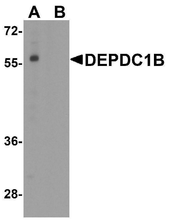
Western blot analysis of DEPDC1B in K562 cell lysate with DEPDC1B antibody at 1 μg/ml in (A) the absence and (B) the presence of blocking peptide.

Click image to see more details
Immunohistochemistry of DEPDC1B in human spleen tissue with DEPDC1B antibody at 5 μg/ml.
Specific Publications For Anti-DEPDC1B Antibody (A11735)
Loading publications
Recommended Resources
Here are featured tools and databases that you might find useful.
- Boster's Pathways Library
- Protein Databases
- Bioscience Research Protocol Resources
- Data Processing & Analysis Software
- Photo Editing Software
- Scientific Literature Resources
- Research Paper Management Tools
- Molecular Biology Software
- Primer Design Tools
- Bioinformatics Tools
- Phylogenetic Tree Analysis
Customer Reviews
Have you used Anti-DEPDC1B Antibody?
Share your experimental results or join a short interview to earn up to $1,000 in product credits or other rewards.
0 Reviews For Anti-DEPDC1B Antibody
Customer Q&As
Have a question?
Find answers in Q&As, reviews.
Can't find your answer?
Submit your question
