Click image to see more details
Product Info Summary
| SKU: | A02249-2 |
|---|---|
| Size: | 100 μg/vial |
| Reactive Species: | Rat |
| Host: | Rabbit |
| Application: | WB |
Customers Who Bought This Also Bought

Product info
Product Name
Anti-DIO2 Antibody Picoband®
SKU/Catalog Number
A02249-2
Size
100 μg/vial
Form
Lyophilized
Description
Boster Bio Anti-DIO2 Antibody Picoband® catalog # A02249-2. Tested in WB applications. This antibody reacts with Rat. The brand Picoband indicates this is a premium antibody that guarantees superior quality, high affinity, and strong signals with minimal background in Western blot applications. Only our best-performing antibodies are designated as Picoband, ensuring unmatched performance.
Storage & Handling
At -20°C for one year from date of receipt. After reconstitution, at 4°C for one month. It can also be aliquotted and stored frozen at -20°C for six months. Avoid repeated freezing and thawing.
Cite This Product
Anti-DIO2 Antibody Picoband® (Boster Biological Technology, Pleasanton CA, USA, Catalog # A02249-2)
Host
Rabbit
Contents
Each vial contains 4 mg Trehalose, 0.9 mg NaCl, 0.2 mg Na2HPO4.
Clonality
Polyclonal
Immunogen
A synthetic peptide corresponding to a sequence at the N-terminus of human DIO2, which shares 95.7% amino acid (aa) sequence identity with both mouse and rat DIO2.
Reactive Species
A02249-2 is reactive to DIO2 in Rat
Calculated molecular weight
30.6 kDa
Background of DIO2
The protein encoded by this gene belongs to the iodothyronine deiodinase family. It catalyzes the conversion of prohormone thyroxine (3,5,3',5'-tetraiodothyronine, T4) to the bioactive thyroid hormone (3,5,3'-triiodothyronine, T3) by outer ring 5'-deiodination. This gene is widely expressed, including in thyroid and brain. It is thought to be responsible for the 'local' production of T3, and thus important in influencing thyroid hormone action in these tissues. It has also been reported to be highly expressed in thyroids of patients with Graves disease, and in follicular adenomas. The intrathyroidal T4 to T3 conversion by this enzyme may contribute significantly to the relative increase in thyroidal T3 production in these patients. This protein is a selenoprotein containing the non-standard amino acid, selenocysteine (Sec), which is encoded by the UGA codon that normally signals translation termination. The 3' UTRs of selenoprotein mRNAs contain a conserved stem-loop structure, designated the Sec insertion sequence (SECIS) element, that is necessary for the recognition of UGA as a Sec codon, rather than as a stop signal. Unlike the other two members (DIO1 and DIO3) of this enzyme family, the mRNA for this gene contains an additional in-frame UGA codon that has been reported (in human) to function either as a Sec or a stop codon, which can result in two isoforms with one or two Sec residues; however, only the upstream Sec (conserved with the single Sec residue found at the active site in DIO1 and DIO3) was shown to be essential for enzyme activity (PMID:10403186). Alternatively spliced transcript variants have been described for this gene.
Antibody Validation
Boster validates all antibodies on WB, IHC, ICC, Immunofluorescence, and ELISA with known positive control and negative samples to ensure specificity and high affinity, including thorough antibody incubations.
Application & Images
Applications
A02249-2 is guaranteed for WB Boster Guarantee
Assay Dilutions Recommendation
The recommendations below provide a starting point for assay optimization. The actual working concentration varies and should be decided by the user.
Western blot, 0.25-0.5 μg/ml, Rat
Validation Images & Assay Conditions
Click image to see more details
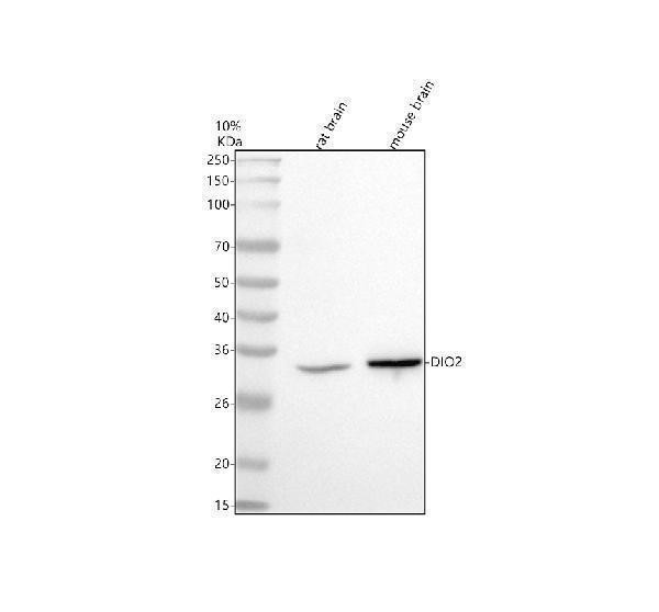
Western blot analysis of DIO2 using anti-DIO2 antibody (A02249-2).
Electrophoresis was performed on a 10% SDS-PAGE gel at 80V (Stacking gel) / 120V (Resolving gel) for 2 hours. The sample well of each lane was loaded with 30 ug of sample under reducing conditions.
Lane 1: rat brain tissue lysates,
Lane 2: mouse brain tissue lysates.
After electrophoresis, proteins were transferred to a nitrocellulose membrane at 150 mA for 50-90 minutes. Blocked the membrane with 5% non-fat milk/TBS for 1.5 hour at RT. The membrane was incubated with rabbit anti-DIO2 antigen affinity purified polyclonal antibody (A02249-2) at 0.5 μg/mL overnight at 4°C, then washed with TBS-0.1%Tween 3 times with 5 minutes each and probed with a goat anti-rabbit IgG-HRP secondary antibody at a dilution of 1:5000 for 1.5 hour at RT. The signal is developed using an ECL Plus Western Blotting Substrate (Catalog # AR1196-200) with Tanon 5200 system. A specific band was detected for DIO2 at approximately 32 kDa. The expected band size for DIO2 is at 31 kDa.
Specific Publications For Anti-DIO2 Antibody Picoband® (A02249-2)
Loading publications
Recommended Resources
Here are featured tools and databases that you might find useful.
- Boster's Pathways Library
- Protein Databases
- Bioscience Research Protocol Resources
- Data Processing & Analysis Software
- Photo Editing Software
- Scientific Literature Resources
- Research Paper Management Tools
- Molecular Biology Software
- Primer Design Tools
- Bioinformatics Tools
- Phylogenetic Tree Analysis
Customer Reviews
Have you used Anti-DIO2 Antibody Picoband®?
Share your experimental results or join a short interview to earn up to $1,000 in product credits or other rewards.
0 Reviews For Anti-DIO2 Antibody Picoband®
Customer Q&As
Have a question?
Find answers in Q&As, reviews.
Can't find your answer?
Submit your question
