Click image to see more details
-

-
-
-
-
+1
Product Info Summary
| SKU: | A03227-3 |
|---|---|
| Size: | 100 μg/vial |
| Reactive Species: | Mouse, Rat |
| Host: | Rabbit |
| Application: | ELISA, Flow Cytometry, WB |
Customers Who Bought This Also Bought

Product info
Product Name
Anti-DR3/Tnfrsf25 Antibody Picoband®
SKU/Catalog Number
A03227-3
Size
100 μg/vial
Form
Lyophilized
Description
Boster Bio Anti-DR3/Tnfrsf25 Antibody Picoband® catalog # A03227-3. Tested in ELISA, Flow Cytometry, WB applications. This antibody reacts with Mouse, Rat. The brand Picoband indicates this is a premium antibody that guarantees superior quality, high affinity, and strong signals with minimal background in Western blot applications. Only our best-performing antibodies are designated as Picoband, ensuring unmatched performance.
Storage & Handling
Store at -20˚C for one year from date of receipt. After reconstitution, at 4˚C for one month. It can also be aliquotted and stored frozen at -20˚C for six months. Avoid repeated freeze-thaw cycles.
Cite This Product
Anti-DR3/Tnfrsf25 Antibody Picoband® (Boster Biological Technology, Pleasanton CA, USA, Catalog # A03227-3)
Host
Rabbit
Contents
Each vial contains 4mg Trehalose, 0.9mg NaCl, 0.2mg Na2HPO4, 0.01mg NaN3.
Clonality
Polyclonal
Isotype
Rabbit IgG
Immunogen
E.coli-derived rat DR3/Tnfrsf25 recombinant protein (Position: E43-E393).
Cross-reactivity
No cross-reactivity with other proteins.
Reactive Species
A03227-3 is reactive to Tnfrsf25 in Mouse, Rat
Observed Molecular Weight
60 kDa
Background of Tnfrsf25
TNFRSF25 (Tumor Necrosis Factor Receptor Superfamily Member 25), also known as LARD, APO3, DR3 or TNFR25, is a protein that in humans is encoded by the TNFRSF25 gene. Members of the mammalian tumor necrosis factor receptor (TNFR) family are cell-surface proteins that interact with a corresponding TNF-related ligand family. By fluorescence in situ hybridization, Marsters et al. (1996) mapped the Apo3 gene to 1p36.3. Marsters et al. (1996) showed that ectopic expression of Apo3 in mammalian cells triggered apoptosis and activated the transcription factor NF-kappa-B. They suggested that, like TNFR1, Apo3 may regulate distinct signaling pathways in different cellular contexts.
Antibody Validation
Boster validates all antibodies on WB, IHC, ICC, Immunofluorescence, and ELISA with known positive control and negative samples to ensure specificity and high affinity, including thorough antibody incubations.
Application & Images
Applications
A03227-3 is guaranteed for ELISA, Flow Cytometry, WB Boster Guarantee
Recommend Dilution
| Application | Dilution | Species |
|---|---|---|
| Western blot | 0.25-0.5μg/ml | Mouse, Rat |
| Flow Cytometry (Fixed) | 1-3μg/1x106 cells | Rat |
| ELISA | 0.1-0.5μg/ml |
Tested application
Suggested blocking solution with 5% non-fat milk or BSA; (*)Recommended protein loading: 20-40 µg per lane
Validation Images & Assay Conditions
Click image to see more details
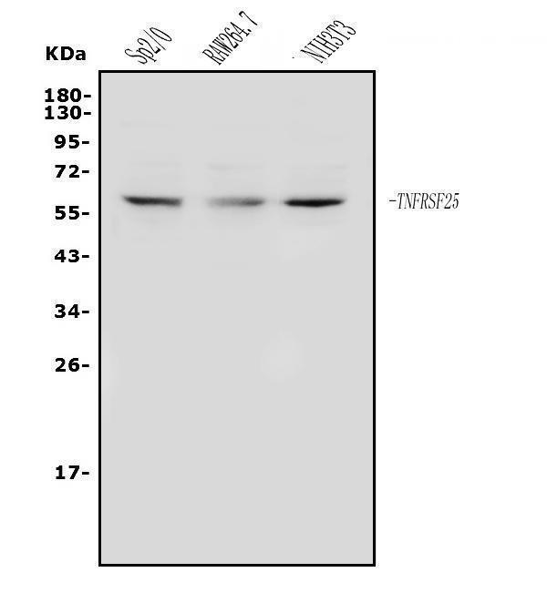
Western blot analysis of DR3/Tnfrsf25 using anti-DR3/Tnfrsf25 antibody (A03227-3).
Electrophoresis was performed on a 5-20% SDS-PAGE gel at 70V (Stacking gel) / 90V (Resolving gel) for 2-3 hours. The sample well of each lane was loaded with 50ug of sample under reducing conditions.
Lane 1: mouse SP2/0 whole cell lysates,
Lane 2: mouse RAW264.7 whole cell lysates,
Lane 3: mouse NIH/3T3 whole cell lysates.
After Electrophoresis, proteins were transferred to a Nitrocellulose membrane at 150mA for 50-90 minutes. Blocked the membrane with 5% Non-fat Milk/ TBS for 1.5 hour at RT. The membrane was incubated with rabbit anti-DR3/Tnfrsf25 antigen affinity purified polyclonal antibody (Catalog # A03227-3) at 0.5 μg/mL overnight at 4°C, then washed with TBS-0.1%Tween 3 times with 5 minutes each and probed with a goat anti-rabbit IgG-HRP secondary antibody at a dilution of 1:5000 for 1.5 hour at RT. The signal is developed using an Enhanced Chemiluminescent detection (ECL) kit (Catalog # EK1002) with Tanon 5200 system. A specific band was detected for DR3/Tnfrsf25 at approximately 60KD. The expected band size for DR3/Tnfrsf25 is at 60KD.

Click image to see more details
Methylating effect of daphnetin and 5-aza-dc on DR3, PDCD5, FasL and p53 in CIA rat synovial cells. Four experimental groups consisting of untreated cell control, or treated with 5-aza-dc at 20 μM, daphnetin at 40 μg/mL, or combination of 5-aza-dc (20 μM) and daphnetin (40 μg/mL). Cells were cultured, treated and harvested as described in Materials and Methods. Total DNA was extracted and used for MSP after bisulphite modification. M: methylated amplification products, U: hypomethylated amplification products.
Index in PubMed under a CC BY license. PMID: 25311560

Click image to see more details
Effect of daphnetin and 5-aza-dc on expression of DR3, PDCD5, FasL, p53 (A) and DNMT1, DNMT3a, DNMT3b (B) in CIA rat synovial cells. Four experimental groups consisting of untreated cell control, or treated with 5-aza-dc at 20 μM, daphnetin at 40 μg/mL, or combination of 5-aza-dc (20 μM) and daphnetin (40 μg/mL). Cells were cultured, treated and harvested as described in Matreials and Methods. Total RNA was extracted and cDNA was synthesized. After reverse transcription, cDNA was used for real time-PCR. Relative quantification of gene expression was performed by the 2-ΔΔCt method. The results show the mean ± S.D. of six independent experiments. ▲P < 0.05 compared with control, ●P < 0.05 compared with 5-aza-dc, ☆P > 0.05 compared with 5-aza-dc, ★P > 0.05 compared with daphnetin, *P < 0.05 compared with daphnetin.
Index in PubMed under a CC BY license. PMID: 25311560

Click image to see more details
Effect of daphnetin on DR3, PDCD5, FasL and p53 protein expression in CIA rats synovial cells. Cells were cultured, treated and harvested as described Materials and Methods. Four experimental groups consisting of untreated cell control, 5-aza-dc at 20 μM, daphnetin at 40 μg/mL, or combination of 5-aza-dc (20 μM) and daphnetin (40 μg/mL). The results show the mean ± S.D. of six independent experiments. ▲P < 0.05 compared with control, ●P < 0.05 compared with 5-aza-dc, ★P > 0.05 compared with 5-aza-dc, *P < 0.05 compared with daphnetin.
Index in PubMed under a CC BY license. PMID: 25311560

Click image to see more details
Flow Cytometry analysis of NRK cells using anti-DR3/Tnfrsf25 antibody (A03227-3).
Overlay histogram showing NRK cells stained with A03227-3 (Blue line). The cells were fixed with 4% paraformaldehyde and blocked with 10% normal goat serum. And then incubated with rabbit anti-DR3/Tnfrsf25 Antibody (A03227-3, 1μg/1x106 cells) for 30 min at 20°C. DyLight®488 conjugated goat anti-rabbit IgG (BA1127, 5-10μg/1x106 cells) was used as secondary antibody for 30 minutes at 20°C. Isotype control antibody (Green line) was rabbit IgG (1μg/1x106) used under the same conditions. Unlabelled sample without incubation with primary antibody and secondary antibody (Red line) was used as a blank control.
Specific Publications For Anti-DR3/Tnfrsf25 Antibody Picoband® (A03227-3)
Loading publications
Recommended Resources
Here are featured tools and databases that you might find useful.
- Boster's Pathways Library
- Protein Databases
- Bioscience Research Protocol Resources
- Data Processing & Analysis Software
- Photo Editing Software
- Scientific Literature Resources
- Research Paper Management Tools
- Molecular Biology Software
- Primer Design Tools
- Bioinformatics Tools
- Phylogenetic Tree Analysis
Customer Reviews
Have you used Anti-DR3/Tnfrsf25 Antibody Picoband®?
Share your experimental results or join a short interview to earn up to $1,000 in product credits or other rewards.
0 Reviews For Anti-DR3/Tnfrsf25 Antibody Picoband®
Customer Q&As
Have a question?
Find answers in Q&As, reviews.
Can't find your answer?
Submit your question
