Click image to see more details
-

-
-
-
-
+12
Product Info Summary
| SKU: | M00023-2 |
|---|---|
| Size: | 100 μl |
| Reactive Species: | Human, Mouse, Rat |
| Host: | Rabbit |
| Application: | Flow Cytometry, IP, IF, IHC, ICC, WB |
Customers Who Bought This Also Bought

Product info
Product Name
Anti-EGFR (ErbB 1) Monoclonal Antibody
SKU/Catalog Number
M00023-2
BM4009 is an alternative SKU for this antibody, used in previous lots.
Size
100 μl
Form
Liquid
Description
Boster Bio Anti-EGFR (ErbB 1) Monoclonal Antibody catalog # M00023-2. Tested in WB, IHC, ICC/IF, IP, Flow Cytometry applications. This antibody reacts with Human, Mouse, Rat.
Storage & Handling
Store at -20°C for one year. For short term storage and frequent use, store at 4°C for up to one month. Avoid repeated freeze-thaw cycles.
Cite This Product
Anti-EGFR (ErbB 1) Monoclonal Antibody (Boster Biological Technology, Pleasanton CA, USA, Catalog # M00023-2)
Host
Rabbit
Contents
Rabbit IgG in stabilizing components, phosphate buffered saline, pH 7.4, 150mM NaCl, 0.02% sodium azide and 50% glycerol.
*This antibody is supplied in a stabilized formulation.
Compatibility with conjugation reactions depends on the chemistry of the conjugation method used.
For conjugation methods that are not compatible with the stabilizing components present in this formulation, a carrier-free antibody format is required.
Clonality
Monoclonal
Clone Number
AEF-5
Isotype
Rabbit IgG
Immunogen
A synthesized peptide derived from human EGFR (ErbB 1)
Reactive Species
M00023-2 is reactive to EGFR in Human, Mouse, Rat
Observed Molecular Weight
175 kDa
Calculated molecular weight
134.3 kDa
Antibody Validation
Boster validates all antibodies on WB, IHC, ICC, Immunofluorescence, and ELISA with known positive control and negative samples to ensure specificity and high affinity, including thorough antibody incubations.
Application & Images
Applications
M00023-2 is guaranteed for Flow Cytometry, IP, IF, IHC, ICC, WB Boster Guarantee
Recommend Dilution
WB 1:1000-5000
IHC 1:50-200
ICC/IF 1:50-200
IP 1:20
FC 1:20
Tested application
Suggested blocking solution with 5% non-fat milk or BSA; (*)Recommended protein loading: 20-40 µg per lane
Use TE buffer pH 9.0 for antigen retrieval; (*) citrate buffer pH 6.0 is an alternative.
Validation Images & Assay Conditions
Click image to see more details
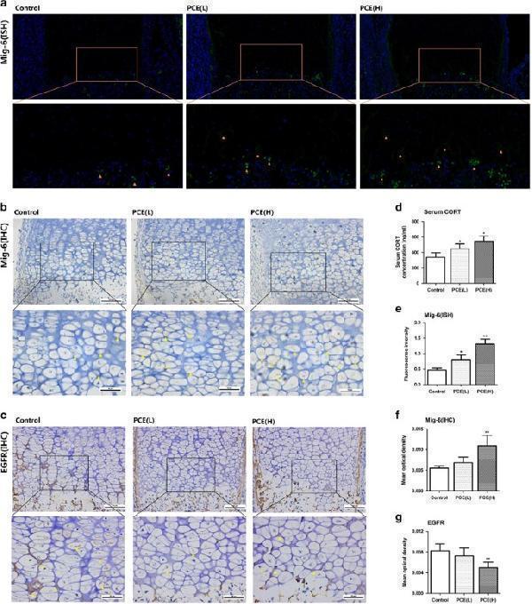
Effects of PCE on the expression changes of Mig-6 and EGFR in fetal long-bone hypertrophic chondrocytes. ( a ) ISH of Mig-6 in hypertrophic chondrocytes. ( b ) Immunostaining of Mig-6 in hypertrophic chondrocytes. ( c ) Immunostaining of EGFR in hypertrophic chondrocytes. ( d ) Serum corticosterone (CORT) concentration of fetal rats (ng/ml). ( e ) Quantification of Mig-6 ISH (fluorescence intensity). ( f ) Quantification of Mig-6 immunostaining (optical density). ( g ) Quantification of EGFR immunostaining (optical density). n =5 per group obtained from different litters. Three random fields/section for quantitative. Data are shown as the mean±S.D. * P <0.05, ** P <0.01 versus control (ANOVA)
Index in PubMed under a CC BY license. PMID: 29072695

Click image to see more details
Effects of corticosterone (250–1250 nM) with/without siRNA (Mig-6, EGFR) for 48 h on rats primary chondrocytes terminal differentiation and apoptosis. ( a and b ) mRNA expression of mitogen-inducible gene 6 (Mig-6) and EGFR after corticosterone and Mig-6 siRNA treatment, ( c ) Protein expression of Mig-6, EGFR, phosphorylated EGFR (P-EGFR), c-Jun N-terminal kinase (JNK) and Phosphorylated JNK (P-JNK) detected by western blotting after corticosterone and Mig-6 siRNA treatment. ( d – h ) Quantification of Mig-6, EGFR, P-EGFR, JNK and P-JNK (relative grayscale). ( i – k ) mRNA expression of runt-related transcription factor 2 (Runx2), collagen type X (Col-X) and matrix metalloproteinases-13 (MMP-13) after corticosterone and Mig-6 siRNA treatment. ( l and m ) Apoptotic analysis detected by Annexin V/PI after corticosterone and Mig-6 siRNA treatment. ( n ) Protein expression of EGFR, JNK and P-JNK detected by western blotting after EGFR siRNA treatment. ( o ) Quantification of Mig-6, EGFR, P-EGFR, JNK and P-JNK (Relative grayscale). ( p ) mRNA expression of Runx2, Col-X and MMP-13 after EGFR siRNA treatment. ( q ) Apoptotic analysis detected by Annexin V/PI after EGFR siRNA treatment. Data are shown as the mean±S.D. of results from three experiments. * P <0.05, ** P <0.01 versus control; # P <0.05, ## P <0.01 versus CORT treatment group. (A-M, ANOVA; O-R, t test)
Index in PubMed under a CC BY license. PMID: 29072695

Click image to see more details
The proposed schematic model of the present study. Col-X, collagen type X; EGFR, epidermal growth factor receptor; JNK, c-Jun N-terminal kinase; Mig-6, mitogen-inducible gene 6; MMP-13, matrix metallopeptidase 13; Runx2, runt-related transcription factor 2
Index in PubMed under a CC BY license. PMID: 29072695

Click image to see more details
Western blot analysis of EGFR using anti-EGFR antibody (M00023-2).
Electrophoresis was performed on a 8% SDS-PAGE gel at 80V (Stacking gel) / 120V (Resolving gel) for 2 hours. The sample well of each lane was loaded with 30 ug of sample under reducing conditions.
Lane 1: human A549- WT whole cell lysates,
Lane 2: human A549-EGFR KO whole cell lysates.
After electrophoresis, proteins were transferred to a nitrocellulose membrane at 150 mA for 50-90 minutes. Blocked the membrane with 5% non-fat milk/TBS for 1.5 hour at RT. The membrane was incubated with rabbit anti-EGFR antigen affinity purified monoclonal antibody (M00023-2) at 0.5 μg/mL overnight at 4°C, then washed with TBS-0.1%Tween 3 times with 5 minutes each and probed with a goat anti-rabbit IgG-HRP secondary antibody at a dilution of 1:5000 for 1.5 hour at RT. The signal is developed using an ECL Plus Western Blotting Substrate (Catalog # AR1196-200) with Tanon 5200 system. A specific band was detected for EGFR at approximately 175 kDa. The expected band size for EGFR is at 134 kDa.

Click image to see more details
Western blot analysis of EGFR using anti-EGFR antibody (M00023-2).
Electrophoresis was performed on a 5-20% SDS-PAGE gel at 70V (Stacking gel) / 90V (Resolving gel) for 2-3 hours. The sample well of each lane was loaded with 30 ug of sample under reducing conditions.
Lane 1: human Hela whole cell lysates,
Lane 2: human A431 whole cell lysates,
Lane 3: human A549 whole cell lysates,
Lane 4: human U-87MG whole cell lysates,
Lane 5: rat liver tissue lysates,
Lane 6: mouse liver tissue lysates.
After electrophoresis, proteins were transferred to a nitrocellulose membrane at 150 mA for 50-90 minutes. Blocked the membrane with 5% non-fat milk/TBS for 1.5 hour at RT. The membrane was incubated with rabbit anti-EGFR antigen affinity purified monoclonal antibody (Catalog # M00023-2) at 1:5000 overnight at 4°C, then washed with TBS-0.1%Tween 3 times with 5 minutes each and probed with a goat anti-rabbit IgG-HRP secondary antibody at a dilution of 1:500 for 1.5 hour at RT. The signal is developed using an Enhanced Chemiluminescent detection (ECL) kit (Catalog # EK1002) with Tanon 5200 system. A specific band was detected for EGFR at approximately 175 kDa. The expected band size for EGFR is at 134 kDa.

Click image to see more details
Immunohistochemical analysis of paraffin-embedded Rat cerebral cortex, using the Antibody at 1:100 dilution.

Click image to see more details
Immunohistochemical analysis of paraffin-embedded Rat stomach, using the Antibody at 1:100 dilution.

Click image to see more details
Immunohistochemical analysis of paraffin-embedded Human thyroid cancer, using the Antibody at 1:100 dilution.

Click image to see more details
Immunohistochemical analysis of paraffin-embedded Human glioblastoma, using the Antibody at 1:100 dilution.

Click image to see more details
Immunohistochemical analysis of paraffin-embedded Mouse intestine, using the Antibody at 1:100 dilution.

Click image to see more details
Immunohistochemical analysis of paraffin-embedded human stomach cancer, using EGFR (ErbB 1) Antibody.

Click image to see more details
Immunofluorescent analysis using the Antibody at 1:50 dilution.

Click image to see more details
Immunofluorescent analysis using the Antibody at 1:50 dilution.

Click image to see more details
Immunofluorescent analysis using the Antibody at 1:150 dilution.

Click image to see more details
Immunofluorescent analysis using the Antibody at 1:500 dilution.

Click image to see more details
Western blot analysis of EGFR using anti-EGFR antibody (M00023-2).
Electrophoresis was performed on a 5-20% SDS-PAGE gel at 70V (Stacking gel) / 90V (Resolving gel) for 2-3 hours. The sample well of each lane was loaded with 30 ug of sample under reducing conditions.
Lane 1-2: human Hela whole cell lysates.
After electrophoresis, proteins were transferred to a nitrocellulose membrane at 150 mA for 50-90 minutes. Blocked the membrane with 5% non-fat milk/TBS for 1.5 hour at RT. The membrane was incubated with rabbit anti-EGFR antigen affinity purified monoclonal antibody (Catalog # M00023-2) at 1:1000 overnight at 4°C, then washed with TBS-0.1%Tween 3 times with 5 minutes each and probed with a goat anti-rabbit IgG-HRP secondary antibody at a dilution of 1:10000 for 1 hour at RT. The signal is developed using an Enhanced Chemiluminescent detection (ECL) kit (Catalog # EK1002) with ChemiDoc MP system. A specific band was detected for EGFR at approximately 175 kDa. The expected band size for EGFR is at 134 kDa.
Specific Publications For Anti-EGFR (ErbB 1) Monoclonal Antibody (M00023-2)
Loading publications
Recommended Resources
Here are featured tools and databases that you might find useful.
- Boster's Pathways Library
- Protein Databases
- Bioscience Research Protocol Resources
- Data Processing & Analysis Software
- Photo Editing Software
- Scientific Literature Resources
- Research Paper Management Tools
- Molecular Biology Software
- Primer Design Tools
- Bioinformatics Tools
- Phylogenetic Tree Analysis
Customer Reviews
Have you used Anti-EGFR (ErbB 1) Monoclonal Antibody?
Share your experimental results or join a short interview to earn up to $1,000 in product credits or other rewards.
1 Reviews For Anti-EGFR (ErbB 1) Monoclonal Antibody
WB analysis using Anti-EGFR antibody (M00023-2) in mouse hippocampal tissue showed a clear band at the expected molecular weight with low background, demonstrating good specificity and reliable performance.
Excellent

| SKU | M00023-2 |
|---|---|
| Application | Western Blot |
| Sample | mouse hippocampal tissue |
| Sample Processing Description | The left hippocampus was dissected from normal mouse brains, and total protein was extracted. |
| Other Reagents | RIPA lysis buffer, Protease inhibitor, Electrophoresis buffer, Transfer buffer, Blocking buffer |
| Primary Antibody | EGFR (ErbB 1) Monoclonal Antibody |
| Primary Incubation | 1:1000, overnight at 4 ℃ |
| Secondary Antibody | HRP-conjugated goat anti-rabbit IgG |
| Secondary Incubation | 1:10000, 1 hour in RT |
| Detection | Substrate: ECL substrate; Image system: ChemiDoc MP |
| Results Summary | EGFR protein is a key molecular switch located on the cell membrane that receives extracellular signals and drives cell proliferation and differentiation through kinase activity. It serves as a central regulator of organ development, tissue homeostasis, and regenerative repair. In this study, hippocampal tissues from two normal mouse brains were used to evaluate the performance of the BDNF antibody. The results showed a clear and correctly positioned target band, indicating that the antibody is functional and works properly. |
Yajun Qiao, Qinghai University
Verified customer
Submitted 2026-05-08
Customer Q&As
Have a question?
Find answers in Q&As, reviews.
Can't find your answer?
Submit your question
