Click image to see more details
-

-
-
-
-
+2
Product Info Summary
| SKU: | PB10059 |
|---|---|
| Size: | 100 μg/vial |
| Reactive Species: | Human |
| Host: | Rabbit |
| Application: | ELISA, Flow Cytometry, IF, IHC, WB |
Customers Who Bought This Also Bought

Product info
Product Name
Anti-EpCAM Antibody Picoband®
SKU/Catalog Number
PB10059
Size
100 μg/vial
Form
Lyophilized
Description
Boster Bio Anti-EpCAM Antibody Picoband® catalog # PB10059. Tested in ELISA, Flow Cytometry, IF, IHC, WB applications. This antibody reacts with Human. The brand Picoband indicates this is a premium antibody that guarantees superior quality, high affinity, and strong signals with minimal background in Western blot applications. Only our best-performing antibodies are designated as Picoband, ensuring unmatched performance.
Storage & Handling
Store at -20˚C for one year from date of receipt. After reconstitution, at 4˚C for one month. It can also be aliquotted and stored frozen at -20˚C for six months. Avoid repeated freeze-thaw cycles.
Cite This Product
Anti-EpCAM Antibody Picoband® (Boster Biological Technology, Pleasanton CA, USA, Catalog # PB10059)
Host
Rabbit
Contents
Each vial contains 4 mg Trehalose, 0.9 mg NaCl and 0.2 mg Na2HPO4.
Clonality
Polyclonal
Isotype
Rabbit IgG
Immunogen
A synthetic peptide corresponding to a sequence in the middle region of human EPCAM, different from the related mouse sequence by fifteen amino acids, and from the related rat sequence by sixteen amino acids.
Cross-reactivity
No cross-reactivity with other proteins
Reactive Species
PB10059 is reactive to EPCAM in Human
Observed Molecular Weight
35-40 kDa
Calculated molecular weight
34.9 kDa
Background of EPCAM
Epithelial cell adhesion molecule (EpCAM) is a transmembrane glycoprotein mediating Ca2+-independent homotypic cell–cell adhesion in epithelia. This gene encodes a carcinoma-associated antigen and is a member of a family that includes at least two type I membrane proteins. This antigen is expressed on most normal epithelial cells and gastrointestinal carcinomas and functions as a homotypic calcium-independent cell adhesion molecule. The antigen is being used as a target for immunotherapy treatment of human carcinomas. Mutations in this gene result in congenital tufting enteropathy.
Antibody Validation
Boster validates all antibodies on WB, IHC, ICC, Immunofluorescence, and ELISA with known positive control and negative samples to ensure specificity and high affinity, including thorough antibody incubations.
Application & Images
Applications
PB10059 is guaranteed for ELISA, Flow Cytometry, IF, IHC, WB Boster Guarantee
Recommend Dilution
| Application | Dilution | Species |
|---|---|---|
| Western blot | 0.1-0.5μg/ml | |
| Immunohistochemistry (Paraffin-embedded Section) | 2-5μg/ml | |
| Immunofluorescence | 5μg/ml | |
| Flow Cytometry (Fixed) | 1-3μg/1x106cells | |
| ELISA | 0.1-0.5μg/ml |
Tested application
Suggested blocking solution with 5% non-fat milk or BSA; (*)Recommended protein loading: 20-40 µg per lane
Use TE buffer pH 9.0 for antigen retrieval; (*) citrate buffer pH 6.0 is an alternative.
Validation Images & Assay Conditions
Click image to see more details
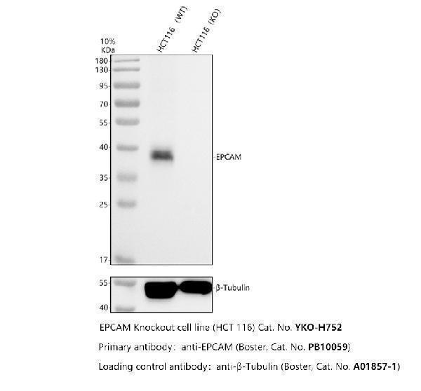
Western blot analysis of CD326/EPCAM using anti-CD326/EPCAM antibody (PB10059).
Electrophoresis was performed on a 10% SDS-PAGE gel at 80V (Stacking gel) / 120V (Resolving gel) for 2 hours. The sample well of each lane was loaded with 30 ug of sample under reducing conditions.
Lane 1: human HCT116- WT whole cell lysates,
Lane 2: human HCT116-EPCAM KO whole cell lysates.
After electrophoresis, proteins were transferred to a nitrocellulose membrane at 150 mA for 50-90 minutes. Blocked the membrane with 5% non-fat milk/TBS for 1.5 hour at RT. The membrane was incubated with rabbit anti-CD326/EPCAM antigen affinity purified polyclonal antibody (PB10059) at 0.5 μg/mL overnight at 4°C, then washed with TBS-0.1%Tween 3 times with 5 minutes each and probed with a goat anti-rabbit IgG-HRP secondary antibody at a dilution of 1:5000 for 1.5 hour at RT. The signal is developed using an ECL Plus Western Blotting Substrate (Catalog # AR1196-200) with Tanon 5200 system. A specific band was detected for CD326/EPCAM at approximately 35-40 kDa. The expected band size for CD326/EPCAM is at 35 kDa.

Click image to see more details
Western blot analysis of EPCAM using anti-EPCAM antibody (PB10059).
Electrophoresis was performed on a 5-20% SDS-PAGE gel at 70V (Stacking gel) / 90V (Resolving gel) for 2-3 hours. The sample well of each lane was loaded with 30 ug of sample under reducing conditions.
Lane 1: human CACO-2 whole cell lysates,
Lane 2: human MCF-7 whole cell lysates.
After electrophoresis, proteins were transferred to a nitrocellulose membrane at 150 mA for 50-90 minutes. Blocked the membrane with 5% non-fat milk/TBS for 1.5 hour at RT. The membrane was incubated with rabbit anti-EPCAM antigen affinity purified polyclonal antibody (Catalog # PB10059) at 0.5 μg/mL overnight at 4°C, then washed with TBS-0.1%Tween 3 times with 5 minutes each and probed with a goat anti-rabbit IgG-HRP secondary antibody at a dilution of 1:5000 for 1.5 hour at RT. The signal is developed using an Enhanced Chemiluminescent detection (ECL) kit (Catalog # EK1002) with Tanon 5200 system. A specific band was detected for EPCAM at approximately 35-40 kDa. The expected band size for EPCAM is at 35 kDa.

Click image to see more details
IHC analysis of EPCAM using anti-EPCAM antibody (PB10059).
EPCAM was detected in a paraffin-embedded section of human colon cancer tissue. Heat mediated antigen retrieval was performed in EDTA buffer (pH 8.0, epitope retrieval solution). The tissue section was blocked with 10% goat serum. The tissue section was then incubated with 2 μg/ml rabbit anti-EPCAM Antibody (PB10059) overnight at 4°C. Peroxidase Conjugated Goat Anti-rabbit IgG was used as secondary antibody and incubated for 30 minutes at 37°C. The tissue section was developed using HRP Conjugated Rabbit IgG Super Vision Assay Kit (Catalog # SV0002) with DAB as the chromogen.

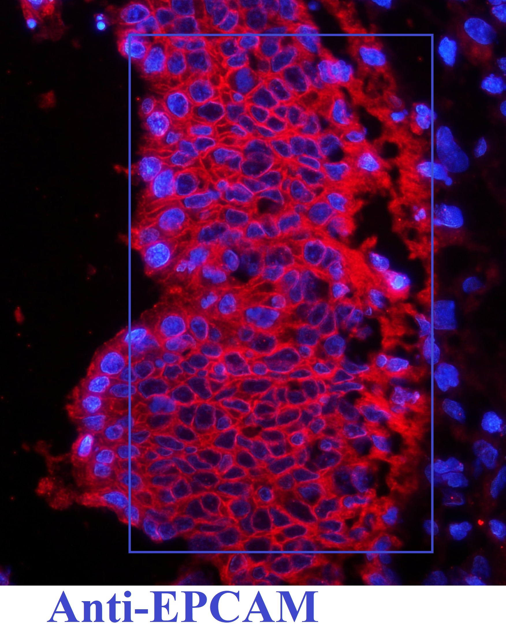
Click image to see more details
IF analysis of EPCAM using anti-EPCAM antibody (PB10059).
EPCAM was detected in a paraffin-embedded section of human rectal cancer tissue. Heat mediated antigen retrieval was performed in EDTA buffer (pH 8.0, epitope retrieval solution). The tissue section was blocked with 10% goat serum. The tissue section was then incubated with 5 μg/mL rabbit anti-EPCAM Antibody (PB10059) overnight at 4°C. DyLight®594 Conjugated Goat Anti-Rabbit IgG (BA1142) was used as secondary antibody at 1:500 dilution and incubated for 30 minutes at 37°C. The section was counterstained with DAPI. Visualize using a fluorescence microscope and filter sets appropriate for the label used.

Click image to see more details
Flow Cytometry analysis of CACO-2 cells using anti-EPCAM antibody (PB10059).
Overlay histogram showing CACO-2 cells stained with PB10059 (Blue line). The cells were fixed with 4% paraformaldehyde and blocked with 10% normal goat serum. And then incubated with rabbit anti-EPCAM Antibody (PB10059, 1 μg/1x106 cells) for 30 min at 20°C. DyLight®488 conjugated goat anti-rabbit IgG (BA1127, 5-10 μg/1x106 cells) was used as secondary antibody for 30 minutes at 20°C. Isotype control antibody (Green line) was rabbit IgG (1 μg/1x106) used under the same conditions. Unlabelled sample without incubation with primary antibody and secondary antibody (Red line) was used as a blank control.
Specific Publications For Anti-EpCAM Antibody Picoband® (PB10059)
Loading publications
Recommended Resources
Here are featured tools and databases that you might find useful.
- Boster's Pathways Library
- Protein Databases
- Bioscience Research Protocol Resources
- Data Processing & Analysis Software
- Photo Editing Software
- Scientific Literature Resources
- Research Paper Management Tools
- Molecular Biology Software
- Primer Design Tools
- Bioinformatics Tools
- Phylogenetic Tree Analysis
Customer Reviews
Have you used Anti-EpCAM Antibody Picoband®?
Share your experimental results or join a short interview to earn up to $1,000 in product credits or other rewards.
1 Reviews For Anti-EpCAM Antibody Picoband®
Immunoflorescence Review for Anti-EpCAM Antibody
Excellent
| SKU | PB10059 |
|---|---|
| Application | Immunofluorescence (IF) |
| Blocking step | 5% BSA as a blocking agent for 30 min at 37°C |
| Sample | Human rectal |
| Fixative | Fixed with 4% paraformaldehyde |
| Primary Ab Incubation | 4°C overnight |
| Primary Ab Incubation diluent | 5% BSA in TBS |
| Primary Ab Concentration | 1ug/ml |
| Secondary Antibody | SABC
kit from Boster Bio, (SA1022 |
| Secondary Ab Dilution | The kit was ready to use, no dilution needed |
| Secondary Ab Incubation | at 37°C for 30 min |
A. Dayal
Verified customer
Submitted 2019-05-25
Customer Q&As
Have a question?
Find answers in Q&As, reviews.
Can't find your answer?
Submit your question
16 Customer Q&As for Anti-EpCAM Antibody Picoband®
Question
Is this PB10059 anti-EpCAM antibody reactive to the isotypes of EPCAM?
Verified Customer
Verified customer
Asked: 2020-03-10
Answer
The immunogen of PB10059 anti-EpCAM antibody is A synthetic peptide corresponding to a sequence in the middle region of human EPCAM (147-189aa ELKHKAREKPYDSKSLRTALQKEITTRYQLDPKFITSILYENN), different from the related mouse sequence by fifteen amino acids, and from the related rat sequence by sixteen ami. Could you tell me which isotype you are interested in so I can help see if the immunogen is part of this isotype?
Boster Scientific Support
Answered: 2020-03-10
Question
I have attached the WB image, lot number and protocol we used for placenta using anti-EpCAM antibody PB10059. Please let me know if you require anything else.
Verified Customer
Verified customer
Asked: 2020-02-28
Answer
Thank you very much for the data. Our lab team are working to resolve this as quickly as possible, and we appreciate your patience and understanding! You have provided everything we needed. Please let me know if there is anything you need in the meantime.
Boster Scientific Support
Answered: 2020-02-28
Question
I was wanting to use your anti-EpCAM antibody for IHC-P for human placenta on frozen tissues, but I want to know if it has been validated for this particular application. Has this antibody been validated and is this antibody a good choice for human placenta identification?
Verified Customer
Verified customer
Asked: 2020-02-21
Answer
It shows on the product datasheet, PB10059 anti-EpCAM antibody has been validated for ELISA, Flow Cytometry, IF, IHC-P, WB on human tissues. We have an innovator award program that if you test this antibody and show it works in human placenta in IHC-frozen, you can get your next antibody for free.
Boster Scientific Support
Answered: 2020-02-21
Question
I see that the anti-EpCAM antibody PB10059 works with IHC-P, what is the protocol used to produce the result images on the product page?
Verified Customer
Verified customer
Asked: 2020-02-17
Answer
You can find protocols for IHC-P on the "support/technical resources" section of our navigation menu. If you have any further questions, please send an email to support@bosterbio.com
Boster Scientific Support
Answered: 2020-02-17
Question
Our lab want to know about to test anti-EpCAM antibody PB10059 on human placenta for research purposes, then I may be interested in using anti-EpCAM antibody PB10059 for diagnostic purposes as well. Is the antibody suitable for diagnostic purposes?
Verified Customer
Verified customer
Asked: 2020-02-13
Answer
The products we sell, including anti-EpCAM antibody PB10059, are only intended for research use. They would not be suitable for use in diagnostic work. If you have the means to develop a product into diagnostic use, and are interested in collaborating with us and develop our product into an IVD product, please contact us for more discussions.
Boster Scientific Support
Answered: 2020-02-13
Question
Is there a BSA free version of anti-EpCAM antibody PB10059 available?
Verified Customer
Verified customer
Asked: 2020-01-07
Answer
Thanks for your recent telephone inquiry. I can confirm that some lots of this anti-EpCAM antibody PB10059 are BSA free. For now, these lots are available and we can make a BSA free formula for you free of charge. It will take 3 extra days to prepare. If you require this antibody BSA free again in future, please do not hesitate to contact me and I will be pleased to check which lots we have in stock that are BSA free.
Boster Scientific Support
Answered: 2020-01-07
Question
We bought anti-EpCAM antibody for IF on placenta a few years ago. I am using human, and We intend to use the antibody for WB next. My question regards examining placenta as well as lung adenocarcinoma in our next experiment. Could you please give me some suggestion on which antibody would work the best for WB?
Verified Customer
Verified customer
Asked: 2019-09-17
Answer
I have checked the website and datasheets of our anti-EpCAM antibody and it seems that PB10059 has been tested on human in both IF and WB. Thus PB10059 should work for your application. Our Boster satisfaction guarantee will cover this product for WB in human even if the specific tissue type has not been validated. We do have a comprehensive range of products for WB detection and you can check out our website bosterbio.com to find out more information about them.
Boster Scientific Support
Answered: 2019-09-17
Question
Is a blocking peptide available for product anti-EpCAM antibody (PB10059)?
Verified Customer
Verified customer
Asked: 2019-09-02
Answer
We do provide the blocking peptide for product anti-EpCAM antibody (PB10059). If you would like to place an order for it please contact support@bosterbio.com and make a special request.
Boster Scientific Support
Answered: 2019-09-02
Question
Would PB10059 anti-EpCAM antibody work on parafin embedded sections? If so, which fixation method do you recommend we use (PFA, paraformaldehyde, other)?
Verified Customer
Verified customer
Asked: 2019-07-29
Answer
It shows on the product datasheet, PB10059 anti-EpCAM antibody as been validated on IHC-P. It is best to use PFA for fixation because it has better tissue penetration ability. PFA needs to be prepared fresh before use. Long term stored PFA turns into formalin, as the PFA molecules congregate and become formalin.
Boster Scientific Support
Answered: 2019-07-29
Question
Does anti-EpCAM antibody PB10059 work for IHC-P with placenta?
F. Dhar
Verified customer
Asked: 2018-05-10
Answer
According to the expression profile of placenta, EPCAM is highly expressed in placenta. So, it is likely that anti-EpCAM antibody PB10059 will work for IHC-P with placenta.
Boster Scientific Support
Answered: 2018-05-10
Question
We appreciate helping with my inquiry over the phone. Here are the WB image, lot number and protocol we used for placenta using anti-EpCAM antibody PB10059. Let me know if you need anything else.
Verified Customer
Verified customer
Asked: 2018-04-10
Answer
We appreciate the data. You have provided everything we needed. Our lab team are working to resolve your inquiry as quickly as possible, and we appreciate your patience and understanding! Please let me know if there is anything you need in the meantime.
Boster Scientific Support
Answered: 2018-04-10
Question
Our team were content with the WB result of your anti-EpCAM antibody. However we have been able to see positive staining in liver lateral cell membrane using this antibody. Is that expected? Could you tell me where is EPCAM supposed to be expressed?
Verified Customer
Verified customer
Asked: 2018-02-15
Answer
From literature, liver does express EPCAM. Generally EPCAM expresses in lateral cell membrane. Regarding which tissues have EPCAM expression, here are a few articles citing expression in various tissues:
Colon carcinoma, Pubmed ID: 2333300
Liver, Pubmed ID: 19159218
Lung adenocarcinoma, Pubmed ID: 2463074, 2469722, 2108441
Lymphoma, Pubmed ID: 8382772
Ovary, Pubmed ID: 15489334
Placenta, Pubmed ID: 2911574
Boster Scientific Support
Answered: 2018-02-15
Question
We are currently using anti-EpCAM antibody PB10059 for human tissue, and we are satisfied with the ELISA results. The species of reactivity given in the datasheet says human. Is it likely that the antibody can work on primate tissues as well?
Verified Customer
Verified customer
Asked: 2018-02-05
Answer
The anti-EpCAM antibody (PB10059) has not been tested for cross reactivity specifically with primate tissues, but there is a good chance of cross reactivity. We have an innovator award program that if you test this antibody and show it works in primate you can get your next antibody for free. Please contact me if I can help you with anything.
Boster Scientific Support
Answered: 2018-02-05
Question
We have been able to see staining in human lung adenocarcinoma. What should we do? Is anti-EpCAM antibody supposed to stain lung adenocarcinoma positively?
Verified Customer
Verified customer
Asked: 2017-11-30
Answer
From literature lung adenocarcinoma does express EPCAM. From Uniprot.org, EPCAM is expressed in jejunal mucosa, lung adenocarcinoma, colon carcinoma, lymphoma, ovary, placenta, liver, among other tissues. Regarding which tissues have EPCAM expression, here are a few articles citing expression in various tissues:
Colon carcinoma, Pubmed ID: 2333300
Liver, Pubmed ID: 19159218
Lung adenocarcinoma, Pubmed ID: 2463074, 2469722, 2108441
Lymphoma, Pubmed ID: 8382772
Ovary, Pubmed ID: 15489334
Placenta, Pubmed ID: 2911574
Boster Scientific Support
Answered: 2017-11-30
Question
My question regarding product PB10059, anti-EpCAM antibody. I was wondering if it would be possible to conjugate this antibody with biotin. I would need it to be without BSA or sodium azide. I am planning on using a buffer exchange of sodium azide with PBS only. Would there be problems for me to conjugate the antibody and store it in -20 degrees in small aliquots?
Verified Customer
Verified customer
Asked: 2017-10-06
Answer
We do not recommend storing this antibody with PBS buffer only in -20 degrees. If you want to store it in -20 degrees it is best to add some cryoprotectant like glycerol. If you want carrier free PB10059 anti-EpCAM antibody, we can provide it to you in a special formula with trehalose and/or glycerol. These molecules will not interfere with conjugation chemistry and provide a good level of protection for the antibody from degradation. Please be sure to specify this in your purchase order.
Boster Scientific Support
Answered: 2017-10-06
Question
I would like using your anti-EpCAM antibody for positive regulation of cell population proliferation studies. Has this antibody been tested with western blotting on hela whole cell lysates? We would like to see some validation images before ordering.
H. Krishna
Verified customer
Asked: 2013-01-09
Answer
I appreciate your inquiry. This PB10059 anti-EpCAM antibody is tested on hela whole cell lysates, a549 whole cell lysates, colon organoid tissue, a431 cells. It is guaranteed to work for ELISA, Flow Cytometry, IF, IHC-P, WB in human. Our Boster guarantee will cover your intended experiment even if the sample type has not been be directly tested.
Boster Scientific Support
Answered: 2013-01-09
