Click image to see more details
Product Info Summary
| SKU: | DZ41317 |
|---|---|
| Size: | 200 μl/vial |
| Reactive Species: | Fruit fly |
| Host: | Rabbit |
| Application: | IF, WB |
Customers Who Bought This Also Bought

Product info
Product Name
Anti-Fruit fly Phb2 Antibody
SKU/Catalog Number
DZ41317
Size
200 μl/vial
Form
Liquid
Description
Boster Bio Anti-Phb2 Antibody catalog # DZ41317. This antibody reacts with Fruit fly.
Storage & Handling
At -20°C for one year, at 4°C for one month. Avoid repeated freezing and thawing.
Cite This Product
Anti-Fruit fly Phb2 Antibody (Boster Biological Technology, Pleasanton CA, USA, Catalog # DZ41317)
Host
Rabbit
Contents
Each vial contains 20mM PBS, 50% glycerol, 0.02% NaN3.
Clonality
Polyclonal
Isotype
Rabbit IgG
Reactive Species
DZ41317 is reactive to Phb2 in Fruit fly
Application & Images
Applications
DZ41317 is guaranteed for IF, WB Boster Guarantee
Recommend Dilution
WB: 1:1000
IF: 1:100
Validation Images & Assay Conditions
Click image to see more details
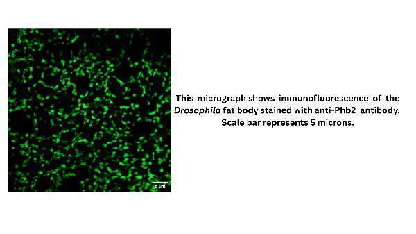
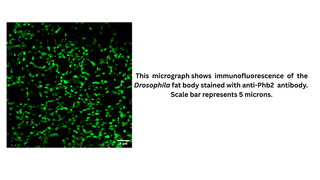
IF analysis of Phb2 using anti-Phb2 antibody (DZ41317).
Phb2 was detected in a paraffin-embedded section of Drosophila fat body tissue. The tissues were fixed with 4% PFA and permeabilized in PBS-TX. And then incubated with 1:100 rabbit anti-Phb2 Antibody (DZ41317) in 2% BSA and 0.1% PBS-TX overnight at 4°C. Anti-Rabbit/Mouse Alexa Fluor 488/Cy3/Cy5 was used as secondary antibody at 1:1000 dilution and incubated for 1 hours at RT. Visualize using Zeiss LSM 900 Confocal Microscope with Airyscan 2 and filter sets appropriate for the label used.

Click image to see more details
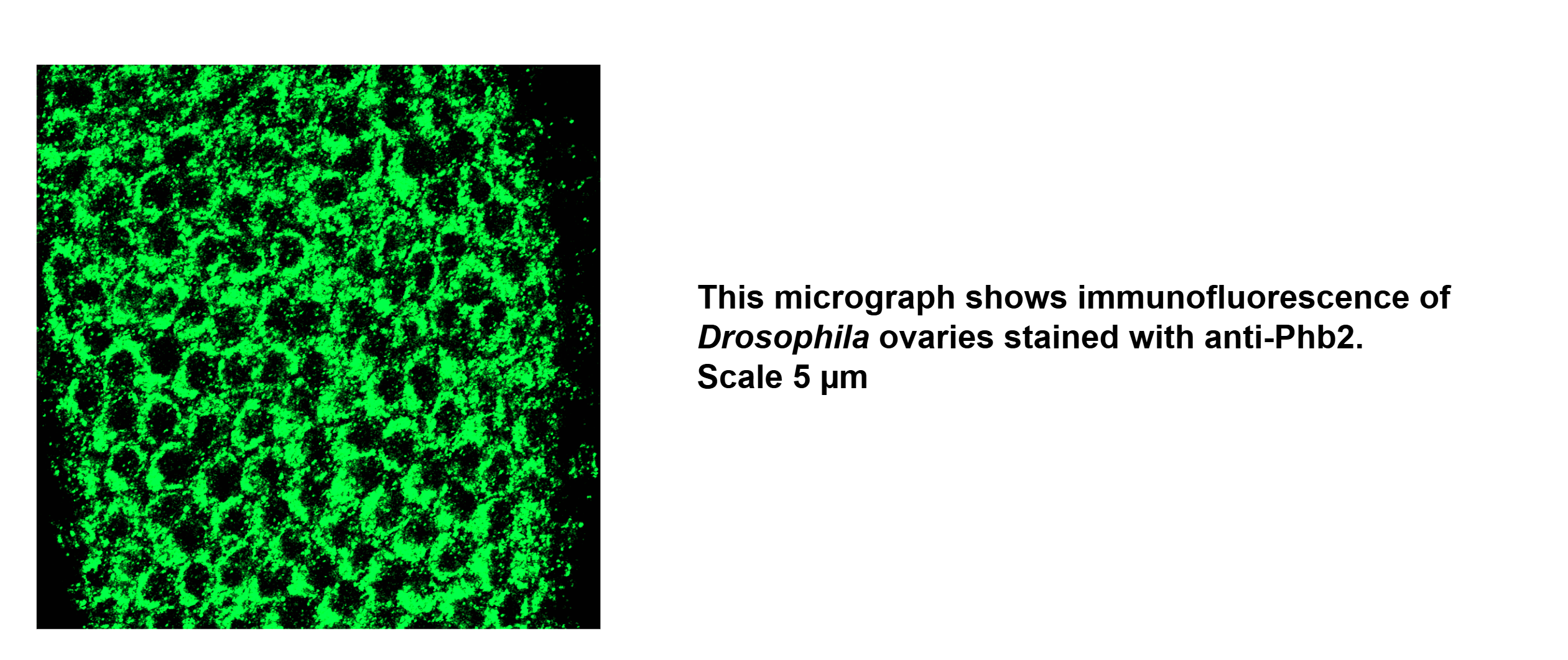
IF analysis of Phb2 using anti-Phb2 antibody (DZ41317).
Phb2 was detected in a paraffin-embedded section of Drosophila ovary tissue. The tissues were fixed with 4% PFA and permeabilized in PBS-TX. And then incubated with 1:100 rabbit anti-Phb2 Antibody (DZ41317) in 2% BSA and 0.1% PBS-TX overnight at 4°C. Anti-Rabbit/Mouse Alexa Fluor 488/Cy3/Cy5 was used as secondary antibody at 1:1000 dilution and incubated for 1 hours at RT. Visualize using Zeiss LSM 900 Confocal Microscope with Airyscan 2 and filter sets appropriate for the label used.

Click image to see more details
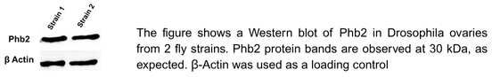
The figure shows a Western blot of Phb2 in Drosophila ovaries from 2 fly strains. Phb2 protein bands are observed at 30 kDa, as expected. β-Actin was used as a loading control.
Specific Publications For Anti-Fruit fly Phb2 Antibody (DZ41317)
Loading publications
Recommended Resources
Here are featured tools and databases that you might find useful.
- Boster's Pathways Library
- Protein Databases
- Bioscience Research Protocol Resources
- Data Processing & Analysis Software
- Photo Editing Software
- Scientific Literature Resources
- Research Paper Management Tools
- Molecular Biology Software
- Primer Design Tools
- Bioinformatics Tools
- Phylogenetic Tree Analysis
Customer Reviews
Have you used Anti-Fruit fly Phb2 Antibody?
Share your experimental results or join a short interview to earn up to $1,000 in product credits or other rewards.
2 Reviews For Anti-Fruit fly Phb2 Antibody
Western blot analysis of Drosophila ovaries from three fly strains showed comparable Phb2 protein levels across all samples. Consistent bands at 30 kDa confirmed expected molecular weight, with β-Actin used as a loading control to verify equal protein loa
Excellent
| SKU | DZ41317 |
|---|---|
| Application | Western Blot |
| Sample | Drosophila Ovaries |
| Sample Processing Description | Dissection, Homogenization, Sample boiling in 1X Lamelli, SDS-PAGE, Standard Western Blotting |
| Primary Antibody | Anti-Fruit fly Phb2 Antibody |
| Primary Incubation | 1:1000 in 5% BSA and 0.1% PBST. Either 2 hours at RT or Overnight at 4 Degrees |
| Secondary Antibody | 1:10000 Anti-rabbit/mouse IgG horseradish peroxidase-conjugated |
| Secondary Incubation | 1 hour at room temperature |
| Detection | Biorad ChemiDoc |
| Results Summary | Western blot analysis of Drosophila ovaries from three fly strains showed comparable Phb2 protein levels across all samples. Consistent bands at 30 kDa confirmed expected molecular weight, with β-Actin used as a loading control to verify equal protein loading. |
Kasturi Mitra
Verified customer
Submitted 2026-01-07
Drosophila fatbody showed good staining of Phb2.
Excellent
| SKU | DZ41317 |
|---|---|
| Application | Immunofluorescence |
| Sample | Drosophila Fat body and Ovaries |
| Sample Processing Description | Dissection, Fixation in 4%PFA, Permeabilization in PBS-TX, Blocking, Primary, Washes,Secondary, Washes,Nuclei Staining, Slide Preparation |
| Primary Antibody | Anti-Fruit fly Phb2 Antibody |
| Primary Incubation | 1:200 in 2%BSA and 0.5% PBS-TX for Ovaries, 1:100 in 2%BSA and 0.1% PBS-TX for Fat body |
| Secondary Antibody | 1:1000 Anti-Rabbit/Mouse Alexa Fluor 488/Cy3/Cy5 |
| Secondary Incubation | 1 hour at room temperature |
| Detection | Zeiss LSM 900 Confocal Microscope with Airyscan 2 |
| Results Summary | Drosophila fatbody showed good staining of Phb2. |
Kasturi Mitra
Verified customer
Submitted 2026-01-07
Customer Q&As
Have a question?
Find answers in Q&As, reviews.
Can't find your answer?
Submit your question
