Click image to see more details
Product Info Summary
| SKU: | A01460-1 |
|---|---|
| Size: | 100 μg/vial |
| Reactive Species: | Human, Rat |
| Host: | Rabbit |
| Application: | ELISA, Flow Cytometry, IP, IHC, WB |
Customers Who Bought This Also Bought

Product info
Product Name
Anti-GALT Antibody Picoband®
SKU/Catalog Number
A01460-1
Size
100 μg/vial
Form
Lyophilized
Description
Boster Bio Anti-GALT Antibody Picoband® catalog # A01460-1. Tested in ELISA, Flow Cytometry, IP, IHC, WB applications. This antibody reacts with Human, Rat. The brand Picoband indicates this is a premium antibody that guarantees superior quality, high affinity, and strong signals with minimal background in Western blot applications. Only our best-performing antibodies are designated as Picoband, ensuring unmatched performance.
Storage & Handling
Store at -20˚C for one year from date of receipt. After reconstitution, at 4˚C for one month. It can also be aliquotted and stored frozen at -20˚C for six months. Avoid repeated freeze-thaw cycles.
Cite This Product
Anti-GALT Antibody Picoband® (Boster Biological Technology, Pleasanton CA, USA, Catalog # A01460-1)
Host
Rabbit
Contents
Each vial contains 4 mg Trehalose, 0.9 mg NaCl and 0.2 mg Na2HPO4.
Clonality
Polyclonal
Isotype
Rabbit IgG
Immunogen
E. coli-derived human GALT recombinant protein (Position: Q188-A379).
Cross-reactivity
No cross-reactivity with other proteins.
Reactive Species
A01460-1 is reactive to GALT in Human, Rat
Observed Molecular Weight
43 kDa
Calculated molecular weight
43.4 kDa
Background of GALT
Galactose-1-phosphate uridyl transferase (GALT) catalyzes the second step of the Leloir pathway of galactose metabolism, namely the conversion of UDP-glucose + galactose-1-phosphate to glucose-1-phosphate + UDP-galactose. The absence of this enzyme results in classic galactosemia in humans and can be fatal in the newborn period if lactose is not removed from the diet. The pathophysiology of galactosemia has not been clearly defined. Two transcript variants encoding different isoforms have been found for this gene.
Antibody Validation
Boster validates all antibodies on WB, IHC, ICC, Immunofluorescence, and ELISA with known positive control and negative samples to ensure specificity and high affinity, including thorough antibody incubations.
Application & Images
Applications
A01460-1 is guaranteed for ELISA, Flow Cytometry, IP, IHC, WB Boster Guarantee
Assay Dilutions Recommendation
The recommendations below provide a starting point for assay optimization. The actual working concentration varies and should be decided by the user.
Western blot, 0.1-0.5μg/ml
Immunohistochemistry(Paraffin-embedded Section), 2-5μg/ml
Immunoprecipitation, 0.5-2 μg/ml, Human
Flow Cytometry(Fixed), 1-3 μg/1x106 cells
ELISA, 0.1-0.5μg/ml
Positive Control
WB: human THP-1 whole cell, human HepG2 whole cell, human Hela whole cell, rat lung tissue, rat kidney tissue
IHC: human colon cancer tissue
FCM: HepG2 cell
IP: THP-1 whole cell
Validation Images & Assay Conditions
Click image to see more details
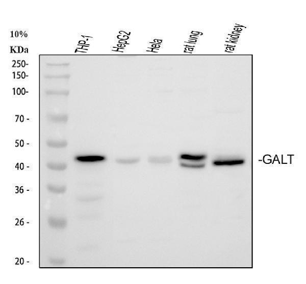
Western blot analysis of GALT using anti-GALT antibody (A01460-1).
Electrophoresis was performed on a 5-20% SDS-PAGE gel at 70V (Stacking gel) / 90V (Resolving gel) for 2-3 hours. The sample well of each lane was loaded with 30 ug of sample under reducing conditions.
Lane 1: human 293T whole cell lysates,
Lane 2: human THP-1 whole cell lysates,
Lane 3: human HepG2 whole cell lysates,
Lane 4: human Hela whole cell lysates,
Lane 5: rat lung tissue lysates,
Lane 6: rat kidney tissue lysates.
After electrophoresis, proteins were transferred to a nitrocellulose membrane at 150 mA for 50-90 minutes. Blocked the membrane with 5% non-fat milk/TBS for 1.5 hour at RT. The membrane was incubated with rabbit anti-GALT antigen affinity purified polyclonal antibody (A01460-1) at 0.5 μg/mL overnight at 4°C, then washed with TBS-0.1%Tween 3 times with 5 minutes each and probed with a goat anti-rabbit IgG-HRP secondary antibody at a dilution of 1:5000 for 1.5 hour at RT. The signal is developed using an Enhanced Chemiluminescent detection (ECL) kit (Catalog # EK1002) with Tanon 5200 system. A specific band was detected for GALT at approximately 43 kDa. The expected band size for GALT is at 43 kDa.

Click image to see more details
IHC analysis of GALT using anti-GALT antibody (A01460-1).
GALT was detected in a paraffin-embedded section of human colon cancer tissue. Heat mediated antigen retrieval was performed in EDTA buffer (pH 8.0, epitope retrieval solution). The tissue section was blocked with 10% goat serum. The tissue section was then incubated with 2 μg/ml rabbit anti-GALT Antibody (A01460-1) overnight at 4°C. Peroxidase Conjugated Goat Anti-rabbit IgG was used as secondary antibody and incubated for 30 minutes at 37°C. The tissue section was developed using HRP Conjugated Rabbit IgG Super Vision Assay Kit (Catalog # SV0002) with DAB as the chromogen.

Click image to see more details
Flow Cytometry analysis of HepG2 cells using anti-GALT antibody (A01460-1).
Overlay histogram showing HepG2 cells stained with A01460-1 (Blue line). To facilitate intracellular staining, cells were fixed with 4% paraformaldehyde and permeabilized with permeabilization buffer. The cells were blocked with 10% normal goat serum. And then incubated with rabbit anti-GALT Antibody (A01460-1, 1 μg/1x106 cells) for 30 min at 20°C. DyLight®488 conjugated goat anti-rabbit IgG (BA1127, 5-10 μg/1x106 cells) was used as secondary antibody for 30 minutes at 20°C. Isotype control antibody (Green line) was rabbit IgG (1 μg/1x106) used under the same conditions. Unlabelled sample without incubation with primary antibody and secondary antibody (Red line) was used as a blank control.

Click image to see more details
Immunoprecipitating GALT in THP-1 whole cell lysate.
Western blot analysis of GALT using anti-GALT antibody (A01460-1);
Lane 1: THP-1 whole cell lysates (30ug);
Lane 2: Rabbit control IgG instead of anti-GALT antibody in THP-1 whole cell lysate;
Lane 3: anti-GALT antibody (2μg) + THP-1 whole cell lysate (500μg).
After electrophoresis, proteins were transferred to a membrane. Then the membrane was incubated with rabbit anti-GALT antigen affinity purified polyclonal antibody (A01460-1) at a dilution of 0.5 μg/mL and probed with a goat anti-rabbit IgG-HRP secondary antibody (Catalog # BA1054). The signal is developed using ECL Plus Western Blotting Substrate (Catalog # AR1196-200). A specific band was detected for GALT at approximately 43 kDa. The expected band size for GALT is at 43 kDa.
Specific Publications For Anti-GALT Antibody Picoband® (A01460-1)
Loading publications
Recommended Resources
Here are featured tools and databases that you might find useful.
- Boster's Pathways Library
- Protein Databases
- Bioscience Research Protocol Resources
- Data Processing & Analysis Software
- Photo Editing Software
- Scientific Literature Resources
- Research Paper Management Tools
- Molecular Biology Software
- Primer Design Tools
- Bioinformatics Tools
- Phylogenetic Tree Analysis
Customer Reviews
Have you used Anti-GALT Antibody Picoband®?
Share your experimental results or join a short interview to earn up to $1,000 in product credits or other rewards.
0 Reviews For Anti-GALT Antibody Picoband®
Customer Q&As
Have a question?
Find answers in Q&As, reviews.
Can't find your answer?
Submit your question
7 Customer Q&As for Anti-GALT Antibody Picoband®
Question
Is this A01460-1 anti-GALT antibody reactive to the isotypes of GALT?
Verified Customer
Verified customer
Asked: 2020-02-06
Answer
The immunogen of A01460-1 anti-GALT antibody is E. coli-derived human GALT recombinant protein (Position: Q188-A379). Could you tell me which isotype you are interested in so I can help see if the immunogen is part of this isotype?
Boster Scientific Support
Answered: 2020-02-06
Question
I see that the anti-GALT antibody A01460-1 works with WB, what is the protocol used to produce the result images on the product page?
Verified Customer
Verified customer
Asked: 2019-09-10
Answer
You can find protocols for WB on the "support/technical resources" section of our navigation menu. If you have any further questions, please send an email to support@bosterbio.com
Boster Scientific Support
Answered: 2019-09-10
Question
Is a blocking peptide available for product anti-GALT antibody (A01460-1)?
Verified Customer
Verified customer
Asked: 2019-07-01
Answer
We do provide the blocking peptide for product anti-GALT antibody (A01460-1). If you would like to place an order for it please contact support@bosterbio.com and make a special request.
Boster Scientific Support
Answered: 2019-07-01
Question
I am looking for to test anti-GALT antibody A01460-1 on mouse right lobe of liver for research purposes, then I may be interested in using anti-GALT antibody A01460-1 for diagnostic purposes as well. Is the antibody suitable for diagnostic purposes?
Verified Customer
Verified customer
Asked: 2019-05-21
Answer
The products we sell, including anti-GALT antibody A01460-1, are only intended for research use. They would not be suitable for use in diagnostic work. If you have the means to develop a product into diagnostic use, and are interested in collaborating with us and develop our product into an IVD product, please contact us for more discussions.
Boster Scientific Support
Answered: 2019-05-21
Question
Will A01460-1 anti-GALT antibody work on parafin embedded sections? If so, which fixation method do you recommend we use (PFA, paraformaldehyde, other)?
Verified Customer
Verified customer
Asked: 2018-07-11
Answer
It shows on the product datasheet, A01460-1 anti-GALT antibody as been validated on WB. It is best to use PFA for fixation because it has better tissue penetration ability. PFA needs to be prepared fresh before use. Long term stored PFA turns into formalin, as the PFA molecules congregate and become formalin.
Boster Scientific Support
Answered: 2018-07-11
Question
Please see the WB image, lot number and protocol we used for right lobe of liver using anti-GALT antibody A01460-1. Please let me know if you require anything else.
Verified Customer
Verified customer
Asked: 2018-01-01
Answer
Thank you very much for the data. Our lab team are working to resolve this as quickly as possible, and we appreciate your patience and understanding! You have provided everything we needed. Please let me know if there is anything you need in the meantime.
Boster Scientific Support
Answered: 2018-01-01
Question
We are currently using anti-GALT antibody A01460-1 for human tissue, and we are satisfied with the ELISA results. The species of reactivity given in the datasheet says human, mouse, rat. Is it true that the antibody can work on zebrafish tissues as well?
C. Johnson
Verified customer
Asked: 2016-09-27
Answer
The anti-GALT antibody (A01460-1) has not been validated for cross reactivity specifically with zebrafish tissues, but there is a good chance of cross reactivity. We have an innovator award program that if you test this antibody and show it works in zebrafish you can get your next antibody for free. Please contact me if I can help you with anything.
Boster Scientific Support
Answered: 2016-09-27
