Click image to see more details
-

-
-
-
-
+2
Product Info Summary
| SKU: | PA1738 |
|---|---|
| Size: | 100 μg/vial |
| Reactive Species: | Human, Mouse, Rat |
| Host: | Rabbit |
| Application: | IHC, WB |
Customers Who Bought This Also Bought

Product info
Product Name
Anti-Granzyme B/GZMB Antibody Picoband®
SKU/Catalog Number
PA1738
BA3719-2 is an alternative SKU for this antibody, used in previous lots.
Size
100 μg/vial
Form
Lyophilized
Description
Boster Bio Anti-Granzyme B/GZMB Antibody catalog # PA1738. Tested in IHC, WB applications. This antibody reacts with Human, Mouse, Rat. The brand Picoband indicates this is a premium antibody that guarantees superior quality, high affinity, and strong signals with minimal background in Western blot applications. Only our best-performing antibodies are designated as Picoband, ensuring unmatched performance.
Storage & Handling
Store at -20˚C for one year from date of receipt. After reconstitution, at 4˚C for one month. It can also be aliquotted and stored frozen at -20˚C for six months. Avoid repeated freeze-thaw cycles.
Cite This Product
Anti-Granzyme B/GZMB Antibody Picoband® (Boster Biological Technology, Pleasanton CA, USA, Catalog # PA1738)
Host
Rabbit
Contents
Each vial contains 4 mg Trehalose, 0.9 mg NaCl and 0.2 mg Na2HPO4.
Clonality
Polyclonal
Isotype
Rabbit IgG
Immunogen
A synthetic peptide corresponding to a sequence at the C-terminus of human Granzyme B.
Cross-reactivity
No cross-reactivity with other proteins
Reactive Species
PA1738 is reactive to GZMB in Human, Mouse, Rat
Observed Molecular Weight
33 kDa
Calculated molecular weight
27.7 kDa
Background of GZMB
Granzyme B is a serine protease that in humans is encoded by the GZMB gene. Granzyme B is expressed by cytotoxic T lymphocytes (CTL) and natural killer (NK) cells. CTL and NK cells share the remarkable ability to recognize specific infected target cells. They are though to protect their host by inducing apoptosis of cells that bear on their surface "nonself" antigens, usually peptides or proteins resulting from infection by intracellular pathogens. The protein encoded by this gene is crucial for the rapid induction of target cell apoptosis by CTL in cell-mediated immune response.
Antibody Validation
Boster validates all antibodies on WB, IHC, ICC, Immunofluorescence, and ELISA with known positive control and negative samples to ensure specificity and high affinity, including thorough antibody incubations.
Application & Images
Applications
PA1738 is guaranteed for IHC, WB Boster Guarantee
Recommend Dilution
| Application | Dilution | Species |
|---|---|---|
| Western blot | 0.1-0.5μg/ml | Human, Mouse, Rat |
| Immunohistochemistry (Paraffin-embedded Section) | 2-5μg/ml | Human, Mouse |
Tested application
Suggested blocking solution with 5% non-fat milk or BSA; (*)Recommended protein loading: 20-40 µg per lane
Use TE buffer pH 9.0 for antigen retrieval; (*) citrate buffer pH 6.0 is an alternative.
Validation Images & Assay Conditions
Click image to see more details
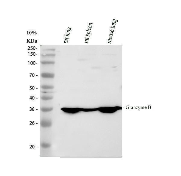
Western blot analysis of GZMB using anti-GZMB antibody (PA1738).
Electrophoresis was performed on a 10% SDS-PAGE gel at 80V (Stacking gel) / 120V (Resolving gel) for 2 hours. The sample well of each lane was loaded with 30 ug of sample under reducing conditions.
Lane 1: rat lung tissue lysates,
Lane 2: rat spleen tissue lysates,
Lane 3: mouse lung tissue lysates.
After electrophoresis, proteins were transferred to a nitrocellulose membrane at 150 mA for 50-90 minutes. Blocked the membrane with 5% non-fat milk/TBS for 1.5 hour at RT. The membrane was incubated with rabbit anti-GZMB antigen affinity purified polyclonal antibody (PA1738) at 0.5 μg/mL overnight at 4°C, then washed with TBS-0.1%Tween 3 times with 5 minutes each and probed with a goat anti-rabbit IgG-HRP secondary antibody at a dilution of 1:5000 for 1.5 hour at RT. The signal is developed using an ECL Plus Western Blotting Substrate (Catalog # AR1196-200) with Tanon 5200 system. A specific band was detected for GZMB at approximately 33 kDa. The expected band size for GZMB is at 27 kDa.

Click image to see more details
Western blot analysis of GZMB using anti-GZMB antibody (PA1738).
Electrophoresis was performed on a 10% SDS-PAGE gel at 80V (Stacking gel) / 120V (Resolving gel) for 2 hours. The sample well of each lane was loaded with 30 ug of sample under reducing conditions.
Lane 1: human NK92 whole cell lysates,
Lane 2: human NK92 whole cell lysates.
After electrophoresis, proteins were transferred to a nitrocellulose membrane at 150 mA for 50-90 minutes. Blocked the membrane with 5% non-fat milk/TBS for 1.5 hour at RT. The membrane was incubated with rabbit anti-GZMB antigen affinity purified polyclonal antibody (PA1738) at 0.5 μg/mL overnight at 4°C, then washed with TBS-0.1%Tween 3 times with 5 minutes each and probed with a goat anti-rabbit IgG-HRP secondary antibody (Catalog # BA1054) at a dilution of 1:5000 for 1.5 hour at RT. The signal is developed using an ECL Plus Western Blotting Substrate (Catalog # AR1196-200) with Tanon 5200 system. A specific band was detected for GZMB at approximately 33 kDa. The expected band size for GZMB is at 27 kDa.

Click image to see more details
IHC analysis of GZMB using anti-GZMB antibody (PA1738).
GZMB was detected in a paraffin-embedded section of human appendix tissue. Heat mediated antigen retrieval was performed in EDTA buffer (pH 8.0, epitope retrieval solution). The tissue section was blocked with 10% goat serum. The tissue section was then incubated with 2 μg/ml rabbit anti-GZMB Antibody (PA1738) overnight at 4°C. Peroxidase Conjugated Goat Anti-rabbit IgG was used as secondary antibody and incubated for 30 minutes at 37°C. The tissue section was developed using HRP Conjugated Rabbit IgG Super Vision Assay Kit (Catalog # SV0002) with DAB as the chromogen.

Click image to see more details
IHC analysis of GZMB using anti-GZMB antibody (PA1738).
GZMB was detected in a paraffin-embedded section of mouse spleen tissue. Heat mediated antigen retrieval was performed in EDTA buffer (pH 8.0, epitope retrieval solution). The tissue section was blocked with 10% goat serum. The tissue section was then incubated with 2 μg/ml rabbit anti-GZMB Antibody (PA1738) overnight at 4°C. Peroxidase Conjugated Goat Anti-rabbit IgG was used as secondary antibody and incubated for 30 minutes at 37°C. The tissue section was developed using HRP Conjugated Rabbit IgG Super Vision Assay Kit (Catalog # SV0002) with DAB as the chromogen.
Specific Publications For Anti-Granzyme B/GZMB Antibody Picoband® (PA1738)
Loading publications
Recommended Resources
Here are featured tools and databases that you might find useful.
- Boster's Pathways Library
- Protein Databases
- Bioscience Research Protocol Resources
- Data Processing & Analysis Software
- Photo Editing Software
- Scientific Literature Resources
- Research Paper Management Tools
- Molecular Biology Software
- Primer Design Tools
- Bioinformatics Tools
- Phylogenetic Tree Analysis
Customer Reviews
Have you used Anti-Granzyme B/GZMB Antibody Picoband®?
Share your experimental results or join a short interview to earn up to $1,000 in product credits or other rewards.
0 Reviews For Anti-Granzyme B/GZMB Antibody Picoband®
Customer Q&As
Have a question?
Find answers in Q&As, reviews.
Can't find your answer?
Submit your question
6 Customer Q&As for Anti-Granzyme B/GZMB Antibody Picoband®
Question
See attached the WB image, lot number and protocol we used for leukocyte using anti-Granzyme B/GZMB antibody PA1738. Please let me know if you require anything else.
A. Krishna
Verified customer
Asked: 2020-04-06
Answer
Thank you very much for the data. Our lab team are working to resolve this as quickly as possible, and we appreciate your patience and understanding! You have provided everything we needed. Please let me know if there is anything you need in the meantime.
Boster Scientific Support
Answered: 2020-04-06
Question
Is there a BSA free version of anti-Granzyme B/GZMB antibody PA1738 available?
Verified Customer
Verified customer
Asked: 2020-03-09
Answer
We appreciate your recent telephone inquiry. I can confirm that some lots of this anti-Granzyme B/GZMB antibody PA1738 are BSA free. For now, these lots are available and we can make a BSA free formula for you free of charge. It will take 3 extra days to prepare. If you require this antibody BSA free again in future, please do not hesitate to contact me and I will be pleased to check which lots we have in stock that are BSA free.
Boster Scientific Support
Answered: 2020-03-09
Question
I see that the anti-Granzyme B/GZMB antibody PA1738 works with WB, what is the protocol used to produce the result images on the product page?
Verified Customer
Verified customer
Asked: 2020-02-24
Answer
You can find protocols for WB on the "support/technical resources" section of our navigation menu. If you have any further questions, please send an email to support@bosterbio.com
Boster Scientific Support
Answered: 2020-02-24
Question
Would PA1738 anti-Granzyme B/GZMB antibody work on parafin embedded sections? If so, which fixation method do you recommend we use (PFA, paraformaldehyde, other)?
Verified Customer
Verified customer
Asked: 2020-01-30
Answer
You can see on the product datasheet, PA1738 anti-Granzyme B/GZMB antibody as been validated on WB. It is best to use PFA for fixation because it has better tissue penetration ability. PFA needs to be prepared fresh before use. Long term stored PFA turns into formalin, as the PFA molecules congregate and become formalin.
Boster Scientific Support
Answered: 2020-01-30
Question
Would anti-Granzyme B/GZMB antibody PA1738 work for WB with leukocyte?
Verified Customer
Verified customer
Asked: 2018-12-31
Answer
According to the expression profile of leukocyte, GZMB is highly expressed in leukocyte. So, it is likely that anti-Granzyme B/GZMB antibody PA1738 will work for WB with leukocyte.
Boster Scientific Support
Answered: 2018-12-31
Question
We are currently using anti-Granzyme B/GZMB antibody PA1738 for human tissue, and we are happy with the WB results. The species of reactivity given in the datasheet says human. Is it true that the antibody can work on primate tissues as well?
B. Singh
Verified customer
Asked: 2017-07-06
Answer
The anti-Granzyme B/GZMB antibody (PA1738) has not been tested for cross reactivity specifically with primate tissues, though there is a good chance of cross reactivity. We have an innovator award program that if you test this antibody and show it works in primate you can get your next antibody for free. Please contact me if I can help you with anything.
Boster Scientific Support
Answered: 2017-07-06
