Click image to see more details
-

-
-
-
-
+1
Product Info Summary
| SKU: | A00506-1 |
|---|---|
| Size: | 100 μg/vial |
| Reactive Species: | Human |
| Host: | Rabbit |
| Application: | IF, IHC, ICC, WB |
Customers Who Bought This Also Bought

Product info
Product Name
Anti-HFE Antibody Picoband®
SKU/Catalog Number
A00506-1
Size
100 μg/vial
Form
Lyophilized
Description
Boster Bio Anti-HFE Antibody Picoband® catalog # A00506-1. Tested in ICC/IF, IHC, WB applications. This antibody reacts with Human. The brand Picoband indicates this is a premium antibody that guarantees superior quality, high affinity, and strong signals with minimal background in Western blot applications. Only our best-performing antibodies are designated as Picoband, ensuring unmatched performance.
Storage & Handling
Store at -20˚C for one year from date of receipt. After reconstitution, at 4˚C for one month. It can also be aliquotted and stored frozen at -20˚C for six months. Avoid repeated freeze-thaw cycles.
Cite This Product
Anti-HFE Antibody Picoband® (Boster Biological Technology, Pleasanton CA, USA, Catalog # A00506-1)
Host
Rabbit
Contents
Each vial contains 4 mg Trehalose, 0.9 mg NaCl and 0.2 mg Na2HPO4.
Clonality
Polyclonal
Isotype
Rabbit IgG
Immunogen
E.coli-derived human HFE recombinant protein (Position: Q82-R199). Human HFE shares 72.2% and 74.6% amino acid (aa) sequence identity with mouse and rat HFE, respectively.
Cross-reactivity
No cross-reactivity with other proteins.
Reactive Species
A00506-1 is reactive to HFE in Human
Observed Molecular Weight
48-50 kDa
Calculated molecular weight
40.1 kDa
Background of HFE
Human hemochromatosis protein also known as the HFE protein is a protein which in humans is encoded by the HFE gene. The HFE gene is located on short arm of chromosome 6 at location 6p21.3. The protein encoded by this gene is a membrane protein that is similar to MHC class I-type proteins and associates with beta2-microglobulin (beta2M). It is thought that this protein functions to regulate iron absorption by regulating the interaction of the transferrin receptor with transferrin. The iron storage disorder, hereditary haemochromatosis, is a recessive genetic disorder that results from defects in this gene.
Antibody Validation
Boster validates all antibodies on WB, IHC, ICC, Immunofluorescence, and ELISA with known positive control and negative samples to ensure specificity and high affinity, including thorough antibody incubations.
Application & Images
Applications
A00506-1 is guaranteed for IF, IHC, ICC, WB Boster Guarantee
Recommend Dilution
| Application | Dilution | Species |
|---|---|---|
| Western blot | 0.1-0.5μg/ml | Human |
| Immunohistochemistry (Paraffin-embedded Section) | 2-5μg/ml | Human |
| Immunocytochemistry/Immunofluorescence | 5 μg/ml | Human |
Tested application
Suggested blocking solution with 5% non-fat milk or BSA; (*)Recommended protein loading: 20-40 µg per lane
Use TE buffer pH 9.0 for antigen retrieval; (*) citrate buffer pH 6.0 is an alternative.
Validation Images & Assay Conditions
Click image to see more details
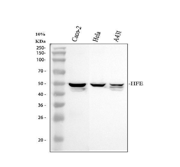
Western blot analysis of HFE using anti-HFE antibody (A00506-1).
Electrophoresis was performed on a 10% SDS-PAGE gel at 80V (Stacking gel) / 120V (Resolving gel) for 2 hours. The sample well of each lane was loaded with 30 ug of sample under reducing conditions.
Lane 1: human CACO-2 whole cell lysates,
Lane 2: human Hela whole cell lysates,
Lane 3: human A431 whole cell lysates.
After electrophoresis, proteins were transferred to a nitrocellulose membrane at 150 mA for 50-90 minutes. Blocked the membrane with 5% non-fat milk/TBS for 1.5 hour at RT. The membrane was incubated with rabbit anti-HFE antigen affinity purified polyclonal antibody (A00506-1) at 0.5 μg/mL overnight at 4°C, then washed with TBS-0.1%Tween 3 times with 5 minutes each and probed with a goat anti-rabbit IgG-HRP secondary antibody at a dilution of 1:5000 for 1.5 hour at RT. The signal is developed using an ECL Plus Western Blotting Substrate (Catalog # AR1196-200) with Tanon 5200 system. A specific band was detected for HFE at approximately 48-50 kDa. The expected band size for HFE is at 40 kDa.

Click image to see more details
IHC analysis of HFE using anti-HFE antibody (A00506-1).
HFE was detected in a paraffin-embedded section of human fallopian tube tissue. Heat mediated antigen retrieval was performed in EDTA buffer (pH 8.0, epitope retrieval solution). The tissue section was blocked with 10% goat serum. The tissue section was then incubated with 2 μg/ml rabbit anti-HFE Antibody (A00506-1) overnight at 4°C. Peroxidase Conjugated Goat Anti-rabbit IgG was used as secondary antibody and incubated for 30 minutes at 37°C. The tissue section was developed using HRP Conjugated Rabbit IgG Super Vision Assay Kit (Catalog # SV0002) with DAB as the chromogen.

Click image to see more details
IHC analysis of HFE using anti-HFE antibody (A00506-1).
HFE was detected in a paraffin-embedded section of human pancrease cancer tissue. Heat mediated antigen retrieval was performed in EDTA buffer (pH 8.0, epitope retrieval solution). The tissue section was blocked with 10% goat serum. The tissue section was then incubated with 2 μg/ml rabbit anti-HFE Antibody (A00506-1) overnight at 4°C. Peroxidase Conjugated Goat Anti-rabbit IgG was used as secondary antibody and incubated for 30 minutes at 37°C. The tissue section was developed using HRP Conjugated Rabbit IgG Super Vision Assay Kit (Catalog # SV0002) with DAB as the chromogen.

Click image to see more details
IF analysis of HFE using anti-HFE antibody (A00506-1).
HFE was detected in an immunocytochemical section of A549 cells. Enzyme antigen retrieval was performed using IHC enzyme antigen retrieval reagent (AR0022) for 15 mins. The cells were blocked with 10% goat serum. And then incubated with 5 μg/mL rabbit anti-HFE Antibody (A00506-1) overnight at 4°C. Cy3 Conjugated Goat Anti-Rabbit IgG (BA1032) was used as secondary antibody at 1:500 dilution and incubated for 30 minutes at 37°C. The section was counterstained with DAPI. Visualize using a fluorescence microscope and filter sets appropriate for the label used.
Specific Publications For Anti-HFE Antibody Picoband® (A00506-1)
Loading publications
Recommended Resources
Here are featured tools and databases that you might find useful.
- Boster's Pathways Library
- Protein Databases
- Bioscience Research Protocol Resources
- Data Processing & Analysis Software
- Photo Editing Software
- Scientific Literature Resources
- Research Paper Management Tools
- Molecular Biology Software
- Primer Design Tools
- Bioinformatics Tools
- Phylogenetic Tree Analysis
Customer Reviews
Have you used Anti-HFE Antibody Picoband®?
Share your experimental results or join a short interview to earn up to $1,000 in product credits or other rewards.
0 Reviews For Anti-HFE Antibody Picoband®
Customer Q&As
Have a question?
Find answers in Q&As, reviews.
Can't find your answer?
Submit your question
4 Customer Q&As for Anti-HFE Antibody Picoband®
Question
Is there a BSA free version of anti-HFE antibody A00506-1 available?
Verified Customer
Verified customer
Asked: 2020-04-17
Answer
We appreciate your recent telephone inquiry. I can confirm that some lots of this anti-HFE antibody A00506-1 are BSA free. For now, these lots are available and we can make a BSA free formula for you free of charge. It will take 3 extra days to prepare. If you require this antibody BSA free again in future, please do not hesitate to contact me and I will be pleased to check which lots we have in stock that are BSA free.
Boster Scientific Support
Answered: 2020-04-17
Question
I have a question about product A00506-1, anti-HFE antibody. I was wondering if it would be possible to conjugate this antibody with biotin. I would need it to be without BSA or sodium azide. I am planning on using a buffer exchange of sodium azide with PBS only. Would there be problems for me to conjugate the antibody and store it in -20 degrees in small aliquots?
Verified Customer
Verified customer
Asked: 2019-02-15
Answer
It is not recommended storing this antibody with PBS buffer only in -20 degrees. If you want to store it in -20 degrees it is best to add some cryoprotectant like glycerol. If you want carrier free A00506-1 anti-HFE antibody, we can provide it to you in a special formula with trehalose and/or glycerol. These molecules will not interfere with conjugation chemistry and provide a good level of protection for the antibody from degradation. Please be sure to specify this in your purchase order.
Boster Scientific Support
Answered: 2019-02-15
Question
Is a blocking peptide available for product anti-HFE antibody (A00506-1)?
Verified Customer
Verified customer
Asked: 2017-11-15
Answer
We do provide the blocking peptide for product anti-HFE antibody (A00506-1). If you would like to place an order for it please contact support@bosterbio.com and make a special request.
Boster Scientific Support
Answered: 2017-11-15
Question
We are currently using anti-HFE antibody A00506-1 for human tissue, and we are satisfied with the WB results. The species of reactivity given in the datasheet says human. Is it true that the antibody can work on horse tissues as well?
W. Patel
Verified customer
Asked: 2015-08-25
Answer
The anti-HFE antibody (A00506-1) has not been validated for cross reactivity specifically with horse tissues, though there is a good chance of cross reactivity. We have an innovator award program that if you test this antibody and show it works in horse you can get your next antibody for free. Please contact me if I can help you with anything.
Boster Scientific Support
Answered: 2015-08-25
