Click image to see more details
Product Info Summary
| SKU: | M01235-2 |
|---|---|
| Size: | 0.1 mg |
| Reactive Species: | Human |
| Host: | Mouse |
| Application: | ELISA, Flow Cytometry, IP, IHC-F, ICC |
Customers Who Bought This Also Bought

Product info
Product Name
Anti-HLA-G Purified Azide Free Monoclonal Antibody
SKU/Catalog Number
M01235-2
Size
0.1 mg
Form
Liquid
Description
Boster Bio Anti-HLA-G Purified Azide Free Monoclonal Antibody (Catalog# M01235-2). Tested in Flow Cytometry, IP, IHC-F, ICC, ELISA application(s). This antibody reacts with Human.
Storage & Handling
Store at 2-8°C. Do not freeze.
Cite This Product
Anti-HLA-G Purified Azide Free Monoclonal Antibody (Boster Biological Technology, Pleasanton CA, USA, Catalog # M01235-2)
Host
Mouse
Contents
Phosphate buffered saline (PBS), pH 7.4, azide-free
Clonality
Monoclonal
Clone Number
MEM-G/9
Isotype
Mouse IgG1
Immunogen
Recombinant human HLA-G refolded with beta2-microglobulin and peptide. The antibody MEM-G/9 reacts with an extracellular epitope on native form of human HLA-G1 on the cell surface as well as with soluble HLA-G5 isoform in its beta2-microglobulin associated form. Reactivity with HLA-G3 was also reported. The antibody MEM-G/9 is standard reagent thoroughly validated during 3rd International Conference on HLA-G (Paris, 2003).
Reactive Species
M01235-2 is reactive to HLA-G in Human
Observed Molecular Weight
42 kDa
Calculated molecular weight
38.2 kDa
Background of HLA-G
Human leukocyte antigen G (HLA-G), belonging to MHC class I glycoproteins, plays important roles in both physiological and pathological immunotolerance. It gives an inhibitory signal to cytotoxic T cells, NK cells, monocytes, and some other immune cells. It also induces regulatory T cells and anti-inflammatory macrophages. HLA-G is important e.g. for maternal tolerance to the fetus, and for immunomodulation in particular adult tissues, such as in cornea, pancreatic islets, thymus and other. On the other hand, it is expressed in many solid and hematologic malignancies, where it contributes to evasion of the immune surveillance. HLA-G expression pattern in cancer is an important prognostic factor regarding a poor clinical outcome. Unlike most other MHC glycoproteins, HLA-G acts as an immune checkpoint molecule rather than as an antigen presenting molecule. It concerns both transmembrane and soluble HLA-G isoforms. Among other, HLA-G can promote Th2 immunological response and downregulate Th1 immunological response. For its benefits regarding allograft tolerance, including embryo implantation, soluble HLA-G (sHLA-G) can be used as a marker of developmental potential of embryos during the process of in vitro fertilization. Similarly, sHLA-G concentrations in maternal serum are decreased in preeclampsia. Transplanted patients with increased sHLA-G serum levels have improved allograft acceptance. On the other hand, increased sHLA-G can also indicate presence of malignant (sometimes also of benign) tumor cells. Another important topic is induction of HLA-G expression (sometimes associated with shedding of HLA-G from the cell surface) by some anti-cancer or anti-viral therapies, which can weaken the therapy effect. Monitoring of HLA-G in patients thus has a wide usage.
Antibody Validation
Boster validates all antibodies on WB, IHC, ICC, Immunofluorescence, and ELISA with known positive control and negative samples to ensure specificity and high affinity, including thorough antibody incubations.
Application & Images
Applications
M01235-2 is guaranteed for ELISA, Flow Cytometry, IP, IHC-F, ICC Boster Guarantee
Recommend Dilution
| Application | Dilution | Species |
|---|---|---|
| ELISA: The antibody MEM-G/9 has been tested as the capture antibody in a sandwich ELISA for analysis of human HLA-G in combination with antibody B2M-01 or with antibody W6/32. Coating antibody 10 μg/ml | detection antibody (biotin or peroxidase conjugate) 1 μg/ml. |
Validation Images & Assay Conditions
Click image to see more details
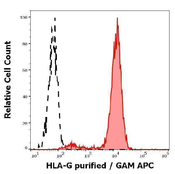
Separation of HLA-G trasnfected LCL cells (red-filled) from K562 cells (black-dashed) in flow cytometry analysis (surface staining) stained using anti-human HLA-G (MEM-G/9) purified antibody (concentration in sample 0.3 µg/ml, GAM APC).

Click image to see more details
Immunoprecipitation of HLA-G from HLA-G1 transfectants (LCL-HLA-G1) by anti-human HLA-G (MEM-G/9) and protein G. HLA-G was detected by anti-human HLA-G (clone 4H84) and goat anti-mouse HRP in cell lysate (Lane 1) and in the immunoprecipitate (Lane 2).
Specific Publications For Anti-HLA-G Purified Azide Free Monoclonal Antibody (M01235-2)
Loading publications
Recommended Resources
Here are featured tools and databases that you might find useful.
- Boster's Pathways Library
- Protein Databases
- Bioscience Research Protocol Resources
- Data Processing & Analysis Software
- Photo Editing Software
- Scientific Literature Resources
- Research Paper Management Tools
- Molecular Biology Software
- Primer Design Tools
- Bioinformatics Tools
- Phylogenetic Tree Analysis
Customer Reviews
Have you used Anti-HLA-G Purified Azide Free Monoclonal Antibody?
Share your experimental results or join a short interview to earn up to $1,000 in product credits or other rewards.
0 Reviews For Anti-HLA-G Purified Azide Free Monoclonal Antibody
Customer Q&As
Have a question?
Find answers in Q&As, reviews.
Can't find your answer?
Submit your question
